Effects of low-frequency repetitive transcranial magnetic stimulation combined with
1
intensive speech therapy on cerebral blood flow in post-stroke aphasia
2 3
Takatoshi Hara,MD;Masahiro Abo, MD, PhD; Kentaro Kobayashi,MD; Motoi Watanabe,
4
PhD; Wataru Kakuda, MD, PhD; Atushi Senoo, PhD
5 6
Department of Rehabilitation Medicine,The Jikei University School of Medicine, 3-25-8,
7
Nishi-Shimbashi, Minato-Ku, Tokyo 105-8461, Japan
8 9
Short title: SPECT study of rTMS and rehabilitation
10 11
Key words: single photon emission computed tomography, repetitive transcranial
12
magnetic stimulation, Aphasia, language therapy, functional magnetic resonance imaging
13 14
Corresponding author:
15
Masahiro Abo, MD, PhD,
16
Department of Rehabilitation, Jikei University School of Medicine,
17
3-25-8, Nishi-Shinbashi, Minato-ku, Tokyo 105-8461, Japan
18
Tel: +81-3-3433-1111
19
Fax: +81-3-5497-4120
20
E-mail: [email protected]
21 22 23 24
25 26
東京慈恵会医科大学
電子署名者 : 東京慈恵会医科大学
DN : cn=東京慈恵会医科大学, o, ou, [email protected], c=JP日付 : 2017.03.17 16:37:37 +09'00'
Abstract
27
We provided an intervention to chronic post-stroke aphasic patients using low frequency
28
repetitive transcranial magnetic stimulation (LF-rTMS) guided by a functional magnetic
29
resonance imaging(fMRI) evaluation of language laterality, combined with intensive speech
30
therapy (ST). We performed a single photon emission computed tomography (SPECT) scan
31
pre- and post-intervention, and investigated the relationship between cerebral blood flow (CBF) and
32
language function. Fifty right-handed chronic post-stroke aphasic patients were enrolled in
33
the study. During their 11-day hospital admission, the patients received a 40-min session of
34
1-Hz LF-rTMS on the left or right hemisphere, according to language localization
35
identified by the fMRI evaluation, and intensive ST daily for 10 days, except for Sunday. A
36
SPECT scan and language evaluation by the Standard Language Test of Aphasia (SLTA) were
37
performed at the time of admission and at 3 months following discharge. We calculated
38
laterality indices (LIs) of regional CBF (rCBF) in 13 language-related
39
Brodmann Area (BA) regions of interest. In patients who received LF-rTMS to the intact
40
right hemisphere (RH-LF-rTMS), the improvement in the total SLTA score was significantly
41
correlated with the pre- and post-intervention change of LI (ΔLI) in BA44. In patients who
42
received LF-rTMS to the lesional left hemisphere (LH-LF-rTMS), this association was not
43
observed. Analyses of the SLTA subscales and rCBF ΔLI demonstrated that in the
44
RH-LF-rTMS group, the SLTA Speaking subscale scores were significantly correlated with ΔLIs in
45
BA11, 20, and 21, and the SLTA Writing subscale scores were significantly correlated with ΔLIs in
46
BA6 and 39. Conversely, in the LH-LF-rTMS group, the SLTA Speaking subscale scores were
47
correlated with ΔLI in BA10, and the SLTA Reading subscale scores were significantly correlated
48
with ΔLIs in BA13, 20, 22, and 44. Our results suggest the possibility that fMRI-guided
49
LF-rTMS combined with intensive ST may affect CBF and contribute to the improvement of
50
language function of post-stroke aphasic patients. LF-rTMS to the non-lesional and lesional
51
hemispheres showed a difference in the associations between language performance and CBF.
52
The results indicate that more effective rTMS intervention needs to be explored for patients
53
who show right hemisphere language activation in an fMRI language evaluation.
54 55 56 57 58 59 60 61 62 63 64 65 66 67 68 69 70 71 72 73 74 75 76 77 78
Introduction
79
Recent studies reported that repetitive transcranial magnetic stimulation (rTMS) improved
80
language function in chronic post-stroke patients with aphasia who sustained an insult to the
81
left hemisphere1,2. These studies postulated that the recovery of language function was due to
82
perilesional compensation in the ipsilateral hemisphere, facilitated by reduced
83
interhemispheric inhibition that resulted from inhibitory low-frequency rTMS (LF-rTMS).
84
However, it is also considered that the contralateral homotopic areas contribute as
85
compensatory regions to the recovery of language function in post-stroke aphasia3-5.
86
Furthermore, in a study using a functional magnetic resonance imaging (fMRI) language task,
87
patients with aphasia demonstrated stronger activation in the right hemisphere relative to
88
healthy control participants6. Therefore, it may be that LF-rTMS to the right hemisphere
89
results in a deterioration of language function in a patient with aphasia whose right
90
hemisphere has already assumeds an important role in language recovery7. On the basis of these
91
studies, we proposed a treatment intervention consisting of fMRI-guided selective LF-rTMS
92
combined with intensive speech therapy (ST). This proposed method utilizes fMRI to identify
93
the language regions of each patient and administers LF-rTMS and intensive ST, to achieve
94
the recovery of activity in these identified regions, based on the principles of
95
interhemispheric inhibition. Our previous study conducted under the same premise found a
96
significant improvement in language function in response to LF-rTMS administered to
97
chronic post-stroke patients with aphasia 8. In addition, previously carried out LF-rTMS Wernicke’s
98
area in fluent aphasia patients and an improvement in the Token Test and subscale scores of the
99
Standard Language Test of Aphasia (SLTA)9.
100
In recent years, the effects of rTMS on cerebral networks in chronic post-stroke patients
101
with aphasia have been reported1,10,11. However, the effects of fMRI-guided selective
102
LF-rTMS combined with intensive ST on language networks of the brain have not been
103
examined fully.
104
Single photon emission computed tomography (SPECT) is an application of scintigraphy
105
that detects gamma rays from a radioisotope delivered into a patient, creating a
106
cross-sectional image of the gamma ray distribution. SPECT imaging of the brain is used
107
widely in the clinical setting as a method to obtain physiological and functional information
108
of the brain. Particularly, in recent years, SPECT imaging has been used widely to evaluate
109
and assess treatment effectiveness in the field of rehabilitation12-14.
110
In this study, we hypothesized that LF-rTMS to the hemisphere contralateral to the language
111
compensation regions identified by fMRI would result in the recovery of language function.
112
We performed language function evaluation and SPECT imaging in chronic post-stroke
113
patients with aphasia, pre- and post-intervention, consisting of LF-rTMS and intensive ST.
114
The purpose of the current study was to investigate the different effects that LF-rTMS may
115
have on cerebral blood flow within the hemispheres with and without a stroke lesion.
116
Furthermore, we investigated the relationship between fMRI activation within the language
117
compensatory regions and improvement in language function resulting from selective
118
LF-rTMS and intensive ST.
119 120
Subjects and Methods
121
Patients and Study Protocol
122
Among the patients with chronic post-stroke aphasia who were admitted to the Tokyo Jikei
123
University Hospital between May 2010 and January 2013, 50 right-handed patients who
124
underwent SPECT scans at the time of admission and at 3 months following discharge were
125
included retrospectively in the current study. None of the patients demonstrated significant
126
language improvement despite receiving outpatient ST for 1–3 months. The clinical
127
backgrounds of these patients are summarized in Table 1. The average age at the time of the
128
intervention was 60.3 years with a standard deviation (SD) of 12.1, ranging from 35 to 82
129
years. Forty patients were men and 10 were women. The stroke subtypes consisted of
130
ischemic in 29 patients, hemorrhagic in 20 patients, and subarachnoid hemorrhage in 1
131
patient. On the basis of the results of the SLTA
132
described in detail below, the patients were categorized into nonfluent or fluent aphasia 15.
133
Twenty-seven patients had nonfluent aphasia, while 23 patients had fluent aphasia. The
134
average duration from the onset of stroke to the intervention was 55.9 months.
135
Twenty-nine patients received LF-rTMS to the right non-lesional hemisphere after the fMRI
136
task identified the left hemisphere as the language compensatory hemisphere (RH-LF-rTMS
137
group). Twenty-one patients received LF-rTMS on the lesional left hemisphere after the
138
fMRI evaluation identified the right hemisphere as the language compensatory hemisphere
139
(LH-LF-rTMS group).
140
The average age of the RH-LF-rTMS group at the time of the intervention was 59.9 years
141
and the group consisted of 22 men and 7 women. Seventeen of these patients had
142
ischemic stroke and 12 had hemorrhagic stroke. Seventeen of these patients had nonfluent
143
aphasia, while 12 had fluent aphasia. The average duration from the onset of stroke to the
144
intervention was 56.2 months.
145
The average age of the LH-LF-rTMS group at the time of the intervention was 60.9 years
146
and the group consisted of 18 men and 3 women. Twelve of these patients had
147
ischemic stroke, 8 had hemorrhagic stroke, and 1 had subarachnoid hemorrhage. Ten of these
148
patients had nonfluent aphasia, while 11 had fluent aphasia. The average duration from the
149
onset of stroke to the intervention was 55.6 months.
150
Patients were excluded if they had alteration of consciousness, neurophysiological signs,
151
neurological symptoms that are considered contraindications to LF-rTMS based on
152
Wasserman’s guidelines16, or evidence of electroencephalographic epileptiform discharges
153
during the duration of the study.
154
Each patient was admitted to the rehabilitation department for 11 days after receiving an
155
outpatient fMRI language evaluation at 1 week prior to admission. The patients received a
156
total of 10 sessions of 40-min 1-Hz LF-rTMS and 60-min intensive ST (i.e., 1 session per day,
157
except for Sunday). During admission, medical and neurological evaluations were conducted
158
before and after each session to monitor adverse effects and worsening of language function.
159
Prior to participation, the attending physician provided a comprehensive explanation of the
160
planned treatment intervention, and written informed consent was obtained from all patients.
161
Furthermore, the current study was conducted following the approval of the Tokyo Jikei
162
University Institutional Review Board.
163 164
Functional Magnetic Resonance Imaging
165
All MRI was performed on a 3T scanner. The fMRI scan was performed using a gradient
166
echo echo-planar sequence (slice thickness = 5 mm, field of view = 240 mm, TR = 5000 ms,
167
TE = 90.5 ms, flip angle = 80°, and matrix = 128 × 128) at 1 week prior to admission. One
168
fMRI run consisted of 12 cycles of 60-s long “repetition” and “rest” periods, and the patient
169
completed 4 runs. During the repetition period, the patient overtly repeated back the words
170
that were played every 5 s through earphones, and the patient’s responses were recorded. If
171
the patient correctly repeated greater than half of the words that were presented, the fMRI
172
data were considered valid. If the patient repeated fewer than half of the words, the fMRI
173
session was repeated. The horizontal and coronal views of a standard T1-weighted image
174
(slice thickness = 2 mm, field of view = 240 mm, TR = 26 ms, TE = 2.4 ms, and matrix = 256
175
168 × 256) were used to register the fMRI image to the structural data in order to localize
176
accurately the activation regions. The fMRI data were analyzed with SPM2 (Wellcome
177
Department of Cognitive Neurology) implemented in MATLAB (MathWorks, Natick, MA,
178
USA), and an alpha of 0.01 was used as a significance threshold for brain activation.
179 180
Evaluation of Language Function
181
Language function was evaluated by the SLTA15. The SLTA is a widely used standardized
182
language test to evaluate the language function of native speakers of the Japanese language.
183
This instrument evaluates various language functions including speaking, reading, naming,
184
repetition, listening, discourse, discourse comprehension, writing, and calculation. In the
185
current study, we evaluated the patients’ language function using 4 of the subscales, i.e.,
186
Speaking, Listening, Reading, and Writing. A total SLTA score below 100 was categorized as
187
severely impaired, 100 - 140 as moderately impaired, and above 140 as mildly impaired.
188
The SLTA was performed at the time of admission and at 3 months following discharge.
189 190
Therapeutic Application of Low-Frequency rTMS
191
LF-rTMS (MagVenture Company, Farum, Denmark) was administered to the patient in a
192
sitting position, using an 8-shaped 70-mm coil and a MagPro R30 stimulator. During
193
admission, each patient received 1 LF-rTMS session every day, except for Sunday, which
194
came to a total of 10 sessions. One session of 1-Hz LF-rTMS lasted for 40 min (2400 total
195
stimulations). Stimulation intensity was at 90% of each individual patient’s motor threshold
196
intensity, with motor threshold intensity defined as the smallest stimulation intensity in the
197
left first dorsal interosseous muscle that could induce a motor evoked potential.
198
In a previous study, this threshold has been shown to be safe according to Wasserman's guidelines16.
199
Prior to each session, this motor threshold intensity was measured by stimulating the primary motor
200
cortex within the right hemisphere. LF-rTMS was performed by the attending physicians, and in the
201
case of adverse events or side effects, the treatment was terminated immediately.
202
The LF-rTMS stimulation site was determined based on the fMRI results and the type of
203
aphasia, as described previously8. Using the fMRI task, we determined the hemisphere that
204
was responsible for compensation of language function, as well as the region that was the
205
most active. Under the aforementioned fMRI scanning conditions, no case exhibited activation of
206
the bilateral cerebral hemispheres, but the activation sites were identified unilaterally on the right
207
or left. Similarly, no case showed multiple active areas.
208
The aphasia types were categorized using the SLTA prior to the intervention.
209
LF-rTMS was administered to the inferior frontal gyrus (IFG) and superior temporal gyrus
210
(STG) for patients with nonfluent and fluent aphasia, respectively. Jennum et al. defined the
211
language areas by the international 10-20 electrode system17 to apply inhibitory LF-rTMS18.
212
In the international 10-20 electrode system, F7/8 and CP5/6 correspond to the IFG and STG,
213
respectively. Therefore, we chose F7/8 as the stimulation target site for the patients with
214
non-fluent aphasia, and CP5/6 for those with fluent aphasia. We adopted the stimulation threshold for
215
them because its efficacy has been proven in previous studies 2,9,26.
216
In the aphasia patients who sustained an insult to the left cerebral cortex due to stroke, the
217
compensatory language region may change over time during the recovery process. Therefore,
218
it is essential to identify accurately the compensatory language regions prior to delivering
219
therapeutic rTMS. We used language task fMRI in order to identify the compensatory
220
language regions that developed in response to the loss of previous language function. We
221
hypothesized that LF-rTMS to the hemisphere contralateral to the identified
222
compensatory language regions combined with concurrent intensive ST should reduce
223
interhemispheric inhibition and facilitate neuronal activity in the compensatory regions,
224
which may result in improved language function. We used LF-rTMS because it has a much
225
lower risk of inducing seizures relative to high-frequency rTMS, and its effects may extend
226
broadly including the hemisphere contralateral to the stimulation site.
227 228
Intensive Speech Therapy
229
A speech therapist provided a one-on-one intensive ST session for 60 min in an individual
230
room. The purpose of this ST was to improve the patient’s expressive modalities including
231
language expression, repetition, naming, and writing. All communication was limited to
232
verbal communication, and communication through gestures and drawing was prohibited.
233
The therapy consisted of 3 main tasks. First, the patient was asked to describe and answer
234
questions about a photograph or a short comic depicting a typical object or situation from
235
everyday life. In addition, the patient was asked to recall the names of objects and scenes
236
presented previously in the photographs and comics. Second, the patient was asked to repeat
237
words and short sentences multiple times that were presented by the therapist. Third, the
238
patient was asked to dictate words and sentences presented by the therapist. During the
239
training, the speech therapist encouraged the patient to make an attempt to work on their
240
communication skills as much as possible. The difficulty level of the training was increased
241
gradually based on the levels of observed improvement of language function during the
242
intensive ST training. During the 3 months following discharge, the patients continued
243
outpatient ST. The skills attained during the intensive ST and their related skills were trained
244
further during this follow-up period. Feedback for attained communication skills was given to
245
the patient on a regular basis in order to reinforce the obtained skills.
246 247
Single Photon Emission Computed Tomography and Laterality Index
248
We used SPECT to measure the regional cerebral blood flow (rCBF) of each patient. SPECT
249
studies were performed at the time of admission and at 3 months following discharge with
250
99mTc-ethyl cysteine dimer (99mTc-ECD) as a tracer. We used a gamma camera with a
251
low-energy high-resolution collimator (MultiSPECT3; Siemens PANA K.K., Tokyo, Japan).
252
SPECT acquisition was performed at 20 min after an intravenous injection of 600 MBq
253
99mTc-ECD while the patient was resting in a supine position with their eyes closed.
254
Attenuation correction of the SPECT images was achieved by Chang’s method19. SPECT
255
acquisition was performed with the following parameters: step-and-shoot acquisition, fan
256
beam collimator, matrix size = 128 × 128, 138 KeV window, 30 s/direction scan time, voxel
257
size = 2.46 mm, Butterworth pre-processing filter (5th order, cutoff frequency 0.3 cycles/cm),
258
and ramp reconstruction filter. Image analyses were carried out by the first author of the
259
current study.
260 261
Statistical Analysis
262
The SPECT images were standardized anatomically and smoothed using SPM5. The count
263
was normalized to the whole brain count, and volume of interest values of the selected
264
regions were calculated20,21. Thirteen language-related Brodmann area (BA) regions (BA8, 9,
265
10, 13, 20, 21, 22, 39, 40, 44, 45, and 46) were selected prior to the analyses. For each of the
266
BA regions, a laterality index (LI; ranging from -1 to +1) was calculated as follows: LI =
267
(lesion side rCBF – non-lesion side rCBF) / (lesion side rCBF + non-lesion side rCBF).
268
Next, using these LIs, the LI change ratio from before to after the intervention
269
(DLI) was calculated. For the denominator, the absolute value of the pre-intervention LI was
270
used. By doing so, a positive DLI would indicate a change toward the lesion hemisphere,
271
while a negative DLI would indicate a laterality change toward the non-lesion hemisphere.
272
For the SLTA total scores and SLTA subscale scores, paired t-tests were performed. In order
273
to investigate associations between the SLTA scores and DLIs, Spearman’s rank correlation
274
coefficients were calculated. These correlation analyses were performed selectively on the 13
275
BA regions associated with aphasia, instead of all BA regions. Therefore, corrections for
276
multiple comparison were not carried out, and an alpha of 0.05 was used21. All statistical
277
analyses were performed with IBM SPSS Statistics 22.
278 279
Results
280
All 50 patients completed the 11-day treatment protocol, and no adverse events were noted
281
during admission. In addition, no adverse events were reported during the 3 months following
282
discharge.
283
Table 2 shows the change of the total SLTA scores over time. The SLTA total mean score
284
improved from 148.8 to 154.7 and 127.0 to 133.6 in
285
the RH-LF-rTMS and LH-LF-rTMS groups, respectively (p < 0.01). When the SLTA
286
subscales were compared, the RH-LF-rTMS group demonstrated a significant improvement
287
in the Speaking (from 59.4 to 61.1 ), Reading (from 34.2 to 34.9), and Writing (25.0 to 26.4 )
288
subscales. The LH-LF-rTMS group
289
demonstrated a significant improvement in the Listening (28.4 to 30.0),
290
Speaking (46.2 to 49.0 ), and Writing (21.0 to 22.6 ) subscales.
291
Correlation analyses between the SLTA total change scores and rCBF DLIs showed that a
292
statistically significant association was found in BA44 in the RH-LF-rTMS group (r = 0.402,
293
p < 0.05, R2 = 0.144, Figure 1). However, the LH-LF-rTMS group did not show any
294
significant association between the SLTA total change scores and rCBF DLIs.
295
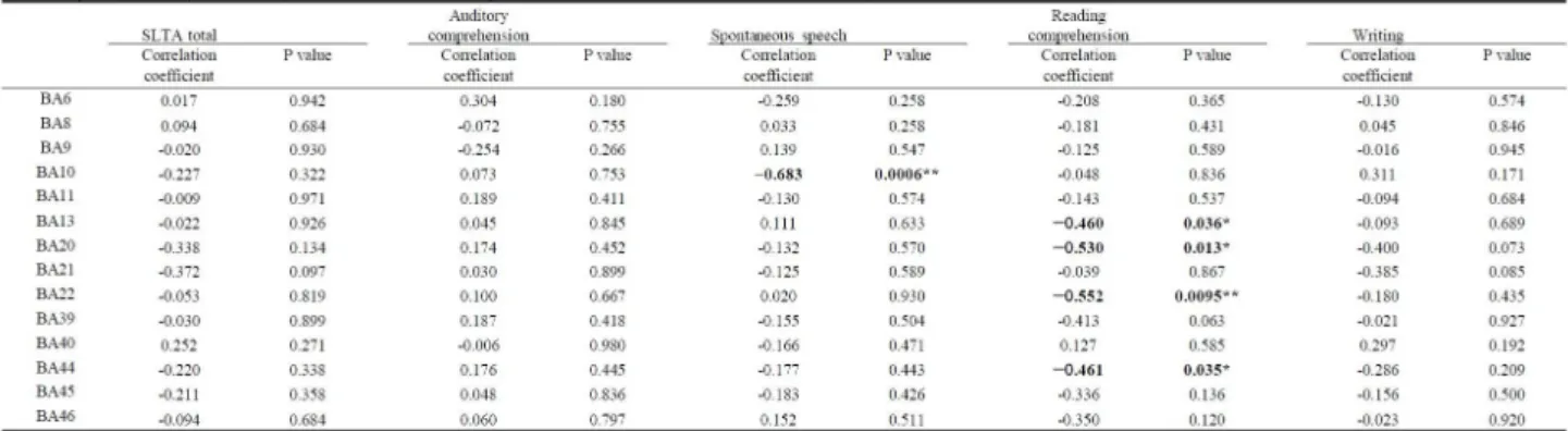
When the SLTA subscale change scores and rCBF DLIs were examined in the
296
RH-LF-rTMS group, statistically significant associations were detected in BA11, 20, and 21
297
for the Speaking subscale (r = 0.456, p < 0.05, R2 = 0.184; r = 0.437, p < 0.05, R2 = 0.112;
298
and r = 0.376, p < 0.05, R2 = 0.089, respectively), and in BA6 and 39 for the Writing subscale
299
(r = 0.574, p < 0.01, R2 = 0.311; and r = 0.384, p < 0.05, R2 = 0.157, respectively). In the
300
LH-LF-rTMS group, significant associations were found in BA10 for the Speaking subscale (r
301
= -0.683, p < 0.01, R2 = 0.353) and in BA13, 20, 22, and 24 for the Reading subscale (r = 291
302
-0.460, p < 0.05, R2 = 0.338; r = -0.530, p < 0.05, R2 = 0.286; r = -0.552, p < 0.01, R2 =
303
0.264; and r = -0.461, p < 0.05, R2 = 0.285, respectively). Tables 3 and 4 show the correlation
304
coefficients between the SLTA scores and rCBF DLIs.
305 306
Discussion
307
Recently, there has been accumulating evidence of the effectiveness of LF-rTMS in patients
308
with post-stroke aphasia1,2,21,23-26. LF-rTMS is often applied to the contralesional homotopic
309
regions based on the principles of reduces interhemispheric inhibition and facilitation of
310
neuronal activity in the compensation regions25. Naeser et al. reported that LF-rTMS to the
311
right hemisphere in patients with post-stroke aphasia led to an improvement of language
312
function23,24. On the basis of their results, the authors speculated that LF-rTMS reduced the
313
interhemispheric inhibition arising from the lesional hemisphere. In addition, Thiel et al.
314
investigated the pre- and post-intervention changes of language activation in response to
315
LF-rTMS to the right pars triangularis in subacute post-stroke patients with aphasia using
316
O-15-water positron emission tomography (PET). The authors demonstrated a significant
317
correlation between the improvement in language performance and changes measured in the
318
PET images. Furthermore, this study also visualized patients’ PET activation regions in
319
comparison to those of healthy controls1.
320
Conversely, other research groups proposed that right hemisphere activity is necessary to
321
compensate for the lost language function observed in chronic post-stroke patients with
322
aphasia8,27-29. For instance, in a review of imaging studies of subacute and chronic post-stroke
323
patients with aphasia, Price et al. discussed one study showing right hemisphere activation
324
was correlated with improved language performance, raising the possibility of a contribution
325
of right hemisphere activation to language recovery in chronic aphasia patients30. Richter et
326
al. reported that greater right hemisphere activation was observed in patients with aphasia
327
than in controls in response to language task fMRI, showing that language performance
328
recovery was associated with a relative reduction in right hemisphere activation, and that
329
changes within the left hemisphere did not correlate with language recovery6. Therefore,
330
these data support a view that right hemispheric activation in chronic post-stroke patients
331
with aphasia may not always be a maladaptive reaction, as proposed by Naeser’s group. In
332
particular, Hamilton et al. pointed out that the degree to which the right hemisphere network
333
contributes to language recovery in post-stroke aphasia may depend on the time course of the
334
injury3. In addition, Heiss and Thiel discussed that the size and location of a lesion within the
335
left hemisphere may determine how the right hemisphere would contribute to language
336
recovery3,4. Heiss and Thiel also suggested that in the case of an insult affecting a broad
337
ipsilateral region, language recovery would have to depend on a very small remaining area in
338
the left hemisphere or on homotopic right hemisphere regions. In such cases, the effects of
339
LF-rTMS to the right hemisphere may remain small. Given these past studies, we utilized an
340
fMRI repetition task to identify the language activation regions in order to guide LF-rTMS
341
intervention8.
342
Thiel’s group calculated LIs of PET activation within both hemispheres in investigations of
343
LF-rTMS in post-stroke patients with aphasia1,26. These studies examined the relationship
344
between pre- and post-intervention LI changes and language recovery. Similarly, we
345
examined pre- and post-intervention changes using SPECT after identifying language-related
346
regions of interest (ROIs), and investigated the change ratios of LIs in each ROI and language
347
performance
348
changes. We believed that this approach would enable us to study the effects of right
349
hemispheric LF-rTMS on the left hemisphere and the effects of left hemispheric LF-rTMS on
350
the right.
351
A SPECT study examining the effects of rTMS on CBF reported a correlation
352
between LI changes in motor regions and upper limb motor function following LF-rTMS to
353
the non-lesional hemisphere of post-stroke patients with upper limb hemiparesis12. We used
354
LIs due to their wide use in investigating changes in neural plasticity and neuromodulation
355
due to LF-rTMS. As the lesions in these patients were extensive and variable, we judged that
356
it was not ideal to subject their whole brain images to group statistical analyses. Therefore,
357
we measured the changes in rCBF LIs by measuring the accumulation of radioisotopes in the
358
language-related regions.
359
Although the number of patients differed between our RH-LF-rTMS and LH-LF-rTMS
360
groups, there was no statistically significant difference between both groups with respect to
361
age and the duration from the onset of aphasia to intervention. However, in terms of the
362
severity of aphasia as measured by the SLTA total scores, a greater proportion of patients
363
were classified as severely impaired in the LH-LF-rTMS group. This may indicate that the
364
insult to the lesional (left) hemisphere was severe, and the reduction of activity in the
365
language areas in the left hemisphere elicited compensatory activity in the non-lesional
366
(right) hemisphere, and that there was no shift of the compensatory regions from the
367
non-lesional (right) hemisphere to the lesional (left) hemisphere3,31.In the current study,
368
we performed LF-rTMS after identifying the language activation areas and observed
369
a significant improvement in language function. Similar to previous PET studies, pre- and
370
post-rTMS LI changes were associated with an improvement in language performance, and
371
the results suggested that right hemisphere LF-rTMS may contribute to language function. It
372
is of note that both the SLTA total and SLTA subscale scores showed associations with LI
373
changes in the language regions in this group. Conversely, in the LH-LF-rTMS group, a
374
significant correlation between LI changes and language performance improvement was
375
limited to the SLTA subscales .
376
This improvement in language function is in line with our previous study
377
demonstrating the effectiveness of LF-rTMS based on the principle of interhemispheric
378
inhibition. In addition, our results suggest that the effects of neuromodulation on language
379
regions via an interhemispheric network may be different between the RH-LF-rTMS and
380
LH-LF-rTMS groups.
381
In the RH-LF-rTMS group, there was an association between the total SLTA score and
382
rCBF ΔLI in BA44 .
383
Left BA44 is part of the dorsal pathway of language that is involved in acoustic speech and is
384
considered to be responsible for articulatory and syntactic processes32,33. A previous PET
385
study on chronic post-stroke aphasia patients found that an increase in rCBF in the left BA44
386
during a repetition task correlated with Western Aphasia Battery scores of spontaneous
387
speech27. Furthermore, a series of studies on language recovery in post-stroke aphasic
388
patients conducted by Naeser’s group suggested that LF-rTMS to the right pars triangularis
389
was effective, and that the pars opercularis was essential in language recovery1,23,24. The
390
results of the current study are in line with these previous studies. With regard to the
391
association between the rCBF ΔLIs of BA11, 20, and 21 and the SLTA Speaking subscale
392
scores, BA11 is connected anatomically
393
via the uncinate fasciculus to the anterior temporal lobe, which is part of the semantic
394
memory network and is involved in lexical retrieval34,35. BA20 is involved in phonological and
395
semantic processing, and BA21 is part of the ventral pathway of language that is responsible
396
for semantic processing and sentence comprehension32,33. In a longitudinal PET study
397
investigating language function in chronic post-stroke aphasia patients during the subacute
398
phase and at 1 year later, left BA20 showed a correlation with improved language
399
performance36. Therefore, these regions are considered to play an essential role in the abilities
400
measured by the SLTA Speaking subscale. With regard to the association between the rCBF
401
ΔLIs and BA6 and 39 and the SLTA Writing subscale scores, BA6 is generally involved in smooth
402
motor programming and motor planning processes, and is a constituent of verbal working
403
memory in language function27,37. In addition, Martin et al. reported fMRI activation in the
404
supplementary motor area (SMA) at the 16th month follow-up of LF-rTMS to the right pars
405
triangularis in chronic post-stroke aphasia patients38. This observation suggests a possibility
406
of neuromodulation within the left SMA during long-term follow-up of rTMS. BA39 is
407
believed to be involved in the auditory short-term memory process that is associated with the
408
“phonological loop,” which consists of a phonological store that is an auditory-motor
409
interface, and the articulatory rehearsal system39. Therefore, the results indicate the
410
possibility that rTMS and intensive rehabilitation resulted in the activation of the left
411
hemisphere regions responsible for executing writing movements by incorporating relevant
412
auditory information, which is an ability associated with what is measured by the SLTA
413
Writing subscale.
414
Conversely, the changes in each of the ROIs and SLTA scores of the LH-LF-rTMS group
415
support the view that our treatment intervention based on the principle of interhemispheric
416
inhibition resulted in the transition of LIs from the originally dominant left hemisphere to the
417
right hemisphere, and this transition was associated with an improvement in language
418
performance. The regions in the right hemisphere that correlated with the SLTA Speaking and
419
Reading subscales are homotopic to the left regions that are responsible for their respective
420
language functions1,33,40. Temporary activation of the homotopic language areas during
421
language recovery and ST has been reported in BA13 and 2241,42. However, the effects of
422
rTMS on these language-related homotopic regions have not been examined fully.
423
In the current study, there was a difference between the RH-LF-rTMS and LH-LF-rTMS
424
groups. With respect to the patients’ clinical background, the LH-LF-rTMS group showed
425
lower mean SLTA scores and greater severity of aphasia relative to the RH-LF-rTMS group.
426
Saur et al. discussed three temporary phases of language recovery that may explain this
427
discrepancy. The authors postulated that patients with an extensive lesion within the left
428
language area may remain at the second phase where the right hemisphere compensates for
429
the lost abilities and may not proceed to the third phase where reactivation of the lesional
430
hemisphere occurs5. An extensive lesion within the left hemisphere would limit compensation
431
by the perilesional areas, and it is possible that activation in the perilesional areas does not
432
occur in response to the fMRI repetition task3. There are two hypotheses
433
regarding the mechanism of the effects of LH-LF-rTMS. The first is that LF-rTMS
434
strengthens right hemisphere compensatory activation through interhemispheric networks.
435
The second is the possibility that LF-rTMS to the left hemisphere prohibits the activation of
436
perilesional regions that would have occurred otherwise. Differential mechanisms of left and
437
right hemisphere LF-rTMS are suggested, and future investigations are warranted.
438
We chose the rTMS stimulation sites based on the past literature, but the regions where CBF
439
LI changes and language performance improvements were observed were broader than the
440
regions to which rTMS was applied. LF-rTMS to chronic post-stroke patients with
441
upper limb paralysis reportedly not only modulated neural connectivity within the hemisphere
442
to which rTMS was applied but also affected distant brain regions9. In addition, it is suggested that
443
the effects observed following rTMS are not so much due to excitation of individual motor
444
regions than they are due to the remodeling of cerebral networks10.
445
It is plausible that similar effects of rTMS on cerebral networks are observed in
446
post-stroke aphasic patients, but future studies are needed to investigate this.
447
The first limitation of the current study is that it was not a randomized controlled trial.
448
Ideally, the current protocol should be compared with conventional ST intervention. However,
449
based on the number of cases, we judged that it would be difficult to conduct 2 sessions of
450
SPECT imaging on patients who were receiving conventional ST; therefore, we did not
451
include them as a comparison. Second, we did not observe an association between the SLTA
452
total improvement and rCBF LI changes in the LH-LF-rTMS group.
453
Recently, one study reported utilizing dual-hemisphere rTMS for subacute
454
post-stroke aphasia43. Khedr et al. discussed that the effects of dual-hemisphere rTMS
455
on patients with complete middle cerebral artery occlusion were not sufficient
456
and that high-frequency rTMS to the right hemisphere may be necessary.
457
Thirdly, we carried out no fMRI after the intervention. Since Thiel et al. had proven the utility of
458
magnetic stimulation therapy for aphasia by PET, we first tried verification by SPECT1,26. We are
459
planning to measure the change in activation before and after the intervention by fMRI.
460
Future studies are needed to investigate whether rTMS should aim to
461
increase the activation of the right hemisphere or to shift language activation to the left
462
hemisphere for those patients whose unaffected (right) hemisphere has significant activation.
463
The method that we used, has not been studied sufficiently. In particular, the number of chronic
464
post-stroke aphasia cases to whom LF-rTMS was applied to the left hemisphere was small and
465
additional studies are warranted.
466
In summary, Our results suggest the possibility that fMRI-guided LF-rTMS combined with
467
intensive ST may affect CBF and contribute to the improvement of language function in
468
post-stroke aphasic patients. LF-rTMS to the non-lesional and lesional hemispheres showed a
469
difference in the associations between language performance and CBF. The results indicate
470
that more effective rTMS intervention needs to be explored for patients who show right
471
hemisphere language activation in an fMRI language evaluation.
472 473
Acknowledgments
474
The authors gratefully acknowledge the participation of the patients in the study. This work
475
was supported by the Grant-in-Aid for Scientific Research from the Japan Society for the
476
Promotion of Science.
477
Conflict of Interest
478
Takatoshi Hara, Masahiro Abo, MD, Kentaro Kobayashi, Motoi Watanabe, Wataru Kakuda
479
and Atushi Senoo declare that they have no conflict of interest.
480
Compliance with Ethics Requirements
481
All procedures followed were in accordance with the ethical standards of the responsible
482
committee on human experimentation (institutional and national) and with the Helsinki
483
Declaration of 1975, as revised in 2008. Informed consent was obtained from all patients for
484
being included in the study.
485 486
References
487
1. Thiel A, Hartmann A, Rubi-Fessen I, et al. Effects of noninvasive brain stimulation
488
on language networks and recovery in early poststroke aphasia. Stroke 2013;44:2240-6. doi:
489
10.1161/STROKEAHA.111.000574. Epub 2013 Jun 27.
490
2. Naeser MA, Martin PI, Theoret H, et al. TMS suppression of right pars triangularis,
491
but not pars opercularis, improves naming in aphasia. Brain Lang 2011;119:206-13. doi:
492
10.1016/j.bandl.2011.07.005. Epub 2011 Aug 23.
493
3. Hamilton RH, Chrysikou EG, Coslett B. Mechanisms of aphasia recovery after
494
stroke and the role of noninvasive brain stimulation. Brain Lang 2011;118:40-50. doi:
495
10.1016/j.bandl.2011.02.005. Epub 2011 Apr 2.
496
4. Heiss WD, Thiel A. A proposed regional hierarchy in recovery of post-stroke
497
aphasia. Brain Lang 2006;98:118-23.
498
5. Saur D, Lange R, Baumgaertner A, et al. Dynamics of language reorganization after
499
stroke. Brain 2006;129:1371-84.
500
6. Richter M, Miltner WH, Straube T. Association between therapy outcome and
501
right-hemispheric activation in chronic aphasia. Brain 2008;131:1391-401.
502
7. Finger S, Buckner RL, Buckingham H. Does the right hemisphere take over after
503
damage to Broca's area? the Barlow case of 1877 and its history. Brain Lang 2003;85:385-95.
504
8. Abo M, Kakuda W, Watanabe M, Morooka A, Kawakami K, Senoo A.
505
Effectiveness of low-frequency rTMS and intensive speech therapy in poststroke patients
506
with aphasia: a pilot study based on evaluation by fMRI in relation to type of aphasia. Eur
507
Neurol 2012;68:199-208. doi: 10.1159/000338773. Epub 2012 Aug 29.
508
9 Kakuda W, Abo M, Uruma G, Kaito N, Watanabe M. Low-frequency rTMS with language
509
therapy over a 3-month period for sensory-dominant aphasia: case series of two post-stroke Japanese
510
patients. Brain injury . 2010 ; 24 :1113-7.
511 512 513
10. Grefkes C, Nowak DA, Wang LE, Dafotakis M, Eickhoff SB, Fink GR. Modulating
514
cortical connectivity in stroke patients by rTMS assessed with fMRI and dynamic causal
515
modeling. Neuroimage 2010;50:233-42. doi: 10.1016/j.neuroimage.2009.12.029. Epub 2009
516
Dec 18.
517
11. Grefkes C, Fink GR. Reorganization of cerebral networks after stroke: new insights
518
from neuroimaging with connectivity approaches. Brain 2011;134:1264-76. doi:
519
10.1093/brain/awr033. Epub 2011 Mar 16.
520
12. Hara T, Kakuda W, Kobayashi K, Momosaki R, Niimi M, Abo M. Regional
521
Cerebral Blood Flow (rCBF) after Low-frequency Repetitive Transcranial Magnetic
522
Stimulation (rTMS) Combined with Intensive Occupational Therapy for Upper Limb
523
Hemiplegia after Stroke : A Study using Single Photon Emission Computed Tomography. The
524
Jpn J Rehabil Med 2013;50:36-42.
525
13. Takekawa T, Kakuda W, Uchiyama M, Ikegaya M, Abo M. Brain perfusion and
526
upper limb motor function: a pilot study on the correlation between evolution of asymmetry
527
in cerebral blood flow and improvement in Fugl-Meyer Assessment score after rTMS in
528
chronic post-stroke patients. J Neuroradiol 2014;41:177-83. doi:
529
10.1016/j.neurad.2013.06.006. Epub 2013 Jul 22.
530
14. Kononen M, Kuikka JT, Husso-Saastamoinen M, et al. Increased perfusion in
531
motor areas after constraint-induced movement therapy in chronic stroke: a single-photon
532
emission computerized tomography study. J Cereb Blood Flow Metabo 2005;25:1668-74.
533
15. Hasegawa T, Kishi H, Shigeno K. A Study on Aphasia Rating Scale. A Method for
534
Overall Assessment of SLTA Results. Higher Brain Function Research 1984;4:638-46.
535
16. Wassermann EM. Risk and safety of repetitive transcranial magnetic stimulation:
536
report and suggested guidelines from the International Workshop on the Safety of Repetitive
537
Transcranial Magnetic Stimulation, June 5-7, 1996. Electroencephalogr Clin Neurophysiol
538
1998;108:1-16.
539
17. Homan RW, Herman J, Purdy P. Cerebral location of international 10-20 system
540
electrode placement. Electroencephalogr Clin Neurophysiol 1987;66:376-82.
541
18. Jennum P, Friberg L, Fuglsang-Frederiksen A, Dam M. Speech localization using
542
repetitive transcranial magnetic stimulation. Neurology 1994;44:269-73.
543
19. Ohnishi T, Matsuda H, Hashimoto T, et al. Abnormal regional cerebral blood flow
544
in childhood autism. Brain 2000;123 ( Pt 9):1838-44.
545
20. Friston K, Holmes A, Worsley K, Poline JB, Frith C, Frackowiak R. Statistical
546
parametric maps in functional imaging : a general linear approach. Human brain mapping
547
1995;2:189-210.
548
21. Ashburner J, Friston KJ. Nonlinear spatial normalization using basis
549
functions.Hum Brain Mapp 1999;7:254-66.
550
22. Vandenbroucke JP, von Elm E, Altman DG, et al. Strengthening the Reporting of
551
Observational Studies in Epidemiology (STROBE): explanation and elaboration.
552
Epidemiology 2007;18:805-35.
553
23. Naeser MA, Martin PI, Nicholas M, et al. Improved naming after TMS treatments
554
in a chronic, global aphasia patient--case report. Neurocase 2005;11:182-93.
555
24. Naeser MA, Martin PI, Nicholas M, et al. Improved picture naming in chronic
556
aphasia after TMS to part of right Broca's area: an open-protocol study. Brain
557
Lang 2005;93:95-105.
558
25. Takeuchi N, Chuma T, Matsuo Y, Watanabe I, Ikoma K. Repetitive transcranial
559
magnetic stimulation of contralesional primary motor cortex improves hand function after
560
stroke. Stroke 2005;36:2681-6.
561
26. Weiduschat N, Thiel A, Rubi-Fessen I, et al. Effects of repetitive transcranial
562
magnetic stimulation in aphasic stroke: a randomized controlled pilot study. Stroke
563
2011;42:409-15. doi: 10.1161/STROKEAHA.110.597864. Epub 2010 Dec 16.
564
27. Ohyama M, Senda M, Kitamura S, Ishii K, Mishina M, Terashi A. Role of the
565
nondominant hemisphere and undamaged area during word repetition in poststroke aphasics.
566
A PET activation study. Stroke 1996;27:897-903.
567
28. Abo M, Senoo A, Watanabe S, et al. Language-related brain function during word
568
epetition in post-stroke aphasics. Neuroreport 2004;15:1891-4.
569
29. Abo M, Takao H, Hashimoto K, Suzuki M, Kaito N. Re-organization of language
570
function within the right hemisphere. Eur J Neurol 2007;14:e7-8.
571
30. Price CJ, Crinion J. The latest on functional imaging studies of aphasic stroke. Curr
572
Opin Neurol 2005;18:429-34.
573
31. Berthier ML, Garcia-Casares N, Walsh SF, et al Recovery from post-stroke
574
aphasia: lessons from brain imaging and implications for rehabilitation and biological
575
treatments. Discov Med 2011;12:275-89.
576
32. Saur D, Kreher BW, Schnell S, et al. Ventral and dorsal pathways for language.
577
Proc Natl Acad Sci U S A. 2008;105:18035-40. doi: 10.1073/pnas.0805234105. Epub 2008
578
Nov 12.
579
33. Vigneau M, Beaucousin V, Herve PY, et al. Meta-analyzing left hemisphere
580
language areas: phonology, semantics, and sentence processing.NeuroImage
581
2006;30:1414-32.
582
34. Papagno C. Naming and the role of the uncinate fasciculus in language function.
583
Current neurology and neuroscience reports 2011;11:553-9. doi: 10.1007/s11910-011-0219-6.
584
35. Duffau H, Gatignol P, Moritz-Gasser S, Mandonnet E. Is the left uncinate
585
fasciculus essential for language? A cerebral stimulation study. Journal of neurology
586
2009;256:382-9. doi: 10.1007/s00415-009-0053-9. Epub 2009 Mar 6.
587
36. de Boissezon X, Demonet JF, Puel M, et al. Subcortical aphasia: a longitudinal PET
588
study. Stroke 2005;36:1467-73.
589
37. Paulesu E, Frith CD, Frackowiak RS. The neural correlates of the verbal
590
component of working memory. Nature 1993;362:342-5.
591
38. Martin PI, Naeser MA, Ho M, et al. Overt naming fMRI pre- and post-TMS: Two
592
nonfluent aphasia patients, with and without improved naming post-TMS. Brain and
593
language 2009;111:20-35. doi: 10.1016/j.bandl.2009.07.007. Epub 2009 Aug 19. Erratum in:
594
Brain Lang. 2010 Feb;112(2):135. Alonso, Miguel [added].
595
39. Dronkers NF, Wilkins DP, Van Valin RD, Jr., Redfern BB, Jaeger JJ. Lesion
596
analysis of the brain areas involved in language comprehension. Cognition 2004;92:145-77.
597
40. Eckert MA, Menon V, Walczak A, et al. At the heart of the ventral attention system:
598
the right anterior insula. Human brain mapping 2009;30:2530-41. doi: 10.1002/hbm.20688.
599
41. Raboyeau G, De Boissezon X, Marie N, et al. Right hemisphere activation in
600
recovery from aphasia: lesion effect or function recruitment? Neurology 2008;70:290-8. doi:
601
10.1212/01.wnl.0000287115.85956.87.
602
42. Specht K, Zahn R, Willmes K, et al. Joint independent component analysis of
603
structural and functional images reveals complex patterns of functional reorganisation in
604
stroke aphasia. NeuroImage 2009;47:2057-63. doi: 10.1016/j.neuroimage.2009.06.011. Epub
605
2009 Jun 11.
606
43. Khedr EM, Abo El-Fetoh N, Ali AM, et al. Dual-hemisphere repetitive transcranial
607
magnetic stimulation for rehabilitation of poststroke aphasia: a randomized, double-blind
608
clinical trial. Neurorehabilitation and neural repair 2014;28:740-50. doi:
609
10.1177/1545968314521009. Epub 2014 Feb 6.
610 611
Figure Legends
612
Fig. 1. BA44: rCBF
DLIs vs. SLTA total change in the RH-LF-rTMS group
613
:In the RH-LF-rTMS group, the increase in SLTA total change scores was positively correlated with
614
a increase in rCBF
DLIs in BA44 (r = 0.402, p < 0.05, R2 = 0.144). A positive DLI indicates a change
615
toward the lesion hemisphere. This suggests that there is relationship between the improvement in
616
language function and rCBF
DLIs toward the lesion hemisphere. The straight line indicates
617
regression. The curved lines indicate the 95% confidence limit.
618 619
620 621
622 623 624 625
626 627
628 629 630 631
632 633