1
Biochimica et Biophysica Acta – proteins and proteomics Regular Paper
Title: Refolding, characterization and crystal structure of (S)-malate dehydrogenase from the hyperthermophilic archaeon Aeropyrum pernix
Authors: Ryushi Kawakamia, Haruhiko Sakurabab, Shuichiro Godac, Hideaki Tsuged, Toshihisa Ohshimae,∗
aAnalytical Research Center for Experimental Sciences, Saga University, 1
Honjo-machi, Saga 840-8502, Japan
bDepartment of Applied Biological Science, Faculty of Agriculture, The University of
Kagawa, 2392 Ikenobe Miki-cho Kita-gun, Kagawa 761-0795, Japan
cDepartment of Applied Chemistry, Faculty of Engineering, Nagasaki University, 1-14
Bunkyo-machi, Nagasaki 852-8521, Japan
dInstitute for Health Science, Tokushima Bunri University, Yamashiro-cho, Tokushima
770-8514, Japan
eMicrobial Genetics Division, Institute of Genetic Resources, Faculty of Agriculture,
Kyushu University, 6-10-1 Hakozaki, Higashi-ku, Fukuoka 812-8581, Japan
Footnote: ∗Corresponding author. Tel: +81-92-642-3053; Fax: +81-92-642-3059; E-mail: [email protected]
Key words: Aeropyrum pernix; [LDH-like] MDH; Malate dehydrogenase;
2 Abstract
Tartrate oxidation activity was found in the crude extract of an aerobic hyperthermophilic archaeon Aeropyrum pernix and the enzyme was identified as (S)-malate dehydrogenase (MDH). The MDH produced in Escherichia coli was mainly obtained as an inactive inclusion body. The inclusion body was dissolved in 6 M guanidine-HCl and successively refolded to the active enzyme through dilution of the denaturant. The purified recombinant enzyme consisted of four identical subunits with a molecular mass of about 110 kDa. NADP was preferred as a coenzyme over NAD for (S)-malate oxidation and, unlike MDHs from other sources, this enzyme readily catalyzed the oxidation of (2S,3S)-tartrate and (2S,3R)-tartrate. The tartrate oxidation activity was also observed in MDHs from the hyperthermophilic archaea
Methanocaldococcus jannaschii and Archaeoglobus fulgidus, suggesting that these
hyperthermophilic MDHs loosely bind to the substrate. The refolded A. pernix MDH was also crystallized, and the structure was determined at a resolution of 2.9 Å. Its overall structure was similar to those of the M. jannaschii, Chloroflexus aurantiacus,
Chlorobium vibrioforme and Cryptosporidium parvum [lactate dehydrogenase-like]
MDHs with root-mean-square-deviation values between 1.4 and 2.1 Å. Consistent with the previous reports, Ala at position 53 was responsible for coenzyme specificity and additionally, the next residue Arg was suggested to be important for NADP binding. Structural comparison revealed that the hyperthermostability of the A. pernix MDH may be attributable to its smaller cavity volume and larger numbers of ion pair and ion-pair networks but the molecular strategy for thermostability may be different for each enzyme.
3 1. Introduction
(S)-Malate dehydrogenases (EC 1.1.1.37, MDHs) catalyze the reversible
conversion between (S)-malate, which known as L-malate, and oxaloacetate with strict substrate specificity in the presence of NAD or NADP. Because these enzymes play crucial roles in the citric acid cycle and malate/aspartate shuttle, the biochemical and structural properties of MDHs from eukaryal, bacterial and archaeal sources have been studied extensively [1-4]. MDHs and lactate dehydrogenases (LDHs) are members in a large family of dehydrogenases, which can be divided into three sub-families [5]: dimeric MDH, tetrameric LDH and [LDH-like] MDH, whose primary and quaternary structures are similar to those of LDHs. Several archaeal MDHs from Sulfolobus
acidocaldarius [6], Thermoplasma acidophilum [7], Haloarcula marismortui [8], Archaeoglobus fulgidus (Af) [9,10],Methanocaldococcus jannaschii (Mj) [10-12] and Pyrobaculum islandicum [13] have been characterized. These enzymes all belong to
the [LDH-like] MDH sub-family and are homotetramers with a subunit molecular mass of 33-37 kDa, except for A. fulgidus MDH, which is a dimer due to the absence of the sequence that mediates the dimer-dimer interaction [10]. Among these enzymes, the crystal structures of those from H. marismortui, A. fulgidus and M. jannaschii have already been determined [14-17].
Aeropyrum pernix is a strict aerobic hyperthermophilic archaeon isolated from a coastal thermal vent in Japan [18]. Its genome was originally sequenced in 1998 [19] and reannotated in 2006 yielding 1,700 open reading frames in the sequence [20]. All enzymes comprising the citric acid cycle, including MDH, are annotated in the genome database, though functional and structural information on these enzymes remains limited. In the beginning of this study, we found an enzyme having strong activity toward (2S, 3S)-tartrate and (2S,3R)-tartrate with NAD(P) and identified it as MDH. Almost all of MDHs have high substrate specificity to (S)-malate, but some of them show the oxidative activity of other substrates. With less rates, MDHs from bovine
4
heart mitochondria, Pseudomonas testosteroni, Moraxella lwoffi and Rhodobacter
capsulatus can oxidize (2S,3R)-tartrate, (2S,3S)-tartrate and/or (S)-2-hydroxyglutarate
[21-24]. In addition, M. jannaschii MDH catalyzes (S)-sulfolactate oxidation with NADP as the electron accepter at the same level as (S)-malate oxidation [12]. Because there have been no previous reports of anMDH having strong catalytic activity toward tartrate, we endeavored to express the gene in Escherichia coli and characterize the enzyme produced. In this paper, we describe the refolding of the A. pernix MDH (ApeMDH) produced as an inclusion body in E. coli and the biochemical properties of the refolded enzyme. In addition, we determined the crystal structure of this enzyme at a resolution of 2.9 Å, analyzed important residues for coenzyme and substrate binding each and compared it with those of other [LDH-like] MDHs to evaluate the structural features responsible for its high thermostability.
2. Materials and Methods
2.1. Screening of dehydrogenase activity from A. pernix
A. pernix was cultivated aerobically for 48 h at 90ºC according to the procedure previously described [25]. The cells were collected by centrifugation (10,000 x g, 10 min), washed with 3% NaCl, suspended in 10 mM phosphate buffer (pH 7.2) and disrupted by sonication. After centrifugation (20,000 x g, 10 min), supernatant was dialyzed against with 10 mM phosphate buffer (pH 7.2) and used as the crude extract. Screening of dehydrogenase activities was performed at 50ºC using microplate reader system (Benchmark, BioRad). Reaction mixture (0.2 ml) containing 50 mM glycine/NaOH buffer (pH 9.0), 10 mM substrate and the crude extract was
pre-incubated for 3 min at 50ºC, the reaction was started by addition of 1 mM NAD(P), and then increase of absorbance at 340 nm was monitored for 30 min.
5
2.2. Gene cloning and expression of MDH from A. pernix
We initially carried out PCR to amplify an ApeMDH gene fragment using the following primer pair: 5’-GGGACAGCATATGATAACAATAC-3’, containing a unique
NdeI restriction site overlapping the 5’ initial codon, and
5’-TGGTTGGATCCTTACTCTCTGAGCTG-3’, containing a unique BamHI
restriction site proximal to the 3’-end of the termination codon. Chromosomal DNA fromA. pernix was isolated as previously described [26] and used as the template.
The amplified 0.9-kb fragment was confirmed from its sequence, digested with NdeI and BamHI and ligated into pET11a expression vector (Novagen) linearized with NdeI and BamHI, yielding pET/ApeMDH.
2.3. Protein expression, refolding and purification of ApeMDH
E. coli BL-21 CodonPlus (DE3)-RIL cells (Stratagene) were transformed with
pET/ApeMDH, after which the transformants were cultivated for 8 h at 37ºC in LB medium supplemented with 50 μg/ml ampicillin. Expression was then induced by adding 1 mM isopropyl-β-D-thiogalacto-pyranoside to the medium, and cultivation was continued for an additional 3 h at 37ºC. The cells were then harvested by
centrifugation (10,000 x g, 10 min), washed with 0.85% NaCl, suspended in 10 mM Tris/HCl (pH 7.5) buffer and lysed by sonication. After sonication, the suspension was centrifuged at 20,000 × g for 10 min at 4 ºC, and the pellet was resuspended in 10 mM Tris/HCl (pH 7.5) buffer containing 1 mM EDTA and 4% Triton X-100, incubated at room temperature for 30 min and centrifuged again. This procedure was repeated twice. The resultant pellet was washed twice with Milli Q water and then solubilized in the 50 mM Tris-HCl (pH 7.5) buffer containing 1 mM EDTA, 0.2 M NaCl and 6 M guanidine-HCl. The solubilized ApeMDH (200 mg of enzyme in 200 ml solution) was added to refolding buffer (1.5 L of 0.1 M Tris-HCl (pH 7.5) containing 2 mM EDTA
6
and 0.4 M L-arginine) and then incubated for 36 h at 4ºC. The resultant enzyme solution (1.7 L) containing the refolded ApeMDH was concentrated to a volume of 50 ml, dialyzed against 50 mM Tris/HCl (pH 7.5) buffer containing 0.2 M NaCl, and then subjected to gel filtration on a Superdex 200 column (2.6 × 60 cm, GE Healthcare) equilibrated with 50 mM Tris-HCl (pH7.5) buffer containing 0.2 M NaCl. The active fractions were pooled and used for biochemical and structural experiments. All solutions used for the refolding procedures were filtered through 0.45-μm membrane filter (Advantec) to remove dust and any other impurities.
2.4. Enzyme assay and determination of protein concentration
MDH activity was determined spectrophotometrically using a Shimadzu UV-1200 spectrophotometer equipped with a thermostat. All assays were performed at 50ºC. The standard reaction mixture (1 ml) consisted of 100 mM Glycine-NaOH (pH 10.0), 50 mM (S)-malate, 1 mM NAD(P) and the enzyme. The appearance of NAD(P)H was monitored from the absorbance at 340 nm (extinction coefficient ε = 6.22 mM-1⋅cm-1). For the kinetic analyses, various concentrations of (S)-malate (0.05 to 2 mM with NAD and 0.02 to 0.5 mM with NADP), (2S,3S)-tartrate (0.5 to 10 mM with NAD and 10 to 200 mM with NADP) and (2S,3R)-tartrate (0.2 to 5 mM with NAD and 2 to 50 mM with NADP) were used in the presence of 4 mM NAD or 1 mM NADP and those of NAD (0.2 to 5 mM) and NADP (0.005 to 0.1 mM) were used in the presence of 2 and 0.5 mM (S)-malate, respectively. The kcat and apparent Km values were estimated from Lineweaver-Burk plots.
Protein concentrations were determined using the Bradford method with bovine serum albumin serving as the standard [27]. The concentration of ApeMDH protein during purification was determined by the absorption at 280 nm (1% absorption coefficient A280 = 6.23 cm-1) calculated from the amino acid sequence of the protein [28].
7
2.5. Electrophoresis and determination of molecular mass
Native-PAGE was carried out on a 7.5% polyacrylamide gel using the method of Davis [29], after which the proteins were stained with Coomassie Brilliant Blue R-250. Active staining was performed at 50ºC using a mixture containing 300 mM Tris/HCl (pH 8.0), 50 mM (S)-malate, 0.1 mM p-iodonitrotetrazorium violet, 0.04 mM phenazine methosulphate and 0.25 mM NAD(P). SDS-PAGE was carried out on a 15%
polyacrylamide gel using the method of Leammli [30] with protein markers from Bio Rad serving as the standards. The molecular mass of the native ApeMDH was determined by Superose 6 gel filtration chromatography using 10 mM potassium phosphate (pH 7.0) and 0.2 M NaCl as the elution buffer. Thyroglobulin (669 kDa), ferritin (440 kDa), aldolase (158 kDa), albumin (67 kDa), chymotrypsinogen A (25 kDa) and ribonuclease A (13.7 kDa) were used as the molecular mass standards (GE Healthcare).
2.6. Crystallization, X-ray data collection, structure determination, refinement and structural analysis
The crystallization conditions were screened using the sitting drop vapor diffusion method with Wizard 1, 2 (Emerald Biosystems) and Crystal Screen 1, 2 (Hampton Research). Drops (2 µl) of protein solution (8.9 mg/ml) were mixed with an equal volume of the reservoir solution and equilibrated against 0.15 ml of reservoir solution at 20ºC. The crystals (ca. 0.1 × 0.2 × 0.4 mm) were grown for 2 weeks in the reservoir solution, which was comprised of 0.1 M CHES (pH9.5) and 40% PEG 600.
The X-ray diffraction data were collected at room temperature on an R-AXIS VII detector system using an in-house rotating copper anode generator (Micromax 007, Rigaku) operating at 40 kV and 20 mA. The crystal belonged to the orthorhombic
8
space group P212121 with the following unit cell parameters: a = 78.17 Å, b = 84.01 Å, c = 216.15 Å, α = β = γ = 90º. A heavy atom derivative was prepared by soaking the crystal in a reservoir solution containing 1mM di-μ-iodobis(ethylendiamine)diplatinum (II) nitrate (PIP). The data were processed using HKL2000 [30]. Native and platinum data sets were used for phase calculation, which was carried out using the single isomorphous replacement with anomalous scattering (SIRAS) method with SOLVE [32]. The SIRAS map at 2.9 Å was subjected to maximum-likelihood
density modification, followed by autotracing using RESOLVE [33]. The initial model was built using Xtalview [34], and refinement to a resolution of 2.9 Å was carried out using Refmac5 [35] and CNS [36]. Then after several cycles of inspection of the 2Fo-Fc and Fo-Fc density maps, the model was rebuilt. The final R and Rfree were calculated to be 20.5 and 24.0 %, respectively. Model geometry was analyzed with PROCHECK [37], and 91.1% of the non-glycine residues were in the most favorable region of the Ramachandran plot and 8.9% were in the additionally allowed region (Table 1). The final coordinates have been deposited in the Protein Data Bank (2D4A).
The Dali server (http://ekhidna.biocenter.helsinki.fi/dali_server/) was used for the structural homology search. Ion pair interactions within the structures were identified using the WHAT IF web server [38] with a cutoff distance of 4.0 Å [39] between
oppositely charged residues. Solvent-accessible surface area (ASA) was defined as the locus of the centre of a probe sphere (representing a solvent molecule) as it rolls over the Van der Waals surface of protein [40] and was calculated using AREAIMOL (ccp4 interface). The overall protein volume and volumes of the cavities within the structure were analyzed using SwissPDBViewer. Molecular graphics figures were created using PyMOL (http://pymol.sourceforge.net/).
9
3.1. Screening of the novel dehydrogenase
We screened the novel dehydrogenases from A. pernix using microplate reader system. About 110 kinds of L- and D-amino acids, organic acids, sugars, alcohols and amines were tested as substrates and the dehydrogenase activity could detected when L-glutamate, (S)-malate, (2S, 3S)-tartrate and (2S, 3R)-tartrate were used. Because L-glutamate and (S)-malate dehydrogenases were well-known enzymes [25], we focused on an enzyme having strong activity toward tartrates. After several chromatographies, we succeeded in purification of the enzyme and identified its encoding gene as APE_0672.1, whose product was predicted to be MDH. MDH activity was also detected with the purified enzyme, indicating that the enzyme is MDH which catalyze the oxidation of tartrate as well as (S)-malate in the presence of NAD(P). Many MDHs so far found have strict substrate specificity to (S)-malate, and in this
study we started characterization of ApeMDH.
3.2. Gene expression and refolding of recombinant ApeMDH
In the E. coli transformants, ApeMDH was present in both the soluble and insoluble fractions, but was found mainly as an inclusion body. The MDH activity in the soluble fraction was unaffected by incubation at 90ºC for 10 min, confirming that APE_0672.1 does indeed encode a hyperthermostable MDH. However, the protein in the soluble fraction migrated as an aggregate in native-PAGE (data not shown). Since this aggregation could not be prevented by changing of cultivation temperature or the conditions under which the cells were disrupted, we decided to carry out refolding of the inclusion body. To accomplish this about 20 g of cells (wet weight) were disrupted by sonication, and the precipitant fraction was washed with buffer containing 4% Triton X-100 as described in Materials and methods. The protein in the washed inclusion body was over 95% pure, as judged from the SDS-PAGE (data not shown), and the
10
amount of the protein solubilized by guanizine-HCl was estimated to be about 200 mg, as judged from the absorbance at 280 nm. The refolded enzyme catalyzed malate dehydrogenation, indicating that the active ApeMDH was successfully recovered from the inclusion body. After further purification by gel chromatography, the mobility of the refolded ApeMDH in native-PAGE was the same as that of the natural ApeMDH (data not shown). With this procedure, about 70 mg of purified ApeMDH were obtained from 2 L of culture.
3.3. Biochemical properties of ApeMDH
The subunit molecular mass of ApeMDH was determined to be about 34 kDa, which coincided with that calculated from the amino acid sequence (33,489 Da). Using gel filtration chromatography, the molecular mass of the native enzyme was determined to be 110 kDa, which indicates the enzyme is a homotetramer, like most other archaeal MDHs (the A. fulgidus enzyme is an exception). When we examined the effect of temperature on the enzyme’s activity, we found that the activity increased as the temperature was increased from 50 to 95 ºC, suggesting that the optimum temperature for activity is above 95ºC. The optimum pH for malate oxidation was determined to be pH 11. The enzyme retained full activity after heating at 100ºC for 10 min, and when heated at 50ºC for 30 min the enzyme showed no loss of activity at pHs between 5.5 and 10.5. Collectively, these findings indicate that the recombinant enzyme was properly refolded and that ApeMDH is a highly stable MDH, like AfMDH and MjMDH [9,11].
We next examined the coenzyme and substrate specificities of ApeMDH. Both NAD and NADP were able to serve as the coenzyme for ApeMDH when (S)-malate was the substrate (Table 2). The coenzyme specificity of several other archaeal MDHs have been reported: AfMDH and P. islandicum MDH are NAD-specific [9,13], whereas MjMDH and S. acidocaldarius MDH have dual coenzyme specificity [6,11]. In this
11
respect, ApeMDH is similar to the latter two enzymes. ApeMDH also catalyzed the oxidation of (2S,3S)-tartrate and (2S,3R)-tartrate using NAD or NADP as the electron acceptor. Notably, the activity toward (2S,3S)-tartrate was three times higher than that toward (S)-malate in the presence of NADP (Table 2). (R)-Malate, (2R,3R)-tartrate and (S)-lactate were inert as substrates. To our knowledge, no other MDHs with strong reactivity toward tartrate have been reported. The MDHs from P. testosteroni, M.
lwoffi and R. capsulatus are known to oxidize (2S,3S)-tartrate and (2S,3R)-tartrate, but
the reaction rates are less than 1% of the (S)-malate oxidation rate [22-24]. Thus, ApeMDH is the first example of an enzyme exhibiting significant reactivity for tartrate oxidation. Furthermore, we examined such reactivity for tertarate oxidation in other archaeal MDH. We constructed the expression systems of AfMDH and MjMDH in E.
coli as described previously [10,11] and partially purified the two enzymes by heat
treatment at 80ºC for 20 min. When we checked the activity for NAD(P)-dependent tartarate dehydrogenation, we found that AfMDH could catalyze both (2S,3S)-tartrate and (2S,3R)-tartrate oxidation, and MjMDH also catalyze (2S,3S)-tartrate oxidation but not (2S,3R)-tartrate. This indicates that at least archaeal MDHs in the [LDH-like] MDH family is not specific to (S)-malate.
We next determined the turnover numbers (kcat) and apparent Km values for each substrate (Table 2). For (S)-malate oxidation, the kcat value (2.6±0.02 sec-1) with NAD was about two times higher than that (1.4±0.04 sec-1) with NADP, but the
apparent Km value (0.12±0.09 mM) for (S)-malate was six times higher in the presence of NAD than with NADP (0.019±0.003 mM). The apparent Km values for NAD and NADP in (S)-malate oxidation were determined to be 0.87±0.05 (kcat: 2.8±0.03 sec-1) and 0.0076±0.0003 mM (kcat: 1.6±0.04 sec-1), respectively, indicating that the enzyme has a higher affinity for NADP than NAD. With NADP, the kcat/Km values for other substrates were quite lower than that for (S)-malate because of the low kcat and high Km values. Thus ApeMDH utilizes (S)-malate and NADP as the preferred substrate and coenzyme, respectively. Consistent with ApeMDH, (S)-malate is the preferred
12
substrate in MjMDH and AfMDH (Table 2). In this study, we found the relative activity for tartrate with A. pernix, M. jannaschii and A. fulgidus MDHs are fairly high, and in particular, the kcat for NADP-dependent (2S,3S)-tartrate oxidation with
ApeMDH and MjMDH were two times higher than those for (S)-malate. By contrast, the kcat/Km values of (2S,3S)-tartrate with the two MDHs were about 1/400 and 1/30 of those for (S)-malate because of much higher Km values for (2S,3S)-tartrate.
Furthermore, we investigated whether these organisms have enzymes related to tartrate metabolism using KEGG database (http://www.genome.jp/) , but could not find any enzyme like oxaloglycolate reductase (decarboxylating, E.C. 1.1.1.92), tartrate dehydrogenase (E.C. 1.1.1.93), meso-tartrate dehydrogenase (E.C. 1.3.1.7), tartrate dehydratase (E.C. 4.2.1.32), tartrate epimerase (E.C. 5.1.2.5) and tartrate epoxidase (E.C. 3.3.2.4). These observations indicate that tartrate oxidation activities by these MDHs are not important biologically. The hyperthermophilic archeal MDHs may exhibit loosely fitted structure for substrate and catalyze the oxidation of tartrate as well as (S)-malate.
3.4. Structure of ApeMDH
We used the molecular replacement method for phase calculation based on the structure of Chloroflexus aurantiacus MDH (PDB ID: 1UXJ) which showed the highest sequence homology with that of ApeMDH, but could not obtain successful data. As the alternative method, SIRAS was used for the determination of the crystal structure of ApeMDH and the structure was refined to a resolution of 2.9 Å (Table 1). The
asymmetric unit consisted of four protein molecules (Fig. 1a) with a solvent content of 53.2%, which corresponds to a Matthew’s coefficient [41] of 2.6 Å3Da-1. In the present model, only subunit D contained all the residues (1-308). Amino acids 79-87 and 308 in subunit A, 79-85 in subunit B, 81-88 and 308 in subunit C were disordered and were not visible in the electron density map. Consequently, the present description
13
of the subunit structure is based on subunit D. Each molecule contained two
domains: an N-terminal dinucleotide binding domain (residues 1-139) with the classical Rossmann fold composed of a parallel six-stranded β-sheet (β1-β6) and four α-helices (α1-α4), and a C-terminal catalytic domain (residues 140-308) with four α-helices (α5-α8) and two antiparallel twisted β-strands (β7 and β8) (Fig. 1b).
When we sent the model for the ApeMDH monomer to the Dali server seeking proteins with a similar structure (as of April 4, 2008), the structure of ApeMDH was found to be quite similar to those of other [LDH-like] MDHs; the
root-mean-square-deviation (RMSD) of the Cα atoms was between 1.4 and 2.2 Å. Unexpectedly, however, ApeMDH showed greater similarity to the eukaryal [LDH-like] MDH from Cryptosporidium parvum (CpMDH; 1.4 Å RMSD with 40% sequence identity; PDB ID: 2hjr) and the bacterial [LDH-like] MDHs from thermophilic
Chloroflexus aurantiacus (CaMDH; 1.5 Å with 39% sequence identity; PDB ID: 1guy)
and mesophilic Chlorobium vibrioforme (CvMDH; 1.4 Å with 39% sequence identity; PDB ID: 1gv1) than to archaeal [LDH-like] MDHs from halophilic H. marismortui (1.9 Å with 32% sequence identity; PDB ID: 1d3a), hyperthermophilic A. fulgidus (1.8 Å with 33% sequence identity; PDB ID: 1ojs) and hyperthermophilic M. jannaschii (2.1 Å with 32% sequence identity; PDB ID: 1hyg). The larger structural similarity of
ApeMDH to Cp, Ca and CvMDHs may be due to the presence of more amino acid insertions and deletions observed among the entire sequences of archaeal MDHs compared with those of Cp, Ca and CvMDHs.
3.5. Coenzyme and substrate binding
Although the present structure was determined with the apo form of the enzyme, structure-based alignment of ApeMDH with the aforementioned MDHs enabled us to compare the amino acid residues involved in coenzyme and substrate binding (Fig. 2). Within the ApeMDH sequence, Ala is present at position 53. This residue is replaced
14
by Gly in MjMDH, and the corresponding residue is replaced by Asp in the other MDHs, which are all NAD-specific. This is to be expected, as Asp or Glu is present at this position in most NAD-specific enzymes to prevent the binding of NADP(H) [42]. It is also consistent with the observation that, like MjMDH [11], ApeMDH shows dual co-enzyme specificity. Beside the substitution at the position 53, Lee et al. have been pointed out that the residues of Ser29B, Arg54, His54B and Ser55 of MjMDH are responsible for binding of NADP [16]. We ascertain that the side chain of Arg54 of ApeMDH was present near the 2’-phosphate group of adenosine ribose moiety of NADP like that of MjMDH (Fig. 3A). In addition, the orientation of side chain hydroxyl group of ApeMDH Thr55 was found to be similar to that of MjMDH Ser55. In the case of other NAD-specific MDHs as shown in Fig. 2, these residues are replaced by non-polar residues like Ile, Val and Ala. This indicates that interactions between these residues, especially Arg54, and the 2’-phosphate group of adenosine ribose moiety of NADP are important to enhance its binding ability. The residues corresponding to Ser29B and His54B of MjMDH were not found in the case of ApeMDH (Fig. 2), suggesting that both the residues are rather less important for NADP binding.
The active site residues [16,43], Arg102, Arg109, Asp168, Arg171 and His195 were completely conserved in ApeMDH (Fig. 2). Among these residues, Arg102 is necessary for binding the dicarboxy substrate and plays a key role in distinguishing the substrate specificities of MDHs and LDHs [43]. In order to analyze how tartrate fit into the active site, we built a substrate binding model of ApeMDH using the structure of human malate-NAD-MDH (PDB ID: 2dfd) (Fig. 3B). The arrangement of four residues (Arg102, Asp168, Arg171 and His195) around malate binding site of the ApeMDH was identical to those of human MDH. Arg109 of ApeMDH were far away 4.3 Å from that of human MDH toward opposite side of malate. This may due to the difference between the apo-form of ApeMDH and the holo-form of human MDH. In spite of the detailed analysis, any significant information as to tartrate binding manner in the substrate binding pocket has not been yet obtained.
15
3.6. Structural features underlying thermostability
We compared the structural features of ApeMDH with those of other tetrameric thermostable MjMDH and CaMDH, and mesophilic CvMDH and CpMDH to evaluate the structural features responsible for its high thermostability. Because the structural features of the H. marismortui MDH likely reflect its resistance to high salt
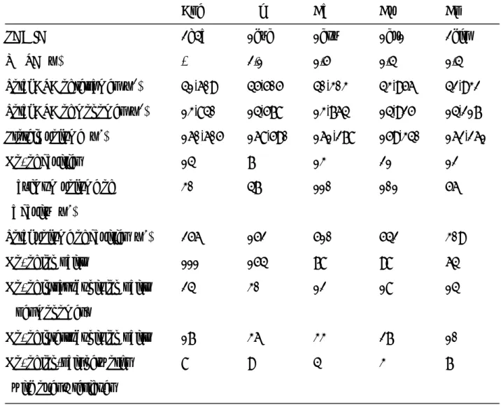
concentrations, and AfMDH forms a dimer, their structures were not included in this analysis. In general, a smaller ASA, a smaller overall volume and a smaller cavity volume each serve to increase the thermostability of a protein [44,45]. Contrary to our expectation, the total ASAs for tetrameric and monomeric ApeMDH were similar to those of CaMDH, CvMDH and CpMDH, as were the volumes of the these enzymes (Table 3) except that these values in MjMDH were larger than these MDHs. By contrast, a large difference between the two hyperthermophilic MDHs and other MDHs was found for the cavity volumes. Although the number of cavities in ApeMDH was not very different from the numbers in CaMDH and CpMDH and somewhat larger than the numbers in MjMDH and CpMDH, the maximum volumes of the ApeMDH and MjMDH cavities were markedly smaller than those of the CaMDH and CvMDH cavities. In addition, the total volume of the cavities for the two hyperthermophilic MDHs was also significantly smaller than those for CaMDH and CvMDH(Table 3). For these hyperthermophilic MDHs, therefore, it appears that a smaller cavity volume makes an important contribution to the overall thermostability.
Recent studies of the structures of hyperthermophilic proteins have shown that the number of ion pairs and the formation of ion-pair networks can also make an important contribution to thermostability [46,47]. Using a cut-off distance of 4.0 Å between oppositely charged residues, we calculated that ApeMDH and MjMDH contain 111 and 154 ion pairs, respectively, whereas CaMDH, CvMDH and CpMDH contain only 78, 98 and 64, respectively. Intrasubunit ion pairs also decreased within these MDHs, but the
16
manner of ion pair formed between subunits was dependent on the kind of MDHs (Table 3). The number of ion-pair networks formed by over four residues was significantly larger in the two hyperthermophilic MDHs than in other moderately thermophilic and mesophilic MDHs. This indicates that the formation of ion pairs undoubtedly strengthens the hyperthermostability of MDHs.
Next, we compared the structural features between the two hyperthermophilic MDHs. Thermal transition between active and inactive MjMDH is reported to start after incubation at 80°C for 15 min [11]. On the other hand, ApeMDH is still stable after incubation even at 100°C for 10 min in this study. This indicates that ApeMDH is more thermostable than MjMDH in spite of different assay conditions. From the comparison of structural features between ApeMDH and MjMDH (Table 3), we noticed that ASAs of tetramer and monomer, protein volume, and maximum volume of a cavity of ApeMDH are rather less than those of MjMDH, respectively. Such differences might be more or less responsible for higher thermostability of ApeMDH, though the total volume of cavities of ApeMDH was larger than that of MjMDH. The total number of ion pairs of ApeMDH was markedly smaller than that of MjMDH, but the number of ion-pair networks of the former enzyme was almost same as that of the latter. In ApeMDH, the most extensively formed ion-pair network (a five-residue network) was observed at the interface between the A-C (or B-D) subunits. With regard to the A-C interface, an ion-pair network was symmetrically observed among:
A-Asp75:C-Arg263:CArg267:C-Glu259:C-Arg43 (Fig. 4). By contrast, no ion-pair interactions could be found in the corresponding region of MjMDH, and the network was mainly formed between the A-B (or C-D) subunits. This indicates that while larger numbers of ion-pair networks may contribute to the thermostability of both hyperthermophilic MDHs, the molecular strategy for increase of thermostability by ion-pair formation may be different for each MDH.
In this study, we determined the functional and structural characteristics of the apo form of ApeMDH. To date, MDHs from various sources have been crystallized,
17
and several structures have been determined in complex with NAD(P)H. Nevertheless, the structural information available on the ternary complex with bound substrate and coenzyme remains very limited [39]. In particular, there is presently no information available on the ternary complex of [LDH-like] MDHs. In this study, we constructed the model of substrate binding to ApeMDH, but could not obtain any informative data about the binding manner of tartrate. The analysis of the ternary structure of
tartarate-NAD(P)- ApeMDH with higher resolution may give us the answer. Our next goal is the structural determination of ApeMDH in complex with a substrate and
coenzyme. We anticipate that this will provide critical information that will further our understanding of the substrate-recognition mechanism of the [LDH-like] MDHs.
Acknowledgement
We are grateful to Prof. Nobuhiko Katunuma for his excellent advice and to Ms. Chizuru Ohtani for her helpful technical assistance. This work was supported in part by the “National Project on Protein Structural and Functional Analysis” prompted by the Ministry of Education, Sciences, Sports, Culture, and Technology of Japan.
18 References
[1] L.J. Banaszak, R.A. Bradshaw, Malate dehydrogenase, in: P.D.E. Boyer (ed.), The enzymes, 3rd ed., Academic Press, New York, 1975, pp. 369-396.
[2] T.K. Sundaram, I.P. Wright, A.E. Wilkinson, Malate dehydrogenase from thermophilic and mesophilic bacteria: Molecular size, subunit structure, amino acid composition, immunochemical homology, and catalytic activity, Biochemistry 19 (1980) 2017-2022.
[3] C.R. Goward, D.J. Nicholls, Malate dehydrogenase: A model for structure, evolution, and catalysis, Prot. Sci. 3 (1994) 1883-1888.
[4] P. Minarik, N. Tomaskova, M. Kollarova, M. Antalik, Malate dehydrogenases- structure and function, Gen. Physiol. Biophys. 21 (2002) 257-65.
[5] D. Madern, Molecular evolution within the L-malate and L-lactate dehydrogenase super-family, J. Mol. Evol. 54 (2002) 825-840.
[6] T. Hartl, W. Grossebuter, H. Gorisch, J.J. Stezowski, Crystalline NAD/NADP dependent-malate dehydrogenase; the enzyme from the thermoacidophilic
archaebacterium Sulfolobus acidocaldarius, Biol. Chem. Hoppe-Seyler 368 (1987) 259-267.
[7] W. Grossebuter, T. Hartl, H. Gorisch, J.J. Stezowski, Purification and properties of malate dehydrogenase from the thermoacidophilic archaebacterium Thermoplasma
acidophilum, Biol. Chem. Hoppe-Seyler 367 (1986) 457-463.
[8] F. Cendrin, J. Chroboczek, G. Zaccai, H. Eisenberg, M. Mevarech, Cloning, sequencing and expression in Escherichia coli of the gene coding for malate dehydrogenase of the extremely halophilic archaebacterium Haloarcula
marismortui, Biochemistry 32 (1993) 4308-4313.
[9] A.S. Langelandsvik, I.H. Steen, N.K. Birkeland, T. Lien, Properties and primary structure of a thermostable L-malate dehydrogenase from Archaeoglobus fulgidus, Arch Microbiol. 168 (1997) 59-67.
19
[10] D. Madern, C. Ebel, H.A. Dale, T. Lien, I.H. Steen, N.K. Birkeland, G. Zaccai, Differences in the oligomeric states of the LDH-like L-MalDH from the
hyperthermophilic archaea Methanococcus jannaschii and Archaeoglobus fulgidus, Biochemistry 40 (2001) 10310-10316.
[11] D. Madern, The putative L-lactate dehydrogenase from Methanococcus jannaschii is an NADPH-dependent L-malate dehydrogenase, Mol. Microbiol. 37 (2000) 1515-1520.
[12] M. Graupner, H. Xu, R.H. White, Identification of an archaeal 2-hydroxy acid dehydrogenase catalyzing reactions involved in coenzyme biosynthesis in methanoarchaea. J. Bacteriol. 182 (2000) 3688-3692.
[13] L.J. Yennaco, Y. Hu, J.F. Holden, Characterization of malate dehydrogenase from the hyperthermophilic archaeon Pyrobaculum islandicum, Extremophiles 11 (2007) 741-746.
[14] S.B. Richard, D. Madern, E. Garcin, and G. Zaccai, Halophilic adaptation: novel solvent protein interactions observed in the 2.9 and 2.6 Å resolution structures of the wild type and a mutant of malate dehydrogenase from Haloarcula marismortui, Biochemistry 39 (2000) 992-1000.
[15] D. Madern, C. Ebel, M. Mevarech, S.B. Richard, C. Pfister, G. Zaccai, Insights into the molecular relationships between malate and lactate dehydrogenases: structural and biochemical properties of monomeric and dimeric intermediates of a mutant of tetrameric L-[LDH-like] malate dehydrogenase from the halophilic archaeon Haloarcula marismortui, Biochemistry 39 (2000) 1001-1010.
[16] B.I. Lee, C. Chang, S.J. Cho, S.H. Eom, K.K. Kim, Y.G. Yu, S.W. Suh, Crystal structure of the MJ0490 gene product of the hyperthermophilic archaebacterium
Methanococcus jannaschii, a novel member of the lactate/malate family of
dehydrogenases, J. Mol. Biol. 307 (2001) 1351-1362.
[17] A. Irimia, F.M. Vellieux, D. Madern, G. Zaccai, A. Karshikoff, G. Tibbelin, R. Ladenstein, T. Lien, N.K. Birkeland, The 2.9 Å resolution crystal structure of
20
malate dehydrogenase from Archaeoglobus fulgidus: mechanisms of
oligomerisation and thermal stabilization, J. Mol. Biol. 335 (2004) 343-356. [18] Y. Sako, N. Nomura, A. Uchida, Y. Ishida, H. Morii, Y. Koga, T. Hoaki, T.
Maruyama, Aeropyrum pernix gen. nov., sp. nov., a novel aerobic
hyperthermophilic archaeon growing at temperatures up to 100 degrees C, Int. J. Syst. Bacteriol. 46 (1996) 1070-1077.
[19] Y. Kawarabayasi, Y. Hino, H. Horikawa, S. Yamazaki, Y. Haikawa, K. Jin-no, M. Takahashi, M. Sekine, S. Baba, H. Kosugi, A. Ankai, H. Kosugi, A. Hosoyama, S. Hukui, Y. Nagai, K. Nishijima, H. Nakazawa, M. Takamiya, S. Masuda, T.
Funahashi, T. Tanaka, Y. Kudoh, J. Yamazaki, N. Kushida, A. Oguchi, K. Aoki, K. Kubota, Y. Nakamura, Y. Nomura, Y. Sako, H. Kikuchi, Complete genome
sequence of an aerobic hyper-thermophilic crenarchaeon, Aeropyrum pernix K1, DNA Res. 6 (1999) 83-101.
[20] S. Yamazaki, J. Yamazaki, K. Nishijima, R. Otsuka, M. Mise, H. Ishikawa, K. Sasaki, S. Tago, K. Isono, Proteome analysis of an aerobic hyperthermophilic crenarchaeon, Aeropyrum pernix K1, Mol. Cell. Proteomics. 5 (2006) 811-823. [21] D.D. Davies, E. Kun, Isolation and properties of malic dehydrogenase from
ox-heart mitochondria, Biochem. J. 66 (1957) 307-316.
[22] K.S. You, N.O. Kaplan, Purification and properties of malate dehydrogenase from
Pseudomonas testosterone, J. Bacteriol. 123 (1975) 704-716.
[23] R. Fernandes, M. Jones, H.K. King, Purification and properties of malate-NAD+ dehydrogenase of Moraxella lwoffi (N.C.I.B. 8250) Biochem. Soc. Trans. 4 (1976) 1080.
[24] T. Ohshima, H. Sakuraba, Purification and characterization of malate
dehydrogenase from the phototrophic bacterium, Rhodopseudomonas capslata, Biochim. Biophys. Acta 869 (1986) 171-177.
[25] M.W. Bhuiya, H. Sakuraba, C. Kujo, N. Nunoura-Kominato, Y. Kawarabayasi, H. Kikuchi, T. Ohshima, Glutamate dehydrogenase from the aerobic
21
hyperthermophilic archaeonAeropyrum pernix K1: enzymatic characterization, identification of the encoding gene, and phylogenetic implications, Extremophiles 4 (2000) 333-341.
[26] J. Sambrook, E.F. Fritsch, T. Maniatis, Molecular Cloning. a Laboratory Manual, 2nd ed., Cold Spring Harbor Laboratory Press, New York, 1989.
[27] M.M. Bradford, A rapid and sensitive method for the quantitation of microgram quantities of protein utilizing the principle of protein-dye binding, Anal. Biochem. 72 (1976) 248-254.
[28] C.N. Pace, F. Vajdos, L. Fee, G. Grimsley, T. Gray, How to measure and predict the molar absorption coefficient of a protein, Protein Sci. 4 (1995) 2411-2423.
[29] B.J. Davis, Disc electrophoresis-2. Method and application to human serum proteins. Ann. N.Y. Acad. Sci. 121 (1964) 404-427.
[30] U.K. Leammli, Cleavage of structural proteins during the assembly of the head bacteriophage T4, Nature 227 (1970) 680-685.
[31] Z. Otwinowski, W. Minor, Processing of X-ray diffraction data collected in oscillation mode, Methods Enzymol. 276 (1997) 307-326.
[32] T.C. Terwilliger, J. Berendzen, Automated MAD and MIR structure solution, Acta Crystallogr. D 55 (1999) 849-861.
[33] T. Terwilliger, SOLVE and RESOLVE: automated structure solution, density modification and model building, J. Synchrotron. Radiat. 11 (2004) 49-52. [34] D.E. McRee, XtalView/Xfit: a versatile program for manipulating atomic
coordinates and electron density, J. Struct. Biol. 125 (1999) 156-165. [35] G.N. Murshudov, A.A. Vagin, E.J. Dodson, Refinement of macromolecular
structures by the maximum-likelihood method, Acta Crystallogr. D 53 (1997) 240-255.
[36] A.T. Brünger, P.D. Adams, G.M. Clore, W.L. DeLano, P. Gros, R.W.
Grosse-Kunstleve, J.S. Jiang, J. Kuszewski, M. Nilges, N.S. Pannu, R.J. Read, L.M. Rice, T. Simonson, G.L. and Warren, Crystallography and NMR system: a
22
new software suite for macromolecular structure determination, Acta Crystallogr. D 54 (1998) 905-921.
[37] R.A. Laskowski, M.W. MacArthur, D.S. Moss, J.M. Thornton, PROCHECK: a program to check the stereochemical quality of protein structures, J. Appl. Cryst. 26 (1993) 283-291.
[38] R. Rodriguez, G. Chinea, N. Lopez, T. Pons, G. Vriend, Homology modeling, model and software evaluation: three related resources, Bioinformatics 14 (1998) 523-528.
[39] D.J. Barlow, J.M. Thornton, Ion-pairs in proteins, J. Mol. Biol. 168 (1983) 867-885.
[40] B. Lee, F.M. Richards, The interpretation of protein structures: estimation of static accessibility, J. Mol. Biol. 55 (1971) 379-400.
[41] B.W. Matthews, Solvent content of protein crystals, J. Mol. Biol. 33 (1968) 491・ 497.
[42] P.J. Baker, K.L. Britton, D.W. Rice, A. Rob, T.J. Stillman, Structural consequences of sequence patterns in the fingerprint region of the nucleotide binding fold. Implications for nucleotide specificity, J. Mol. Biol. 228 (1992) 662–671. [43] A.D.M. Chapman, A. Cortes, T.R. Dafforn, A.R. Clarke, R.L. Brady, Structural
basis of substrate specificity in malate dehydrogenases: crystal structure of a ternary complex of porcine cytoplasmic malate dehydrogenase, α-ketomalonate and tetrahydroNAD, J. Mol. Biol. 285 (1999) 703–712.
[44] M.K. Chan, S. Mukund, A. Kletzin, M.W. Adams, D.C. Rees, Structure of a hyperthermophilic tungstopterin enzyme, aldehyde ferredoxin oxidoreductase, Science 267 (1995) 1463-1469.
[45] B. Dalhus, M. Saarinen, U.H. Sauer, P. Eklund, K. Johansson, A. Karlsson, S. Ramaswamy, A. Bjork, B. Synstad, K. Naterstad, R. Sirevag, H. Eklund, Structural basis for thermophilic protein stability: structures of thermophilic and mesophilic malate dehydrogenases, J. Mol. Biol. 318 (2002) 707-721.
23
[46] K.S. Yip, T.J. Stillman, K.I. Britton, P.J. Artymiuk, P.J. Baker, S.E. Sedelnikova, P.C. Engel, A. Pasquo, R. Chiaraluce, V. Consalvi, The structure of Pyrococcus
furiosus glutamate dehydrogenase reveals a key role for ion-pair networks in
maintaining enzyme stability at extreme temperatures, Structure 3 (1995) 1147-1158.
[47] M. Henning, B. Darimont, R. Sterner ,K. Kirschner, J.N. Jansonius, 2.0 Å structure of indol-3-glycerol phosphate synthase from the hyperthermophile Sulfolobus
solfataricus: possible determinants of protein stability, Science 3 (1995)
1295-1306.
[48] W. Eventoff, M.G. Rossmann, S.S. Taylor, H.-J. Toff, H. Meyer, W. Keil, H.-H. Kiltz, Structural adaptations of lactate dehydrogenase isozymes, Proc. Natl. Acad. Sci. USA 74 (1977) 2677-2681.
24 Figure Legends
Fig. 1 Crystal structure of ApeMDH. (A) Overall structure. The A, B, C and D subunits are shown in green, cyan, magenta and yellow, respectively. (B) Subunit structure of ApeMDH. The rainbow drawing shows the N terminus in blue and the C terminus in red. α-Helices are numbered from H1 to H8,
β-strands are numbered from S1 to S8, and short helices are numbered from G1 to G3.
Fig. 2 Structure-based amino acid sequence alignment of MDHs. MDHs from
Chlorobium vibrioforme, Chloroflexus aurentiacus, Cryptosporidium parvum, Aeropyrum pernix, Archaeoglobus fulgidus, Methanocaldococcus jannaschii and Haloarcula marismortui are represented by Cv, Ca, Cp, Ape, Af, Mj and Hm,
respectively. The sequence of ApeMDH is shown in boldface. The active site residues and the residue involved in coenzyme selectivity are boxed. The secondary structural assignments for ApeMDH are shown above the alignment. β-Strands, α-helices and short helices are labeled as S1-S8, H1-H8 and G1-G3, respectively. To be consistent with most of publications about LDH and [LDH-like] MDH, the residue numbering by Eventoff [48] was shown under the alignment. The residues between 209 and 227 could not be numbered due to disagreement between both structures. Amino acid numbers mentioned in the text were also shown by the same numbering system.
Fig. 3 (A) Stereo view of the structural model for NADP binding. The residues of ApeMDH and MjMDH were shown in green and blue, respectively, and NADP was in yellow. (B) Stereo view of the structural model for substrate binding. The structures of ApeMDH and human MDH were shown in green and blue, respectively. Malate and NADP was in pink stick and yellow line, respectively.
25
Subunits A and C are shown in green and cyan, respectively. Oxygen and nitrogen atoms are shown in red and blue, respectively. The final σA-weighted 2fo-fc electron density map for the residues is shown at the 1 σ level. The ion pair network is depicted with dotted lines.
26
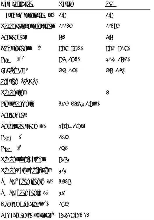
Table 1 X-ray data collection and refinement statistics
Data collection Native PIP Maximum resolution (Å) 2.9 2.9 No. of unique reflections 33305 33095
Redundancy 7.0 6.7 Completeness (%)1 99.8 (98.0) 99.3 (93.8) Rsym (%)1, 2 9.6 (28.0) 13.1 (27.2) <I/Sigma I>1 5.4 (3.5) 5.7 (2.6) Phasing (SIRAS) No. of sites 5 Figure of merit 0.35 (45.6 – 2.9 Å) Refinement Resolution range (Å) 19.9 – 2.9 Å Rcryst (%)3 20.5 Rfree (%)4 24.0
No. of protein atoms 9171 No. of water molecules 121 RMSD5 bond length (Å) 0.007 RMSD bond angle (°) 1.2 Average B-factor (Å2) 38.4 Ramachandran statistics6 91.1/ 8.9/ 0 / 0
1Numbers in parentheses refer to the respective highest resolution data shell in each dataset.
27 2R sym =
( )
( )
( )
∑∑
∑∑
− h i h i h I h I i h I ,, where I ,
( )
h i is the scaled intensity of the ithobservation of reflection h, I
( )
h is the mean value, and the summation is over all measurements. 3R cryst =∑
∑
− l k h obs l k h obs calcF
F
F
k , , , , , whereF
obsandF
calc are the observed and calculatedstructure factors, k is a weighting factor, and the summation is over 95% of the reflections in the working set.
4R free =
∑
∑
− l k h obs l k h obs calcF
F
F
k , , , ,is the summation over 5% of the reflections chosen
randomly.
5RMSD = root mean square deviation.
6Ramachandran statistics indicate the fraction of residues in the most favoured,
additionally allowed, generously allowed and disallowed regions of the Ramachandran diagram as defined by PROCHECK [37].
28
1 mM except that 4 mM NAD was used when the all substrates of ApeMDH and (2S,3S)-tartrate of MjMDH were used. N.D.; not detected.
NAD NADP
Substrate kcat Km kcat /Km kcat Km kcat /Km (sec-1) (mM) (sec-1M-1) (sec-1) (mM) (103 sec-1M-1) (S)-Malate 2.6±0.02 0.12±0.09 22,000 1.4±0.04 0.019±0.003 74,000 ApeMDH (2S,3S)-Tartrate 0.37±0.03 1.2±0.14 310 4.1±0.07 23±0.3 180 (2S,3R)-Tartrate 0.16±0.05 0.2±0.02 800 0.2±0.03 5.8±0.55 34 (S)-Malate 0.33±0.014 0.41±0.06 800 0.069±0.012 0.084±0.012 820 MjMDH (2S,3S)-Tartrate 0.2±0.09 11±0.5 18 0.15±0.006 5.1±0.21 29 (2S,3R)-Tartrate N.D. - - N.D. - - (S)-Malate 5.7±0.32 0.074±0.015 77,000 N.D. - - AfMDH (2S,3S)-Tartrate 1.2±0.09 0.63±0.023 2,300 N.D. - - (2S,3R)-Tartrate 0.33±0.006 0.40±0.024 830 N.D. - -
29
Table 3 Comparison of the structural features of ApeMDH with those of MjMDH, CaMDH, CvMDH and CpMDH
Ape Mj Ca Cv Cp PDB ID 2d4a 1hyg 1guy 1gv1 2hjr RMSD (Å) - 2.1 1.5 1.4 1.4 Total ASA of tetramer (Å2) 41,609 45,505 40,303 43,956 42,932 Total ASA of monomer (Å2) 13,840 14,578 13,764 14,925 14,217 Protein volume (Å3) 160,605 168,590 161,278 159,340 162,261 No. of cavities 14 7 13 21 12 Maximum volume of 30 47 110 101 56
a cavity (Å3)
Total volume of cavities (Å3) 256 152 510 542 309 No. of ion pairs 111 154 78 98 64 No. of intrasubunit ion pairs 24 30 12 18 14
(per monomer)
No. of intersubunit ion pairs 17 36 33 27 10 No. of ion-pair networks 8 9 4 3 7