Regulation of HGF-induced hepatocyte
proliferation by the small GTPase Arf6 through
the PIP2-producing enzyme PIP5K1A
著者
Tsai Meng-Tsz
year
2017
その他のタイトル
低分子量G蛋白質Arf6 によるPIP2合成酵素PIP5K1A
を介したHGF依存肝細胞増殖の制御
学位授与大学
筑波大学 (University of Tsukuba)
学位授与年度
2017
報告番号
12102甲第8413号
URL
http://doi.org/10.15068/00152449
筑波大学
Regulation of HGF-induced hepatocyte
proliferation by the small GTPase Arf6 through
the PIP
2
-producing enzyme PIP5K1A
(低分子量 G 蛋白質 Arf6 による PIP
2合成酵素
PIP5K1A を介した HGF 依存肝細胞増殖の制御)
2017
筑波大学大学院博士課程人間総合科学研究科
蔡 孟詞
1 List of contents
Abbreviations………..………..3
Introductions……….……….……...….…...5
1. Hepatocyte growth factor (HGF) and hepatocyte proliferation…..…..………..5
2. Small GTPases………..…………...6
2.1. ADP-ribosylation factor (Arf) family……….6
2.2. Arf6 activation cycle…...………....7
2.3. Arf GEF………...……7 2.4. Effectors of Arf6………...8 (1) Phospholipid-metabolizing enzymes……….……….8 (2) Arf6 GAPs………..8 (3) Other proteins………....…………...9 3. PIP5K1………..……...10
3.1. Localization of PIP5K1 isozymes……….…..………...10
3.2. Regulation by phosphorylation………..……...11
3.3. Structural differences of PIP5K1 isozymes………..11
3.4. Proteins regulating PIP5K1………...………...12
Rationale……….14
Experimental procedures………....14
1. Reagents and antibodies………..….15
2. Mice………..16
3. Primary hepatocyte isolation and culture……….….17
4. Cell culture, transfection and HGF treatment………..……….17
2
6. Assay for cell proliferation………...………….18
7. Analysis for activation of Arf6………..…19
8. Lipids extraction and quantification of PIP3………...………..……….19
9. Immunofluorescent staining……….……….20 10. Partial hepatectomy………..…….21 11. Statistic analysis…...21 Results…………...………..23 Discussions………..29 Conclusion………..33 Acknowledgements……….…34 References……….…..35 Figures………..……….………44
3 Abbreviations
Arf, ADP-ribosylation factor
ARNO, Arf nucleotide-binding site opener AST, Aspartate aminotransferase
ALT, Alanine transaminase BSA, Bovine Serum Albumin CCl4, Carbon-tetrachloride
DMEM, Dulbecco's modified Eagle's medium EFA6, Exchange factor for Arf6
FBS, Fetal bovine serum
GEF, Guanine nucleotide exchange factor
GEP100, Guanine nucleotide exchange protein 100
GGA3, Golgi-localized, gamma adaptin ear-containing, ARF-binding; Grp1, General receptor for 3-phosphoinositides 1
GST, Glutathione S-transferase HGF, Hepatocyte growth effector p-Akt, Phosphorylated Akt
p-Erk, Phosphorylated Erk PH, Partial hepatectomy PBS, Phosphate buffered saline
PDK, Phosphoinositide-dependent kinase
PIP5K, Phosphatidylinositol 4-phosphate 5-kinase PIP2, Phosphatidylinositol (4,5)-bisphosphate
4
PIP3, Phosphatidylinositol (3,4,5)-trisphosphate
PI3K, Phosphoinositide 3-kinase siRNA, Small interfering RNA WT, Wild type
5 Introduction
1. Hepatocyte proliferation and hepatocyte growth factor (HGF)
Hepatocyte proliferation is a fundamental process for fetal liver development and regeneration, the unique property of this organ in mammals, which is induced in response to liver mass loss caused by physical, infectious or toxic injury. Precious review indicates, the hepatocytes have a unique stem-cell likely proliferate ability comparing to differentiated parenchymal cells, which features the remarkable regeneration capacity of liver1. In the liver regeneration process, the hepatocyte proliferation is required for repair the damage liver, and the partial hepatectomy (PH) is the most common model for examining liver regeneration capacity in vivo. In adult liver, hepatocytes are normally stayed in the G0 phase, and enter the cell cycle for proliferation following the injury to restore the loss mass in a quick manner. After PH in the rat or mice, the great stimulation of DNA replication (peak at 24 h) in hepatocytes is occurred, and the original liver mass then repaired within 5-10 days2. The hepatocyte proliferation is tightly regulated by various growth factors and cytokines, including the hepatocyte growth factor (HGF)3.
HGF was originally identified from the serum and platelets of rat in three independent studies, which were investigating the factor regulating liver regeneration of partially hepatectomized rat4-6. It is now known that HGF is a potent mitogen acting on various cell types to regulate their cellular growth, morphogenesis and motility through activation of the tyrosine kinase receptor c-Met. It has been shown that HGF/c-Met signaling is essential for embryonic organ development, adult organ regeneration and wound healing7. The activated c-Met triggers multiple
6
signaling pathways including phosphoinositide 3-kinase (PI3K)/Akt and Ras/Raf/ERK cascades to regulate multiple cellular functions8. In addition to these downstream targets, our lab and others have reported that the small Guanosine triphosphatase (GTPase) ADP-ribosylation factor 6 (Arf6) is involved in HGF-stimulated signaling pathways to regulate epithelial tubule development9, glioma cell invasion10, tumor angiogenesis11 and hepatocyte cord formation12.
2. Small GTPases
Small GTPases are group of proteins with molecular masses of 20-40 kDa, which can bind and hydrolyze guanosine triphosphate (GTP). There are more than 100 proteins in the small GTPase superfamily, which are classified into five families based on their primary structures: Ras, Rho, Rab, Ran and Arf13. These five families of small GTPases play fundamental and conserved roles in various cell functions, including cell proliferation, differentiation, reorganization of the actin cytoskeleton, cell polarity development and vesicular trafficking13.
2.1. Arf family
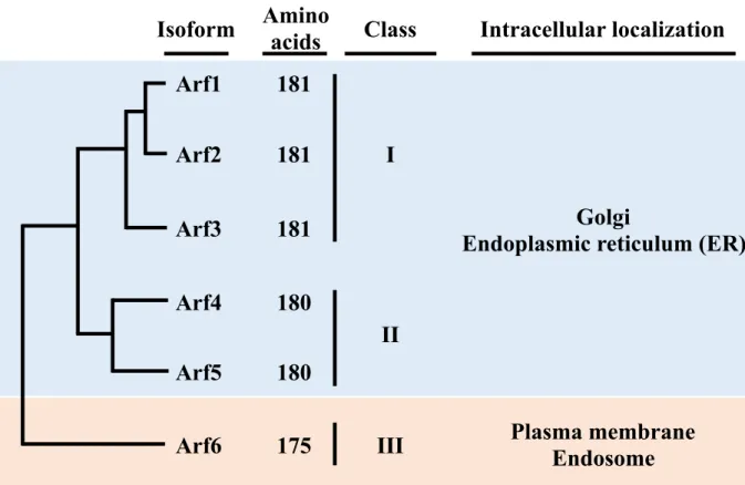
In mammals, the Arf family contains six members, Arf1-6, which are divided into three classes based on their amino acid sequence similarities: class I contains Arf1 to Arf3, class II Arf4 and Arf5, and class III Arf614 (Fig.1). Both Class I and II of Arfs localize at the endoplasmic reticulum (ER) and the Golgi apparatus, which mainly regulate vesicular trafficking between the perinuclear area and endosomes15. In contrast, Arf6, the sole member of class III, primarily localizes at the plasma membrane and endosomal compartments
7
to regulate multiple cellular events, including phosphoinositide metabolism, intracellular membrane trafficking and actin cytoskeleton reorganization15.
2.2. Arf6 activation cycle
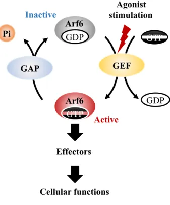
Arf6 cycles between GTP-bound active and GDP-bound inactive forms to function as a molecular switch in the signal transduction (Fig. 2). At the resting state of cells, Arf6 exists as the GDP-bound inactive form. Upon agonist [e.g. HGF and vascular endothelial growth factor (VEGF)] stimulation of the cell, GDP is exchanged to GTP by the action of GEFs, resulting in activation of Arf6. This exchange induces conformational change of Arf6 and increases its affinity to various effector proteins16, through which Arf6 regulates a wide variety of cellular functions. Thereafter, GTP bound to Arf6 is hydrolyzed by the intrinsic GTPase activity of Arf6 with the support of GTPase-activating proteins (GAPs), thereby Arf6 returns to the inactive form17.
2.3. Arf6 GEF
As described above, activity of Arf GTPases is regulated by a large family of GEFs. In human genome, 15 Arf GEFs have been identified, which can be classified into five families by sequence similarity and the presence of functional domains: Golgi brefeldin A (BFA)-resistance factor1/BFA-inhibited GEF (GBF/BIG), Arf nucleotide binding site opener (ARNO)/cytohesin, exchange factor for Arf6 (EFA6), brefeldin-resistant Arf GEF (BRAG), and F-box only protein 8 (Fbx) (Fig. 3). Of these Arf GEFs, seven GEFs have been demonstrated to be involved in Arf6 activation: ARNO/cytohesin2,
8
Grp1/cytohesin 3, EFA6A-D, and BRAG217,18.
2.4. Effectors of Arf6
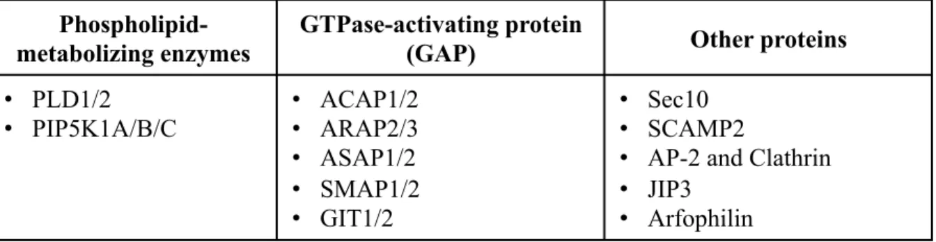
Arf6 regulates various cellular functions through the interaction with its downstream effectors listed below (see also Table 1).
(1) Phospholipid-metabolizing enzymes
Arf6 has been shown to activate two phospholipid-metabolizing enzymes, phospholipase D (PLD)19,20 and phosphatidylinositol 4-phosphate 5-kinase (PIP5K1)21. PLD hydrolyzes the major component of cellular membranes, phosphatidylcholine, to produce phosphatidic acid (PA), which is involved in multiple physiological processes, such as membrane trafficking, cytoskeletal reorganization, receptor-mediated endocytosis, exocytosis, and cell migration. PIP5K1 phosphorylates phosphatidylinositol 4-phosphate [PI(4)P] at the D5 position of the inositol ring to produce the versatile lipid second messenger phosphatidylinositol 4,5-bisphosphate [PI(4,5)P2 or PIP2],
which is involved in a wide variety of cellular events22. Interestingly, PLD and PIP5K1 signaling pathways are closely interconnected by their products: PA generated by PLD activates PIP5K1, and PIP2 generated by PIP5K1 activates
PLD23. Therefore, orchestrated regulation of PLD and PIP5K1 by Arf6 plays pivotal roles in various cellular events.
(2) Arf6 GAPs
9
GTP-hydrolyzing activity, thereby, turning off the Arf6 activity. In addition to the GAP function, several Arf6 GAPs have been also suggested to function as effectors of Arf624. Of the ten Arf6 GAPs identified in human, ACAP1, ARAP2, SMAP1/2 and ASAP1 have been shown to act as Arf6 effectors25-31. These Arf6 GAPs are suggested to recruit their binding proteins to the activated Arf6. For examples, the SMAP1/2 as Arf6 GAPs could directly interact with clathrin through clathrin heavy chain to regulate clathrin-depedent endocytosis, the studies concluded the possibility of SMAP1/2-clathrin-Arf6 complex30,31. A unique Arf6-ARAP2-leucine zipper motif (APPL) endosomal compartment was identified in the previous study, which ARAP2 associated with Arf6-APPL positive compartment through forming complex with APPL1, and this complex controlled the traffic of integrins from APPL endosomes25. , Another previous study indicated the effector function of ACAP1 in Arf6 Arf6-regulated actin cytoskeleton remodeling upon aluminum fluoride (AIFx) stimulation, that ACAP1 formed a complex with GDP-Arf6 and AIFx.
(3) Other proteins
In addition to Afr6 effectors described above, several proteins have also been suggested to function as effectors of Arf6. The previous study demonstrated that GTP-bound Arf6 interacts with an exocyst complex subunit, Sec10, through which Arf6 controls endocytic membrane recycling to the dynamic region of the plasma membrane32. Secretory carrier membrane protein 2 (SCAMP2) mediates the formation of fusion pores during the process of exocytosis through the interaction with Arf6 and PLD in neuroendocrine cells33.
10
Arf6 recruits AP-2, the adaptor protein of clathrin-coated vesicles, to the plasma membrane to regulate clathrin-dependent endocytosis34. The signaling scaffold protein JIP3 acts as a downstream effector of Arf6 in mouse cortical neurons to regulate neurite morphogenesis35. Arfophilin has been reported to serve as a downstream effector of both Arf6 and Arf536, and functions in neuronal migration and breast cancer cell motility37,38. These multiplicity of downstream effectors enables Arf6 to regulate versatile cellular events.
3. PIP5K1
Our group has previously identified PIP5K1 as a direct downstream effector of Arf621. In mammals, three isoforms of PIP5K1 have been identified to date, namely PIP5K1A (corresponding to human PIP5Kα and mouse PIP5Kβ), PIP5K1B (corresponding to human PIP5Kβ and mouse PIP5Kα) and PIP5K1C (corresponding to human and mouse PIP5Kγ) (Fig. 4), all of which catalyze phosphorylation of PI(4)P to generate the pleiotropic lipid messenger PIP2 (Fig. 5).
PIP2 directly binds to and regulates various target proteins, which are involved in
multiple cellular events22. PIP2 also serves as a precursor of lipid second
messengers. It is hydrolyzed by phospholipase C to generate two second messengers, diacylglycerol and inositol 1,4,5-triphosphate (Fig. 6). PIP2 is also
phosphorylated by phosphoinositide 3-kinase (PI3K) to yield phosphatidylinositol 3,4,5-trisphosphate (PIP3), which recruits the protein kinase Akt to the plasma
membrane and promotes its activation, thereby activates downstream signaling pathways required for cell proliferation, growth and survival39,40 (Fig. 7). Therefore, Arf6 regulates various cellular functions through the activation of PIP5K1.
11 3.1. Localization of PIP5K1 isozymes
Each PIP5K1 isozyme localizes at different subcellular compartments. PIP5K1A localizes at the nucleus and the cytosol, PIP5K1B localizes to punctate structures in the perinuclear region, and PIP5K1Clocalizes to adherens junctions in epithelial cells and to focal adhesions41-43. When cells are stimulated, both PIP5K1A and PIP5K1B translocate to the plasma membrane21,44. The different localization of each PIP5K1 isozyme is suggested to be responsible for the generation of PIP2 in different compartments of the cell, thereby PIP5K1
isozymes regulate various cellular functions22.
3.2. Regulation by phosphorylation
Activities of PIP5K1 isozymes are known to be regulated by phosphorylation of their Ser/Thr and Tyr residues. Park et al. have suggested that PIP5K1A, B and C are phosphorylated and suppressed by PKA45. They also demonstrated that Ser214 of PIP5K1A is phosphorylated by PKA and dephosphorylated by PP1: phosphorylation decreases its lipid kinase activity, while dephosphorylation increases it45. A tyrosine residue of PIP5K1A is also phosphorylated: H2O2 stimulation induces tyrosine phosphorylation of PIP5K1A,
which leads to inhibition of PIP5K1A lipid kinase activity and translocation of PIP5K1A away from its substrate at the plasma membrane46. Phosphorylation of PIP5K1C at Ser645 suppresses the binding of PIP5K1C to Talin and AP-2, both of which are activators of PIP5K1C47. Phosphorylation of PIP5K1C at Ser264,
12
which is in the kinase homology domain, also decreases its lipid kinase activity48. Conversely, phosphorylation of Tyr644 by the Src kinase activates PIP5K1C47.
3.3. Structural differences of PIP5K1 isozymes
All PIP5K1 isozymes contain a highly conserved kinase core domain (KCD) at their center, which catalyzes phosphorylation of PI(4)P49 (Fig. 5). In addition to the KCD domain, they possess the N- and C-terminal domains, which are specific for each PIP5K1 isozyme50. N- and C-terminal domains are suggested to modulate kinase activity and localization of each isozyme, thereby conferring isozyme-specific activation and functions.
The previous study has suggested the model for conformational change of PIP5K1C which modulates its activation by Arf6: Arf6 binding region of PIP5K1C located in the KCD is masked by its N-terminal domain, and stimulation of the cell induces conformational change to release this masking, which allows binding of and activation by Arf651.
3.4. Proteins regulating PIP5K1
Activity of PIP5K1 is regulated by several small GTPases. Rho family proteins, RhoA, Rac1 and Cdc42, which regulate actin cytoskeleton reorganization, have been shown to activate lipid kinase activities of all three PIP5K1 isozymes52-54. As described above, Arf6 directly activates all PIP5K1 isozymes21,22,48,55.
In addition to small GTPases, several proteins serve as binding partners and activators of PIP5K156. Talin specifically binds to C-terminal tail of PIP5K1C
13
and activates its lipid kinase activity. Interestingly, Talin binding to PIP5K1C depends on phosphorylation/dephosphorylation of PIP5K1C at Ser645: Talin can only bind to and activate dephosphorylated PIP5K1C57. Similarly, AP-2, the adaptor protein of clathrin-coated vesicles, also binds to C-terminal tail of PIP5K1C dephosphorylated at Ser645 and activates it in mouse hippocampal neurons58. Phosphorylation and dephosphorylaiton of Ser645 are catalyzed by cyclin-dependent kinase 5 (Cdk5) and calcineurin, respectively. PIP5K1 can be also activated by the co-stimulatory receptor CD28 in T lymphocytes: stimulation of CD28 at the immunological synapse induces recruitment of PIP5K1A to CD28 with the aid of the Rho GEF Vav1, and there PIP5K1A is activated to produce PIP2, which induces lipid raft clustering through actin cytoskeleton remodeling
and is also converted to PIP3 by PI3K to activate Akt signaling, thereby
14 Rationale
As described above, the pleiotropic growth factor HGF regulates multiple cellular events through the activation of Arf6 in various types of cell. In the previous study in our laboratory, it has been demonstrated that Arf6-knockout mice exhibit embryonic lethality with a severe defect in liver development. Furthermore, it has also been shown that HGF-dependent in vitro cord formation by primary cultured hepatocytes is significantly impaired in Arf6-deleted hepatocytes12. These observations strongly suggest that Arf6 plays a crucial role in the HGF signaling pathway in hepatocytes. However, the molecular mechanisms of how Arf6 regulates HGF signaling in hepatocytes have not yet been fully understood. In this study, I investigated functions of Arf6 and its downstream effector PIP5K1 in HGF-stimulated hepatocytes.
15 Experimental procedures
1. Reagents and antibodies
Reagents used:
Protein G/protein A sepharose beads: GE healthcare Japan. Anti-Flag affinity gel: SIGMA.
Protease inhibitor cocktail: Nacalai tesque. COSMOGEL GST-Accept: Nacalai tesque. DMEM (4.5g/l glucose): Nacalai tesque. FBS: Gibco.
Penicillin-Streptomycin Mixed solution: Nacalai tesque. OPTI-MEM: Gibco.
Lipofactamine 2000 (1 mg/ml): Invitrogen. siRNA Arf6: Invitrogen.
siRNA human PIP5K1A: GE Healthcare Dharmacon Inc. TRIzol reagent: Invitrogen.
SuperScript III Reverse Transcriptase: Invitrogen. Blend Taq DNA polymerase: TOYOBO.
Antibodies for Western blot analysis and immunofluorescence staining:
Rabbit anti-AKT: Cell Signaling Technologies.
Rabbit anti-phospho-AKT (S473): Cell Signaling Technologies.
Rabbit anti-phospho-Erk1/2 (Thr202/Tyr204): Cell Signaling Technologies. Rabbit anti-Erk1/2: Cell Signaling Technologies.
16
Rabbit anti-phospho-c-Met: Cell Signaling Technologies. Rabbit anti-Actin: SIGMA.
Mouse anti-α-tubulin: SIGMA. Mouse anti-Flag: SIGMA. Mouse anti-BrdU: SIGMA. Rabbit anti-Ki67: Abcam. Rabbit anti-c-Met: Santa Cruz. Goat anti-PIP5K1B: Santa Cruz. Goat anti-PIP5K1C: Santa Cruz. Goat anti-Albumin: Bethyl.
Mouse anti-PIP3: Echelon Biosciences.
Mouse anti-PIP2: Echelon Biosciences.
Rabbit anti-Arf6, rabbit anti-PIP5K1A and rabbit anti-PIP5K1B: Self-prepared antibodies 61.
Horseradish peroxidase-conjugated anti-rabbit secondary antibody: Cell Signaling . Horseradish peroxidase-conjugated anti-mouse secondary antibody: Cell Signaling. Alexa488-conjugated anti-rabbit IgG: Invitrogen.
Alexa488-conjugated anti-mouse IgG: Invitrogen. Alexa647- conjugated anti-goat IgG: Invitrogen. Cy3-conjugated anti-mouse IgM; Invitrogen.
2. Mice
Generation of Pip5k1a-/- mice with C57B1/6 background is described
17
were kept under a controlled humidity and lighting schedule as 12 h dark with free access to food and water. All experiments with mice were conducted according to the Guidelines for Proper Conduct of Animal Experiments, Science Council of Japan, and protocols were approved by the Animal Care and Use Committee, University of Tsukuba.
3. Isolation and culture of primary hepatocytes
Hepatocytes of wild type and Pip5k1a -/- mice (10-12 weeks old) hepatocytes
were isolated as described previously63. Isolated hepatocytes showed >80% cell viability assessed by trypan blue exclusion. Cells were plated on collagen-coated plates and cultured in Dulbecco's modified Eagle's medium (DMEM) (Invitrogen, Carlsbad, CA, USA) containing 10% fetal bovine serum (FBS) and 2 % antibiotic solution (20,000 units/ml penicillin and 20 mg/ml streptomycin) in an atmosphere of 5% CO2 at 37 °C. The primary hepatocytes were stimulated with 10 ng/ ml HGF
for indicated time.
4. Cell culture, plasmid and siRNA transfection and HGF treatment of the Cell The human hepatocellular cell line HepG2 cells were maintained in DMEM supplemented with 10 % FBS and 1% antibiotic solution (10,000 units/ml penicillin and 10 mg/ml streptomycin) in an atmosphere of 5% CO2 at 37°C. They were
transfected with plasmid DNAs or siRNA using Lipofactamine 2000 (Invitrogen) according to the manufacturer’s instructions. For siRNA-mediated knock down experiments, HepG2 cells were transfected with 10 nM siRNA. After 48 h incubation, the cells were serum-starved for 12 h, and then treated with 10 ng/mL
18
HGF for the indicated time. For overexpression experiments, FLAG-tagged Arf6 or an Arf6 mutant (Q67L or T44N; kind gifts provided by Dr. K. Nakayama from Kyoto University) was transfected into HepG2 cells. After 48 h incubation, cells were harvested and subjected to immunoprecipitation.
5. Immunopreciptaiton and Western Blotting
Cells were lysed in lysis buffer (25 mM Tris-HCl, pH7.5, 1% Triton X-100, 10 mM NaCl, 1 mM EGTA and 5 mM MgCl2) containing protease inhibitor cocktail
(Nacalai) at 4 °C for 30 min, and centrifuged at 10,000 ×g for 10 min. The cell lysates were incubated with the indicated antibodies and protein G/protein A sepharose beads (GE healthcare) or Anti-Flag affinity gel (SIGMA) at 4 °C for 4 h. The immune complexes captured on beads were washed with lysis buffer and eluted with the SDS-PAGE sample buffer [4x sample buffer: 25% 0.5M Tris-HCl (pH 6.8), 8% sodium dodecyl sulfate, 20% 2-mercaptoethanol, 0.04% bromophenol blue, and 40% glycerol] by boiling. Eluted proteins were separated by SDS-PAGE, transferred onto a PVDF membrane, and then detected by specific primary antibodies and horseradish peroxidase-conjugated secondary antibodies. Images were quantified using the Image J software (http://rsb.info.nih.gov/ij/).
6. Assay for measuring the cell number
The number of viable cells was assessed by the trypan blue exclusion assay. Cells were seeded in 12 well plates (5 × 10⁴ cells/well) and transfected with siRNA. After 48 h incubation, cells were treated with HGF for the indicated time, trypsinized and stained with trypane blue. The number of viable cells was
19
counted with a hemocytometer. Five counts were performed per well for three independent experiments.
7. Analysis for activation of Arf6
Activation of Arf6 was assessed by the Arf6-GTP pulldown assay as described in the previous report by Santy et al (2001)64. Briefly, HepG2 cells were seeded on 3.5 cm dishes at 3 × 10⁵ cells/dish and incubated overnight. After 12 h starvation, cells were stimulated with or without 10 ng/mL HGF for 10 min. Cells were harvested in lysis buffer [50 mM Tris-HCl (pH 7.5), 100 mM NaCl, 2 mM MgCl2,
0.1% sodium dodecyl sulfate, 0.5% sodium deoxycholate, 1% Triton X-100, 10% glycerol, 1 µg/ml aprotinin, and 1 µg/ml leupeptin] and lysed at 4°C for 30 min. The cell extracts were mixed with glutathione S-transferase (GST)-GGA3-conjugated glutathione-Sepharose beads and incubated for 30 min with gentle rotation. The beads were washed three times with the washing buffer [50 mM Tris-HCl (pH 7.5), 100 mM NaCl, 2 mM MgCl2, 1% NP-40, 10% glycerol,
1 µg/ml aprotinin, and 1 µg/ml leupeptin]. The Arf6-GTP bound to GST-GGA3 beads was eluted by SDS sample buffer, and detected by Western blotting.
8. Lipids extraction and quantification of PIP3
Cell Lipids were extracted by the method reported by Gray et al.65. Cells at 5×10⁶ cells/15 cm dish were transfected with siRNA and treated with HGF as described above. Cells were incubated with ice-cold 0.5 M TCA for 5 min, scraped, and centrifuged at 10,000 ×g for 5 min. The pellet was resuspended in 5% TCA/1
20
mM EDTA and centrifuged, then the supernatant was removed. Neutral lipids were extracted from the pellet with MeOH : CHCl3 (2:1) and the supernatant was
discarded. Acidic lipids were then extracted from the pellet with MeOH : CHCl3 : 12 N HCl (80:40:1). After centrifugation, chloroform and 0.1 M HCl were added to the supernatant and vortexed, followed by centrifugation to separate the organic and aqueous phases. The organic phase was collected and dried in a vacuum dryer. To detect PIP3 levels, the extracted lipids were dissolved in 20% DMSO, and spotted
on nitrocellulose membranes. The dot membranes were blocked in TBS + 0.05% Tween-20 with 5% BSA for 1 h at room temperature and further incubated with anti-PIP3 antibody (1:50 dilution; MBL international) for 1 h at room temperature,
followed by incubation with horseradish peroxidase-conjugated secondary antibodies. Chemiluminescence reagents (Nacalai and Thermo Scientific) were used for detection and the level of PIP3 was quantified by the Image J software.
9. Immunocytochemistry
HepG2 cells or primary hepatocytes at 5×10⁴ cells/12 cm dish were seeded on gelatin-coated coverslips in the dish and transfected with siRNAs. After incubation at 37°C for 48 h, cells were stimulated with HGF as described above, fixed with 4% paraformaldehyde and permeabilized with 0.5% Triton-X100. Cells were then blocked with 1% BSA in PBS and immunostained with indicated primary antibodies. The fluorescent-labeled secondary antibodies and DAPI were used to detect target proteins and the nucleus, respectively.
21 10. Partial Hepatectomy
Eight weeks old female mice were used for PH. Mice were anesthetized with isoflurane and subjected to 70% liver resection of the median and left lateral lobes as previously described66. The mice were euthanized at days after PH described in the figure 18 and 19. Livers were isolated from the mice, and liver weight/body weight was determined. A part of the liver tissue was fixed in 4% paraformaldehyde/PBS and then embedded in O.C.T. (Tissue-Tek®). For measuring serum aspartate aminotransferase (AST) and alanine transaminase (ALT) activities, blood was collected from the submandibular vein before sacrifice. AST and ALT activities were measured by the AmpliteTM fluorimetric assay kit (AAT Bioquest, Inc.).
To measure hepatocyte proliferative activity, the livers removed from mice in the indicated times (0, 2 and 5 days) were fixed in 4% paraformaldehyde/PBS at 4°C overnight, embedded in O.C.T., and sliced into 10 µm sections. Sections were permeabilized and blocked with 0.1% Triton-X/5% BSA/PBS for 30 min at room temperature, and then stained with anti-Ki-67 antibody (Abcam, 1:150 in 5%BSA/PBS) and anti-albumin antibody (Bethyl, 1:150 in 5%BSA/PBS) at 4°C overnight. After washing with PBS, sections were stained with secondary antibodies and DAPI. The Ki-67-positive cells were counted and analyzed using the BZ-II Analyzer (KEYENCE).
11. Statistical analyses
All quantified data were expressed as means ± SEM and analyzed by Student
22
23 Results
Arf6 is essential for Akt activation to promote HGF-dependent proliferation of HepG2 cells
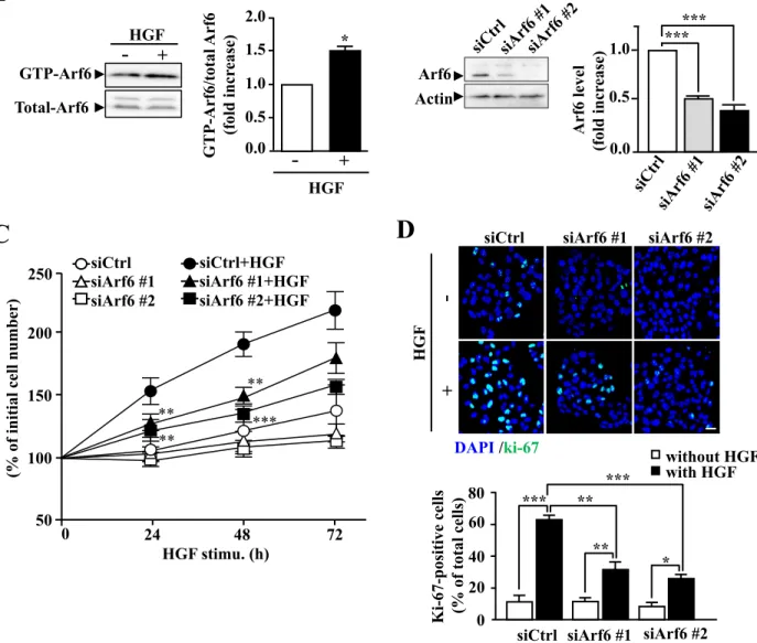
To investigate the role of Arf6 in HGF-dependent cell functions of hepatocytes, I employed the human hepatocellular carcinoma cell line HepG2 cells as a model system. As our lab has previously demonstrated with fetal mouse hepatocytes12, Arf6 in HepG2 cells was also activated in response to HGF stimulation (Fig. 8A). Knockdown of Arf6 in these cells attenuated HGF-stimulated cell proliferation as assessed by counting cell number and immunostaining the proliferation marker Ki-67 (Fig. 8B-D). These results demonstrate that Arf6 mediates HGF signaling to regulate cell proliferation in HepG2 cells.
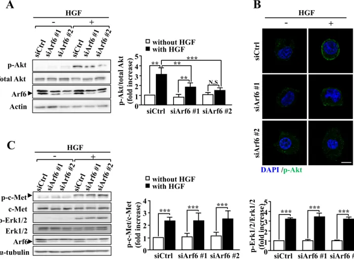
The PI3K/Akt axis is a key pathway of HGF-dependent cell proliferation39. Since Akt is recruited to the plasma membrane and phosphorylated by phosphoinositide-dependent kinase 1 to be activated, I examined the involvement of Arf6 in this process. HGF-stimulated Akt phosphorylation and increase in the phosphorylated-Akt (p-Akt) level at the plasma membrane were significantly suppressed by knockdown of Arf6 (Fig. 9A, B). On the other hand, HGF-dependent phosphorylation of Erk was not affected by knockdown of Arf6 (Fig. 9C), ruling out the involvement of Arf6 in the Ras/Raf/Erk pathway, another key pathway downstream of c-Met. The inhibition of Akt phosphorylation is unlikely to be attributable to the suppression of c-Met: the HGF-stimulated phosphorylation level of c-Met was not affected by Arf6 knockdown (Fig. 9C). These results, taken together, demonstrate that
24
Arf6 regulates the HGF-dependent Akt recruitment to the plasma membrane and its subsequent activation to promote hepatocyte proliferation.
Arf6 promotes PIP2 and PIP3 generation by activating PIP5K1A upon HGF
stimulation
Since the recruitment of Akt to the plasma membrane is mediated by PIP3 generated
in response to various agonists including HGF67, I examined whether Arf6 is involved in the HGF-dependent PIP3 generation. HGF stimulation of HepG2 cells markedly
increased the PIP3 production, which was almost completely suppressed by knockdown
of Arf6 (Fig. 10A), suggesting that Arf6 is involved in HGF-dependent PIP3 production.
PIP3 is generated by phosphorylation of PIP2 by PI3K. Our lab have previously
demonstrated that the PIP2-generating enzyme PIP5K1 is directly activated by Arf621.
These observations led me to speculate that Arf6 activates PIP5K1 to generate the PI3K substrate PIP2 upon HGF stimulation, thereby contributing to the increase in the PIP3
levels. To address this assumption, effects of Arf6 knockdown on the PIP2 level were
analyzed by immunocytochemistry with the PIP2-specific antibody. As was expected,
HGF stimulation of HepG2 cells increased the PIP2 level at the plasma membrane, and
knockdown of Arf6 significantly suppressed the production of PIP2 (Fig. 10B).
Furthermore, it was found that PIP5K1A, but not PIP5K1B and PIP5K1C, interacted with Arf6 upon HGF stimulation of HepG2 cells (Fig. 11). These results, taken together with the result shown in Fig. 8A, suggest that Arf6 activated by HGF stimulation interacts with and positively regulates PIP5K1A to produce the PI3K substrate PIP2 at
25
Arf6 activated by HGF stimulation forms a complex with PIP5K1A and c-Met
The results obtained above, taken together with the report that Arf6 recruits its effector protein GGA3 (golgi-localized, gamma adaptin ear-containing, Arf-binding 3) to c-Met68, raised a possibility that activated Arf6 forms a complex with PIP5K1A and c-Met. To address this issue, interaction of c-Met with PIP5K1A and Arf6 was assessed by immunoprecipitation assay. As expected, interaction between c-Met and PIP5K1A was markedly enhanced by HGF stimulation, and knockdown of Arf6 drastically inhibited this interaction (Fig. 12A). Interestingly, under these conditions, Arf6 constitutively interacted with c-Met (Fig. 12A). Consistent with the results obtained by immunoprecipitation assay, PIP5K1A predominantly locating in the cytosol in the resting state of the cell translocated to the plasma membrane and colocalized with c-Met upon HGF stimulation, which was again attenuated by knockdown of Arf6 (Fig. 12B).
As Arf6 was activated by HGF stimulation as shown in Fig. 8A, the results shown above indicate that Arf6 activated by HGF stimulation is responsible for the PIP5K1A recruitment to the plasma membrane to form a complex with c-Met. To test this assumption, Q67L and T44N mutants of Arf6, which mimic GTP-bound active and GDP-bound inactive forms of Arf669
, respectively, were expressed in HepG2 cells and their effects on the interaction of PIP5K1A with c-Met were analyzed. The interaction was observed in the cell overexpressed with Q67L but not with T44N (Fig. 13), supporting the notion that the active form of Arf6 recruits PIP5K1A to c-Met. Consistent with the result shown in Fig. 12A, Q67L and T44N mutants were both found to interact with c-Met (Fig. 13).
26
PIP5K1A is required for HGF-dependent Akt activation and subsequent proliferation of hepatocytes
To confirm the notion that PIP5K1A is involved in the PIP2 and PIP3 generation, Akt
phosphorylation and cell proliferation through the production of the PI3K substrate PIP2
upon HGF stimulation, I employed siRNAs that efficiently knocked down PIP5K1A in HepG2 cells (Fig. 14A). Knockdown of PIP5K1A significantly suppressed the HGF-dependent PIP2 and PIP3 production (Fig. 14B, C) suggesting that PIP5K1A is the
major enzyme to provide the PI3K substrate PIP2. Consistent with our notion, Akt
phosphorylation and the accumulation of p-Akt at the plasma membrane induced by HGF stimulation were significantly suppressed in PIP5K1A-knocked-down cells, while phosphorylation of c-Met and Erk were not affected (Fig.15A, B). Finally, HGF-dependent proliferation of HepG2 cells, which was assessed by cell number (Fig. 16A) and Ki-67 staining (Fig. 16B), was also inhibited by PIP5K1A knockdown. Thus, these results strongly support our notion described above.
To investigate whether the results obtained above with HepG2 cells are also the case in hepatocytes, I isolated hepatocytes from adult Pip5k1a-/- mice and analyzed Akt
phosphorylation and proliferation stimulated by HGF. Consistent with the results obtained with HepG2 cells, these HGF-dependent phenomena were impaired in
Pip5k1a-/- primary hepatocytes, whereas levels of p-c-Met and p-Erk1/2 were comparable to those of control cells (Fig. 17A, B). These results provide evidence that PIP5K1A plays an important role in HGF-dependent hepatocyte proliferation through the activation of Akt.
27
PIP5K1A is involved in liver regeneration after partial hepatectomy.
Arf6-/- mice exhibit severe defect in liver development12, while Pip5k1a-/- mice did
not show obvious defects in embryonic liver development62. Nevertheless, the results obtained above demonstrate that PIP5K1A functions as a downstream effector of Arf6 in the HGF-dependent cellular signaling pathway regulating hepatocyte proliferation. This conclusion and the fact that HGF plays a pivotal role in liver regeneration70,71, which absolutely requires hepatocyte proliferation, led me to speculate that PIP5K1A is involved in liver regeneration after liver injuries, but not in embryonic liver development. To address this issue, partial hepatectomy was employed to examine the significance of PIP5K1A in liver regeneration. In control mice, the liver weight recovered to 77.7 ± 1.57% of the non-resected liver weight after 5 days of 70% hepatectomy, while the recovery was slower in Pip5k1a-/- mice (Fig. 18A): the liver
weight at 5 days after hepatectomy was 55.6 ± 3.86% of the non-resected liver weight. Analyses of the levels of alanine transaminase (ALT) and aspartate aminotransferase (AST), the liver injury markers released from hepatocytes into the serum, also revealed that repair of the injured liver was slower in Pip5k1a-/- mice (Fig. 18B, C): AST and
ALT activities in sera of control mice were elevated at 1 day after partial hepatectomy and gradually decreased, reaching the basal level after 5 days, while they were significantly higher at 2 days and higher levels sustained till 5 days in Pip5k1a-/- mice. Finally, the increase in the number of proliferating hepatocytes observed after partial hepatectomy was significantly suppressed in Pip5k1a-/- mice (Fig. 19). Nevertheless,
28
29 Discussions
In the present study, I provide evidence that Arf6 directly or indirectly bound to c-Met is activated by HGF stimulation of hepatocytes, and activated Arf6 recruits PIP5K1A to c-Met and activates it to produce the PI3K substrate PIP2, which is converted to PIP3 to
activate Akt, thereby stimulating the HGF-dependent hepatocyte proliferation (Fig. 20). This model for the HGF signaling pathway mediated by Arf6 and PIP5K1A in hepatocyte proliferation gives insight into the molecular mechanism for liver regeneration after the liver injury.
Recruitment of PIP5K1A to c-Met
In this HGF signaling pathway, I speculated that Arf6, which constitutively interacts with c-Met, recruits PIP5K1A to c-Met, when it is activated by HGF stimulation. Although the active form of Arf6 can directly activate PIP5K121, I cannot totally exclude a possibility that another protein(s), which functions as a downstream effector of c-Met, facilitates or mediates the PIP5K1A recruitment to c-Met. This possibility is supported by the report that Arf6 and the c-Met adaptor protein Crk, which is involved in c-Met signaling by interacting with multiple downstream signaling molecules68, cooperate to recruit the Arf6 effector protein GGA3 to c-Met. GGA3 is an adaptor protein, which belongs to GGA family. In previous study, the HGF-dependent Arf6 activation was suppressed by transfecting GGA3-N194A mutant that unable to interact with Arf6-GTP68. Further, the study also showed the Crk binds with GGA3 in an HGF-independent manner, and this binding is required for GGA3 to bind with c-Met.
30
Thus, in the present study, it's possible an unidentified adaptor protein of c-Met may be a key determinant to recruit PIP5K1A to HGF-stimulated c-Met. Arf6 is capable of activating all PIP5K1 isozymes in vitro21. If our assumption is true and there exist several adaptor proteins of c-Met, each of which specifically interacts and recruits each of three PIP5K1 isozymes to cellular compartments in cooperation with Arf6, adaptor proteins determine PIP5K1 isozymes to be spatiotemporally activated by Arf6 in an agonist type-dependent manner. It is of interest to identify whether there exist adaptor proteins, which specifically interact with PIP5K1 isozymes to regulate their translocation dependently upon the types of agonists.
Potential GEFs activating Arf6 upon HGF stimulation
Another question raised in this study is how Arf6 is activated by HGF stimulation. Arf6 activation upon agonist stimulation is precisely regulated by Arf6-specific GEFs, which promote the exchange of GDP on Arf6 for GTP. Our lab have recently suggested that Grp1, EFA6B and EFA6D, of 7 Arf6-specific GEFs so far identified in mammalian cells, spatiotemporally activate Arf6 in the HGF-stimulated vascular endothelial cell to regulate β1 integrin recycling that is the critical cellar event for the HGF-induced tumor angiogenesis11. Attar et al., have shown that ARNO activates Arf6 in HGF-stimulated epithelial cells to regulate cell migration72. In addition, inhibition of cytohesins such as ARNO and Grp1 by the specific cytohesin inhibitor SecinH3 revealed that these cytohesin family members are required for the Arf6 activation in the signaling pathway for the HGF-mediated renal recovery after acute kidney injury73. Thus, several Arf6-sepcific GEFs are the possible candidates for the Arf6 activation in the signaling
31
pathway of HGF-stimulated hepatocyte proliferation. Further investigation, e.g., effects of knockdown of Arf6-specific GEFs on HGF-stimulated hepatocyte proliferation and analysis of their subcellular localization by immunocytochemistry, would identify an Arf6-specific GEF involved in the activation of Arf6 in response to the HGF stimulation of hepatocytes.
Hepatocyte proliferation regulated by the Arf6-PIP5K1A axis might be a limited event for liver regeneration
HGF-stimulated hepatocyte proliferation through the Arf6-PIP5K1A axis is strongly suggested to be a crucial cell event for liver regeneration as demonstrated in this study (Fig. 18). Although Arf6-/- mice exhibit embryonic lethality with a severe
defect in liver development12, Pip5k1a-/- mice did not show obvious histological
abnormality in the fetal liver up to 12 months62. These observations, taken together with the results obtained in this study, suggest that the Arf6-PIP5K1A axis is an important signaling pathway downstream of HGF/c-Met for liver regeneration in adult mice, but not for the fetal liver development: Arf6 activated by HGF stimulation of hepatocytes or hepatoblasts utilizes another unidentified downstream molecule(s) in the fetal liver development. This idea is supported by observations obtained in this study that before the liver injury by partial hepatectomy, weight, histological structure and proliferating cells of the liver in Pip5k1a-/- mice did not show obvious differences from those of
control mice (Fig.18A and 19). Thus, it is plausible that hepatocyte proliferation regulated by the Arf6-PIP5K1A axis downstream of HGF/c-Met might be a limited and
32
crucial event for regeneration after liver damages, but not for fetal liver development. Our lab have previously demonstrated that fetal hepatocytes isolated from Arf6 -/-embryos exhibit a defect in in vitro cord formation, whereas no defect was observed in HGF-stimulated proliferation12. On the other hand, I found that knockdown of Arf6 or its downstream effector PIP5K1A and deletion of PIP5K1A significantly suppressed HGF-stimulated cell proliferation of HepG2 cells (Fig. 8C and D, and Fig. 16A and B) and hepatocytes isolated from adult mice (Fig. 17B), respectively. These observations suggest that Arf6 and PIP5K1A are both dispensable for HGF-regulated cell proliferation of fetal hepatocytes, while HGF-dependent cell proliferation of adult hepatocytes absolutely requires the Arf6-PIP5K1A axis for liver regeneration. It is of interest to clarify a downstream effector of Arf6 functioning in the fetal liver development; its clarification would provide insight into the molecular mechanism of liver development.
HGF is a critical growth factor regulating liver regeneration after liver injuries as well as liver development. Here, I defined a novel HGF signaling pathway regulating adult hepatocyte proliferation that is an essential cell phenomena for the recovery from liver damage. Our findings could provide insight into molecular mechanisms of liver regeneration and into novel therapeutic strategies for hepatic injury caused by infections, toxic materials and surgical resection. In addition, it is of interest to investigate the involvement of c-Met-Arf6-PIP5K1A signaling in hepatocellular carcinoma, since dysregulation of HGF/c-Met is highly related to this disease8.
33 Conclusions
In this study, I demonstrated that the Ar6-PIP5K1A axis plays an important role in HGF-stimulated hepatocyte proliferation. I found that depletion of Arf6 or PIP5K1A in HepG2 cells suppresses HGF-stimulated proliferation, PIP2 and PIP3 generation, and
activation of Akt. Similar phenomena were observed in Pip5k1a-/- mouse hepatocytes. I also showed that Arf6 recruits PIP5K1A to c-Met in response to HGF stimulation. Finally, I demonstrated that hepatocyte proliferation and liver regeneration after acute liver injury was impaired in Pip5k1a-/- mice. From the result obtained, I proposed the
model shown in Figure 20: Arf6 activated by HGF stimulation recruits PIP5K1A to c-Met and activates it to produce PIP2 and PIP3, which in turn activates Akt to promote
hepatocyte proliferation, thereby facilitating liver regeneration after liver injury. The present study provides novel insights into the hepatic response to HGF stimulation and its molecular mechanism. Furthermore, since HGF is involved in liver regenerations after acute liver failures, this work may provide novel therapeutic strategies for promoting liver regeneration after hepatic injury.
34 Acknowledgement
I am grateful to Dr. K. Nakayama (Kyoto University, Japan) for kindly providing cDNAs encoding mouse Arf6 and its mutants. I sincerely appreciate Dr. N. Ohkohchi and K. Iwasaki for technical advises on the partial hepatectomy experiments. I appreciate all the kind help and adivices from my professors Dr. Y. Kanaho, Dr. S-T. Ding and Dr. Y. Funakoshi, and all my lab members in University of Tsukuba and National Taiwan University.
35 References
1 Fausto, N. Liver regeneration. J Hepatol 32, 19-31 (2000).
2 Rozga, J. Hepatocyte proliferation in health and in liver failure. Med Sci Monit 8, RA32-38 (2002).
3 Patijn, G. A., Lieber, A., Schowalter, D. B., Schwall, R. & Kay, M. A. Hepatocyte growth factor induces hepatocyte proliferation in vivo and allows for efficient retroviral-mediated gene transfer in mice. Hepatology 28, 707-716, doi:10.1002/hep.510280317 (1998).
4 Nakamura, T., Nawa, K. & Ichihara, A. Partial purification and characterization of hepatocyte growth factor from serum of hepatectomized rats. Biochem
Biophys Res Commun 122, 1450-1459 (1984).
5 Michalopoulos, G., Houck, K. A., Dolan, M. L. & Leutteke, N. C. Control of hepatocyte replication by two serum factors. Cancer Res 44, 4414-4419 (1984). 6 Russell, W. E., McGowan, J. A. & Bucher, N. L. Biological properties of a
hepatocyte growth factor from rat platelets. J Cell Physiol 119, 193-197, doi:10.1002/jcp.1041190208 (1984).
7 Daniele, B. et al. Met as Prognostic Factor and Therapeutic Target in Pretreated Hepatocellular Carcinoma (Hcc): Final Results of a Randomized Controlled Phase 2 Trial (Rct) with Tivantinib (Arq 197). Ann Oncol 23, 225-225 (2012). 8 Giordano, S. & Columbano, A. Met as a therapeutic target in HCC: Facts and
hopes. J Hepatol 60, 442-452 (2014).
9 Tushir, J. S. & D'Souza-Schorey, C. ARF6-dependent activation of ERK and Rac1 modulates epithelial tubule development. EMBO J 26, 1806-1819,
36
doi:10.1038/sj.emboj.7601644 (2007).
10 Hu, B. et al. ADP-ribosylation factor 6 regulates glioma cell invasion through the IQ-domain GTPase-activating protein 1-Rac1-mediated pathway. Cancer
Res 69, 794-801, doi:10.1158/0008-5472.CAN-08-2110 (2009).
11 Hongu, T. et al. Arf6 regulates tumour angiogenesis and growth through HGF-induced endothelial beta1 integrin recycling. Nat Commun 6, 7925, doi:10.1038/ncomms8925 (2015).
12 Suzuki, T. et al. Crucial role of the small GTPase ARF6 in hepatic cord formation during liver development. Mol Cell Biol 26, 6149-6156, doi:10.1128/MCB.00298-06 (2006).
13 Takai, Y., Sasaki, T. & Matozaki, T. Small GTP-binding proteins. Physiol Rev 81, 153-208 (2001).
14 D'Souza-Schorey, C. & Chavrier, P. ARF proteins: roles in membrane traffic and beyond. Nat Rev Mol Cell Biol 7, 347-358, doi:10.1038/nrm1910 (2006). 15 Donaldson, J. G. & Jackson, C. L. ARF family G proteins and their regulators:
roles in membrane transport, development and disease. Nature Reviews
Molecular Cell Biology 12, 362-375, doi:Doi 10.1038/Nrm3117 (2011).
16 Pasqualato, S., Menetrey, J., Franco, M. & Cherfils, J. The structural GDP/GTP cycle of human Arf6. EMBO Rep 2, 234-238, doi:10.1093/embo-reports/kve043 (2001).
17 Hongu, T. & Kanaho, Y. Activation machinery of the small GTPase Arf6. Adv
Biol Regul 54, 59-66, doi:10.1016/j.jbior.2013.09.014 (2014).
18 Donaldson, J. G. & Jackson, C. L. ARF family G proteins and their regulators: roles in membrane transport, development and disease. Nat Rev Mol Cell Biol 12,
37
362-375, doi:10.1038/nrm3117 (2011).
19 Brown, H. A., Gutowski, S., Moomaw, C. R., Slaughter, C. & Sternweis, P. C. ADP-ribosylation factor, a small GTP-dependent regulatory protein, stimulates phospholipase D activity. Cell 75, 1137-1144 (1993).
20 Hiroyama, M. & Exton, J. H. Localization and regulation of phospholipase D2 by ARF6. J Cell Biochem 95, 149-164, doi:10.1002/jcb.20351 (2005).
21 Honda, A. et al. Phosphatidylinositol 4-phosphate 5-kinase alpha is a downstream effector of the small G protein ARF6 in membrane ruffle formation.
Cell 99, 521-532 (1999).
22 Funakoshi, Y., Hasegawa, H. & Kanaho, Y. Regulation of PIP5K activity by Arf6 and its physiological significance. J Cell Physiol 226, 888-895, doi:10.1002/jcp.22482 (2011).
23 van den Bout, I. & Divecha, N. PIP5K-driven PtdIns(4,5)P2 synthesis: regulation and cellular functions. J Cell Sci 122, 3837-3850, doi:10.1242/jcs.056127 (2009).
24 Inoue, H. & Randazzo, P. A. Arf GAPs and their interacting proteins. Traffic 8, 1465-1475, doi:10.1111/j.1600-0854.2007.00624.x (2007).
25 Chen, P. W., Luo, R., Jian, X. & Randazzo, P. A. The Arf6 GTPase-activating proteins ARAP2 and ACAP1 define distinct endosomal compartments that regulate integrin alpha5beta1 traffic. J Biol Chem 289, 30237-30248, doi:10.1074/jbc.M114.596155 (2014).
26 Jackson, T. R. et al. ACAPs are arf6 GTPase-activating proteins that function in the cell periphery. J Cell Biol 151, 627-638 (2000).
38
both Arf and Rho GTPases, by selective capture on phosphoinositide affinity matrices. Mol Cell 9, 95-108 (2002).
28 Klein, S., Franco, M., Chardin, P. & Luton, F. Role of the Arf6 GDP/GTP cycle and Arf6 GTPase-activating proteins in actin remodeling and intracellular transport. J Biol Chem 281, 12352-12361, doi:10.1074/jbc.M601021200 (2006). 29 Randazzo, P. A. & Hirsch, D. S. Arf GAPs: multifunctional proteins that
regulate membrane traffic and actin remodelling. Cell Signal 16, 401-413 (2004).
30 Tanabe, K. et al. A novel GTPase-activating protein for ARF6 directly interacts with clathrin and regulates clathrin-dependent endocytosis. Mol Biol Cell 16, 1617-1628, doi:10.1091/mbc.E04-08-0683 (2005).
31 Natsume, W. et al. SMAP2, a novel ARF GTPase-activating protein, interacts with clathrin and clathrin assembly protein and functions on the AP-1-positive early endosome/trans-Golgi network. Mol Biol Cell 17, 2592-2603, doi:10.1091/mbc.E05-10-0909 (2006).
32 Prigent, M. et al. ARF6 controls post-endocytic recycling through its downstream exocyst complex effector. J Cell Biol 163, 1111-1121, doi:10.1083/jcb.200305029 (2003).
33 Liu, L. et al. SCAMP2 interacts with Arf6 and phospholipase D1 and links their function to exocytotic fusion pore formation in PC12 cells. Mol Biol Cell 16, 4463-4472, doi:10.1091/mbc.E05-03-0231 (2005).
34 D'Souza-Schorey, C., Li, G., Colombo, M. I. & Stahl, P. D. A regulatory role for ARF6 in receptor-mediated endocytosis. Science 267, 1175-1178 (1995).
39
the small GTPase ARF6 to regulate neurite morphogenesis of cortical neurons.
FEBS Lett 584, 2801-2806, doi:10.1016/j.febslet.2010.05.020 (2010).
36 Shin, O. H., Couvillon, A. D. & Exton, J. H. Arfophilin is a common target of both class II and class III ADP-ribosylation factors. Biochemistry 40, 10846-10852 (2001).
37 Jing, J., Tarbutton, E., Wilson, G. & Prekeris, R. Rab11-FIP3 is a Rab11-binding protein that regulates breast cancer cell motility by modulating the actin cytoskeleton. Eur J Cell Biol 88, 325-341, doi:10.1016/j.ejcb.2009.02.186 (2009).
38 Kim, Y. et al. ADP-ribosylation factor 6 (ARF6) bidirectionally regulates dendritic spine formation depending on neuronal maturation and activity. J Biol
Chem 290, 7323-7335, doi:10.1074/jbc.M114.634527 (2015).
39 Carnero, A., Blanco-Aparicio, C., Renner, O., Link, W. & Leal, J. F. The PTEN/PI3K/AKT signalling pathway in cancer, therapeutic implications. Curr
Cancer Drug Targets 8, 187-198 (2008).
40 Testa, J. R. & Tsichlis, P. N. AKT signaling in normal and malignant cells.
Oncogene 24, 7391-7393, doi:10.1038/sj.onc.1209100 (2005).
41 Di Paolo, G. et al. Recruitment and regulation of phosphatidylinositol phosphate kinase type 1 gamma by the FERM domain of talin. Nature 420, 85-89, doi:10.1038/nature01147 (2002).
42 Ling, K., Doughman, R. L., Firestone, A. J., Bunce, M. W. & Anderson, R. A. Type I gamma phosphatidylinositol phosphate kinase targets and regulates focal adhesions. Nature 420, 89-93, doi:10.1038/nature01082 (2002).
40
adherens junction and E-cadherin trafficking via a direct interaction with mu 1B adaptin. J Cell Biol 176, 343-353, doi:10.1083/jcb.200606023 (2007).
44 Doughman, R. L., Firestone, A. J. & Anderson, R. A. Phosphatidylinositol phosphate kinases put PI4,5P(2) in its place. J Membr Biol 194, 77-89, doi:10.1007/s00232-003-2027-7 (2003).
45 Park, S. J., Itoh, T. & Takenawa, T. Phosphatidylinositol 4-phosphate 5-kinase type I is regulated through phosphorylation response by extracellular stimuli. J
Biol Chem 276, 4781-4787, doi:10.1074/jbc.M010177200 (2001).
46 Halstead, J. R. et al. A role for PtdIns(4,5)P2 and PIP5Kalpha in regulating stress-induced apoptosis. Curr Biol 16, 1850-1856, doi:10.1016/j.cub.2006.07.066 (2006).
47 Ling, K. et al. Tyrosine phosphorylation of type Igamma phosphatidylinositol phosphate kinase by Src regulates an integrin-talin switch. J Cell Biol 163, 1339-1349, doi:10.1083/jcb.200310067 (2003).
48 Aikawa, Y. & Martin, T. F. ARF6 regulates a plasma membrane pool of phosphatidylinositol(4,5)bisphosphate required for regulated exocytosis. J Cell
Biol 162, 647-659, doi:10.1083/jcb.200212142 (2003).
49 Ishihara, H. et al. Type I phosphatidylinositol-4-phosphate 5-kinases. Cloning of the third isoform and deletion/substitution analysis of members of this novel lipid kinase family. J Biol Chem 273, 8741-8748 (1998).
50 Kwiatkowska, K. One lipid, multiple functions: how various pools of PI(4,5)P(2) are created in the plasma membrane. Cell Mol Life Sci 67, 3927-3946, doi:10.1007/s00018-010-0432-5 (2010).
41
isozymes by the small GTPase ARF6. Adv Enzyme Regul 50, 72-80, doi:10.1016/j.advenzreg.2009.11.001 (2010).
52 Weernink, P. A. et al. Activation of type I phosphatidylinositol 4-phosphate 5-kinase isoforms by the Rho GTPases, RhoA, Rac1, and Cdc42. J Biol Chem 279, 7840-7849, doi:10.1074/jbc.M312737200 (2004).
53 Tolias, K. F. et al. Type Ialpha phosphatidylinositol-4-phosphate 5-kinase mediates Rac-dependent actin assembly. Curr Biol 10, 153-156 (2000).
54 Vidal-Quadras, M. et al. Rac1 and calmodulin interactions modulate dynamics of ARF6-dependent endocytosis. Traffic 12, 1879-1896, doi:10.1111/j.1600-0854.2011.01274.x (2011).
55 Hernandez-Deviez, D. J., Roth, M. G., Casanova, J. E. & Wilson, J. M. ARNO and ARF6 regulate axonal elongation and branching through downstream activation of phosphatidylinositol 4-phosphate 5-kinase alpha. Mol Biol Cell 15, 111-120, doi:10.1091/mbc.E03-06-0410 (2004).
56 Porciello, N., Kunkl, M., Viola, A. & Tuosto, L. Phosphatidylinositol 4-Phosphate 5-Kinases in the Regulation of T Cell Activation. Front Immunol 7, 186, doi:10.3389/fimmu.2016.00186 (2016).
57 Lee, S. Y. et al. Regulation of the interaction between PIPKI gamma and talin by proline-directed protein kinases. J Cell Biol 168, 789-799, doi:10.1083/jcb.200409028 (2005).
58 Nakano-Kobayashi, A. et al. Role of activation of PIP5Kgamma661 by AP-2 complex in synaptic vesicle endocytosis. EMBO J 26, 1105-1116, doi:10.1038/sj.emboj.7601573 (2007).
42
Recruitment of Lipid Rafts into the Immunological Synapse. J Immunol 196, 1955-1963, doi:10.4049/jimmunol.1501788 (2016).
60 Muscolini, M. et al. Phosphatidylinositol 4-phosphate 5-kinase alpha activation critically contributes to CD28-dependent signaling responses. J Immunol 190, 5279-5286, doi:10.4049/jimmunol.1203157 (2013).
61 Hasegawa, H. et al. Phosphatidylinositol 4-phosphate 5-kinase is indispensable for mouse spermatogenesis. Biol Reprod 86, 136, 131-112, doi:10.1095/biolreprod.110.089896 (2012).
62 Sasaki, J. et al. Regulation of anaphylactic responses by phosphatidylinositol phosphate kinase type I {alpha}. J Exp Med 201, 859-870, doi:10.1084/jem.20041891 (2005).
63 Baumann, H., Jahreis, G. P. & Gaines, K. C. Synthesis and regulation of acute phase plasma proteins in primary cultures of mouse hepatocytes. J Cell Biol 97, 866-876 (1983).
64 Santy, L. C. & Casanova, J. E. Activation of ARF6 by ARNO stimulates epithelial cell migration through downstream activation of both Rac1 and phospholipase D. J Cell Biol 154, 599-610, doi:10.1083/jcb.200104019 (2001). 65 Gray, A., Olsson, H., Batty, I. H., Priganica, L. & Peter Downes, C.
Nonradioactive methods for the assay of phosphoinositide 3-kinases and phosphoinositide phosphatases and selective detection of signaling lipids in cell and tissue extracts. Anal Biochem 313, 234-245 (2003).
66 Mitchell, C. & Willenbring, H. A reproducible and well-tolerated method for 2/3 partial hepatectomy in mice. Nat Protoc 3, 1167-1170, doi:10.1038/nprot.2008.80 (2008).
43
67 Vivanco, I. & Sawyers, C. L. The phosphatidylinositol 3-Kinase AKT pathway in human cancer. Nat Rev Cancer 2, 489-501, doi:10.1038/nrc839 (2002).
68 Parachoniak, C. A., Luo, Y., Abella, J. V., Keen, J. H. & Park, M. GGA3 functions as a switch to promote Met receptor recycling, essential for sustained ERK and cell migration. Dev Cell 20, 751-763, doi:10.1016/j.devcel.2011.05.007 (2011).
69 Svensson, H. G. et al. A role for ARF6 in dendritic cell podosome formation and migration. Eur J Immunol 38, 818-828, doi:10.1002/eji.200737331 (2008). 70 Michalopoulos, G. K. & DeFrances, M. C. Liver regeneration. Science 276,
60-66 (1997).
71 Birchmeier, C., Birchmeier, W., Gherardi, E. & Vande Woude, G. F. Met, metastasis, motility and more. Nat Rev Mol Cell Biol 4, 915-925, doi:10.1038/nrm1261 (2003).
72 Attar, M. A., Salem, J. C., Pursel, H. S. & Santy, L. C. CNK3 and IPCEF1 produce a single protein that is required for HGF dependent Arf6 activation and migration. Exp Cell Res 318, 228-237, doi:10.1016/j.yexcr.2011.10.018 (2012). 73 Reviriego-Mendoza, M. M. & Santy, L. C. The cytohesin guanosine exchange
factors (GEFs) are required to promote HGF-mediated renal recovery after acute kidney injury (AKI) in mice. Physiol Rep 3, doi:10.14814/phy2.12442 (2015).
Figure 1. Arf isoforms and their intracellular localization. Arf family proteins are classified into
three classes: class I Arf1 to Arf3, class II Arf4 and Arf5, and class III Arf6. Class I and class II of Arfs localize at the golgi apparatus and the endoplasmic reticulum. Arf6, the sole member of class III localizes at the plasma membrane end endosomes.
Arf1
181
Arf2
181
Arf3
181
Arf4
180
Arf5
180
Arf6
175
I
II
III
Golgi
Endoplasmic reticulum (ER)
Plasma membrane
Endosome
Intracellular localization
Class
GEF GAP Inactive Active Cellular functions Effectors Pi Agonist stimulation Arf6 Arf6 GTP GDP GTP GDP
Figure 2. A model for the activation-inactivation cycle of Arf6. GDP-bound inactive Arf6 is
activated by the exchange of GDP to GTP. This process is promoted by guanine nucleotide exchange factors (GEFs). Activated Arf6 binds to effector proteins to regulate various cellular functions. Thereafter, GTP bound to Arf6 is hydrolyzed by the intrinsic GTPase activity of Arf6 with the aid of GTPase-activating proteins (GAPs).
Arf GEF families
(15 Arf GEFs)
• GBF/FIG
• ARNO/Cytohesin
• EFA6
• BRAG
• Fbx
• ARNO/Cytohesin2
• Grp1/Cytohensin3
• EFA6A-D
• BRAG2
7 Arf6 GEFs
Figure 3. The list of Arf GEF families. There have been 15 Arf GEFs identified in human, which
Phospholipid-metabolizing enzymes
GTPase-activating protein
(GAP) Other proteins
• PLD1/2 • PIP5K1A/B/C • ACAP1/2 • ARAP2/3 • ASAP1/2 • SMAP1/2 • GIT1/2 • Sec10 • SCAMP2
• AP-2 and Clathrin • JIP3
• Arfophilin
Table 1. Arf6 effector proteins. Arf6 regulates various cellular functions through interacting with
Figure 4. Schematic diagram of primary structures of PIP5K1 isozymes and their nomenclatures. There are three isozymes of PIP5K1 and several splicing variants of PIP5K1C in
mouse and human. Indicated numbers correspond to the amino acid positions in the mouse or human isozymes.
N-terminal domain
Kinase Core Domain (KCD) C-terminal domain Highly conserved (>80% identity) Specific to Each Isozyme Specific to Each Isozyme PIP5K1A 58 438 546 aa 18 399 539 aa 68 477 635 aa 661 aa 687 aa Mouse 700 aa 707 aa Human 68 478 PIP5Kα PIP5Kβ Nomenclature in this study
PIP5K1B PIP5Kβ PIP5Kα
PIP5K1C PIP5Kγ640 PIP5Kγ635 PIP5Kγ668 PIP5Kγ661 PIP5Kγ687 PIP5Kγ700 PIP5Kγ707
Cell membrane
Phosphatidylinositol 4-Phosphate (PI(4)P)PIP5K1
O=C
O
O
C=O
4HO
HO
OH
O
-
P=O
O
O
5 1 2-
-
6 1 3CH
22-
-
CH
-
-
CH
22OH
Phosphatidylinositol 4,5-bisphosphate [PI(4,5)P2]O=C
O
O
C=O
4HO
HO
OH
O
-
P=O
O
O
5 1 2-
-
6 1 3CH
22-
-
CH
-
-
CH
22 Phosphorylation P PFigure 5. Reaction catalyzed by PIP5K1. PIP5K1 catalyzes phosphorylation of
phosphatidylinositol 4-phosphate [PI(4)P] at the D5 position of the inositol ring to produce phosphatidylinositol 4,5-bisphosophate [PI(4,5)P2 or PIP2]. PIP2 binds to effector proteins to regulate various cellular functions.
P
O
O
-:
O
-O
-=
Effector Proteins
P
P
P
P
Phospholipase C (PLC) PI(4,5)P2 O=C O O C=O 4 HO HO OH O= P=O O O 5 1 2 - 6 3 CH 22 - - CH - - CH 22 O=C O O C=O OH CH 22 - - CH - - CH 22 4 HO HO OH 5 1 2 6 3 Diacylglycerol Inositol 1,4,5-triphosphate I(1,4,5)P3 Hydrolysis P P P P P
Cell membrane
Protein kinase CCa2+ release from the
endoplasmic reticulum
Figure 6. PIP2 is hydrolyzed by Phospholipase C. Phospholipase C hydrolyses PIP2 to produce
diacylglycerol and Inositol 1,4,5-triphosphate, which activates Protein kinase C and induces Ca2+
release from the endoplasmic reticulum, respectively.
P
P
P
P
P
AKT
O=C O O C=O 4 HO HO OH O= P=O O O 5 1 2 - 6 3 CH 22 - - CH - - CH 22 PI(4,5)P2PI3K
O=C O O C=O 4 HO OH O= P=O O O 5 1 2 - 6 3 CH 22 - - CH - - CH 22 Target proteins Cellular Functions Phosphorylation P P P P P PCell
membrane
Phosphatidylinositol 3,4,5-triphosphate [PI(3,4,5)P3]Figure 7. PIP2 is phosphorylated by Phosphatidylinositol 3-kinase (PI3K). PI3K phosphorylates
PIP2 to produce Phosphatidylinositol 3,4,5-triphosphate [PI(3,4,5)P3 or PIP3], which recruits Akt to the plasma membrane. Akt is then phosphorylated by PDK1/2. Phosphorylated Akt activates downstream target proteins to regulate various cellular functions.


![Figure 5. Reaction catalyzed by PIP5K1. PIP5K1 catalyzes phosphorylation of phosphatidylinositol 4-phosphate [PI(4)P] at the D5 position of the inositol ring to produce phosphatidylinositol 4,5-bisphosophate [PI(4,5)P 2 or PIP 2](https://thumb-ap.123doks.com/thumbv2/123deta/8498120.922836/52.744.13.714.56.717/reaction-catalyzed-catalyzes-phosphorylation-phosphatidylinositol-phosphate-phosphatidylinositol-bisphosophate.webp)

![Figure 7. PIP 2 is phosphorylated by Phosphatidylinositol 3-kinase (PI3K). PI3K phosphorylates PIP 2 to produce Phosphatidylinositol 3,4,5-triphosphate [PI(3,4,5)P 3 or PIP 3 ], which recruits Akt to the plasma membrane](https://thumb-ap.123doks.com/thumbv2/123deta/8498120.922836/54.744.28.722.122.705/phosphorylated-phosphatidylinositol-phosphorylates-produce-phosphatidylinositol-triphosphate-recruits-membrane.webp)