(Original) zar"7Is tw:}]'i: 25 : 124--134, 1999
key vvords : skeletal Class I - skeletal ClassM -Arch form, Basal arch
A Morphological Study on the +Relationship between
Arch Form and Craniofacial Structures in
Skeletal Class I and Class III Japanese Patient
ROBERTA MIYOE NARUZAWA, YASUHIRO MINOSHIMA, TORU KAGEYAMA,
TOSHIO DEGUCHI, and SABURO KURIHARA
Department ofOrthodontics, Matsumoto Dental University School ofDentisti yi(Chief: Prof S. Kurihara)
Department ofOrthodontics, Eastman Dental Institute, University College
London, Visiting Professor2 (Chief: Prof N. Hunt)
Summary
Available studies about differences among races, related to prevalence ofmalocclusion or
morphology ofhead and dental arches, suggest a hypothesis that the high prevalence of
skeletal ClassMmalocclusion in Asian ancestry populations could be correlated with a ten-dency toward a brachycephalic head form and larger arches widths.
The purpose of the present study was to evaluate anteroposterior relationship of upper and lowerjaws associated to form of dental arches, maxilla, mandible, face and head.
Materials in this research consisted of pretreatment lateral and posteroanterior
cephalo-metric radiographs and orthodontic models ofJapanese females with skeletal Class I and
ClassM.
Strong correlations between head form and jaws anteroposterior relationship could not be found. However, results indicated that skeletal ClassMhave statistically significant
smaller length ofmaxi11a and greater length ofmandible, than the skeletal Class I group.
Moreover, basal arch length and width of mandible were significantly bigger in skeletal
ClassMgroup.
'Ihese results suggest that skeletal ClassM, at least in this sample, might be associated to local malformation factors.
Introduction
It is important to consider the high prevalence ofmandibular prognathism in patients ofAsian an-cestry, in contrast to its low prevalence in Caucasians.
if}Jlitw:i!]},t` 25(2)•Åq3) 1999 125
According to Lew et alr'., among Chinese students, the prevalence of ClassMmalocclusion is ap-proximately 129o. Endo2' and Susami et al.3', in studies of frequencies of anterior crossbite and edge-to-edge incisal relationships in Japaneses reported ranges from 2.39o to 139o and 2.79o to 7.4% re-spectively. However, among patients submitted to orthodontic treatment, the prevalence of
mandi-bular prognathism becomes 389o in males and 359o in females, being the most frequent
malocclu-sion, according to Kawahara`).
On the other hand, in Americans, Graber5' reported that mandibular protrusion is rare, represent-ing only 2-39o of the patients that undergo treatment, while the incidence of mandibular retrusion is high, representing about 2/3 of the patients.
Head form and occlusion could have some correlation. According to Enlow6', in individuals with dolichocephalic head form, the forward basicranial rotation, and also, the horizontally Ionger ante-rior and middle segments of cranial floor, would result in a forward placement of the maxi11a and backward placement of the mandibular corpus, positioning the molars in a tendency toward a Class
ll position.
On the other hand, in individuals with brachycephalic head form, the horizontal length of the na-somaxi11ary complex is also relatively short and because the brachycephalized basicranium is wider but less elongated in the anteroposterior dimension, the middle and anterior cranial fossae are
cor-respondingly foreshortened, resulting in a relative placement of the entire mandible, causing a
greater tendency toward a prognathic profile and a Class M relationship.
As observed by Graber5', some correlation among the form of head, face and arches could exist. Dolichocephalic individuals trend to have long narrow faces and relatively narrow dental arches,
while brachycephalic individuals trend to have very broad and relatively short faces and broad,
round dental arches. Mesocephalic individuals would fit somewhere in between these two.
Those data suggest the hypothesis that the high prevalence of skeletal CIass M malocclusion in
Asian ancestry populations could be correlated with a tendency toward a brachycephalic head form and larger arches widths.
Despite the several investigations in either head form'-iO' and arches dimensionsi"i6', few data is
found in Japanese individuals. Furthermore, most of them were undertaken on normal occlusion samples.
The present study was undertaken for evaluate the correlation between the anteroposterior
posi-tion ofupper and lowerjaws, and the morphology of coronal and basal arches, maxi11a, mandible,
face and head, in Japanese females with skeletal Class I and skeletal ClassM.
Materials and Methods
Materials
Sets ofpretreatment recordings of30 patients were selected from the clinics at Department of Or-thodontics, Matsumoto Dental University. Each set consisted oflateral and anteroposterior cephalo-metric projections and orthodontic models.
Samples consisted of female individuals between ages of 12yOm and 16yllm (average age of 15y8 m), which comprises the period after peak and before completion of growth. 15 individuals were skeletal Class I (12yO-16y2m) patients and 15, skeletal ClassM (14y6-16yllm).
Classification ofskeletal I and M was based on cephalometric analysis, considering ANB angle
126 NARUZAWA et al. : Relationship to Arch Form and Craniofacial Structures
Casts exhibiting severe crowding, missing or not fu11y erupted permanent teeth (second and third molars not included), evidence of tongue thrusting, teeth with obvious abnormality ofsize or shape, or ectopically erupted teeth were excluded from the sample.
Methods
Cephalometric linear measurements were taken from lateral and posteroanterior cephalogram
tracings ofthe subjects as showed in Fig.1 and 2, respectively.
1. Cephalometricanalysis 1) Lateral cephalogram
Length ofmaxilla {A-Ptm (FH)År : distance from A to Ptm, parallel to Frankfurt Horizontal plane (FH).
Length ofmandible ÅqPog-Ar (FH), : distance from Pog to Ar, parallel to FH. Facial length {S-Or (FH)År : distance from S to Or, parallel to FH.
Head length {G-BaÅr : the linear distance from G to Ba. Usually, in cephalic index evaluation, linear distance from Ba to Op craniofacial surface landmarks is taken, but the limited size of available lateral cephalometric projection films did not permit visualization ofOp point.
Anteroposterior displacement ofjaws {A-B (FH), : distance between A and B points,
dicular to the Frankfort Plane.
2 ) Posteroanterior cephalogram (P-A)
Width ofmaxiIla {Mxl-Mxr) : linear distance between Mx points ofleft and right sides. Width ofthe mandible ÅqGol-Gor} : linear distance between Go points ofleft and right sides. Facial width {Lol-Lor} : linear distance between intersection points of major wing of
sphe-Eur Mxt s Eul Mx1
0
Gor 9 Gol Fig.1 1.Headlength:G-Ba 2.Eaeiallength:S-OreH) 3.Lengthofmaxilla:A-Ptm{F}D4. Length ofmandible : Pog-Ar (FID 5. Anteroposterior displacement ofjaws : A-B (FH)
Measurement variables for Iateral
cephalogram
Fig.2
6.Hesdwidth:Eul-Eur 7. Faedal vidth : Lol-Lor 8.Widthofmaxil1a:Mxl-Mxr 9.Widthefthemsndible:Gol-Gor
Measurement variables teriorcephalogram
posteroan-lt';L,4-Åql:i'I'"tt': 2to2'3 1999 127
2
noid bone and orbita contour, ofleft and right sides, named Lo point in Sassounii" analysis.
Head width `Eul-Eur) : linear distanee between the outermost points in cranium skeleton
contour, regarding midsag.qital plane. Usually, to evaluate the cephalie index, it is used Eu-Eu craniofacial surface landmarks. Midsagittal plane was determined at crista galli.
. Model analysis
Sagittal and transverse measurements in horizont•al plane of coronal and basal arches were
taken directly on casts as shoxKrn in Fig.3 and 4, respectively, using calipers readings at the near-est O.5mm and Otsubo's sliding calipers]'L
1
} Coronal arch measurements
Tooth material (TM) : suin of mesio-distal diameters of 12 teeth comprised between
nent first molars (ineisors, cuspids, bicuspids and first molars}.
Coronal arch length (CL) : distanee between the midincisal edge Cbuccal side ) of central sors and the line tangent to the distal face ofpermanent first molars, measured parallel to tal suture ; in case of a minimum central incisors erowding, it• was used the middle point tween their midineisal edges.
Coronal arch width (.CW) : distance between summits ofbuccal cusps of first bicuspids,
2 ) Basal arch measurements
Basal arch len.qth (BL) : distance from the innermost point at central incisors alveolus (point A in maxiIla and point B in mandible) to the line tangent• to the distal face of permanent first
molars.
Basal arch width {,BW) : distance between the mucogingival junctions below• buccal cusp tips offirstbicuspids,
Fig.3 Measurement xvidth and length in nal archs
Fig.4 Measurement width and length in
l28 NARUZAWA et al. : Relationship to Arch Form and Craniofacial Structures
3) Upper and lower coronal arches
ship
Mesial Step (MS) : the distance between the mesial faces of upper first molar and lower fist molars (Fig.5);it was used the
mean of right and left sides measurements. It represents a form of evaluation of Angle maloeclusion classification.
In cases with first molars mesio-distal
asymmetric position, basal arch length and coronal arch length were determined as the
mean ofright and left sides measurements,
paraliel to palatal suture.
Ratios between Iengthlwidth
ments were calculated as percentage.
Fig.5 Measurement in upper and lower coronal
archs relationship
Measurements analysis were carried out into two parts :
A sequential analysis of anteroposterior displacement ofjaws, using A-B CFH ) measurement as parameter l
A comparison between mean values ofClass I and CIass III groups.
Decrease of the A-B CFH) reading means a increasing tendency toward a skeletal Class M malocclusion, while increased readings represents a increasing tendency toward a skeletal Class ll malocclusion. A-B {FH} measurement was selected as a parameter due to its larger range ofvariation, compared with ANB angle, for example. Furthermore, ANB angle can be
verely affected by position variation ofpoint NL""2i'.
3. Statisticalanalysis
Statistic analysis was conducted as follows :
A two-sided test of sigtiificance (t tesO was used to compare means of cephalometric and model
measurements in skeletal Class I and ClassM groups,
Using a Pearson's correlation coeffieient• at a significance level of 959E , data were evaluated in a
sequential anteroposterior positional change ofmaxilla and mandible, using A-B {FH) ment as parameter.
Results
Results ofdescriptive statistics for the measurements obtained from cephalograms and models are shown in Table 1 and 2, respectively.
1 . Class I Å~ ClassM gr'oups(ttesO
Relevant findings oftwo-sided t test are summarized below. 1 ) Cephalometricanalysis
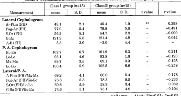
A, Lateral cephalogram measurements
Skeletal ClassM individuals had smaller length ofmaxilla (A-Ptm measuremenO, with
nificance at the 1"/( level,
B. Posteroanterior cephalogram (P. A.) measurements
kE}Zgtw-pF 25(2)•(3) 1999 129
Table1
Cephalometric measurements of skeletal Class I and ClassM groupsClassIgroup(n=15) ClassMgroup(n=15)
Measurement
mean
S.D.mean
S.D. tvalue rvalueLateral Cephalogram
A-Ptm (FH)
Pog-Ar (FH) S-Or (FH)G-Ba
A.B (FH) P. A. CephalogramEu-Eu
Lo-Lo
Mx-Mx
Go-Go
Lateral/P. A.A-Ptm (FH)/Mx-Mx
Pog-Ar (FH)/Go-Go S-Or (FH)ILo-Lo G-Ba (FH)/Eu-Eu 48.1 77.0 56.3 121.25.3
162.7 93.1 69.7 100.4 69.2 76.8 60.6 74.6 2.1 5.4 5.1 5.5 3.0 6.7 4.6 3.5 5.9 4.1 5.8 5.9 5.1 45.4 79.9 54.7 121.4 -2.0 161.9 92.8 69.1 102.5 66.0 78.3 58.9 75.1 1.9 5.8 2.8 4.8 4.4 6.3 1.8 3.3 6.6 5.4 8.5 3.4 4.9 ** O.398 -O.491 -O.O09 O.044 O.211 -O.123 O.125 -O.259 O.178 -O.233 O.062 -O.104unit : mm ttest:**pÅqO.Ol; *pÅqO.05
Table 2 Model measurements of skeletal Class I and ClassM groups
ClassIgroup(n=15) ClassMgroup(n=15)
Measurement
mean
S.D.mean
S.D. tvalue rvalueCoronal Arch
MaxillaCL
CW
cucw
TM
CIrrM
CWII]M
Mandible
CL
CW
cucw
TM
CIA]M
CWII]M
Basal ArchMaxi11a
BL
BW
BLXBW
BIA]M
BWA]M
Mandible
BL
BW
BIVBW
BurM
BWII]M
Mesial StepIM-IM
37.8 42.3 89.7 89.9 42.1 47.1 33.6 34.4 97.9 82.2 41.0 41.9 33.0 44.5 74.3 36.7 49.6 31.5 39.8 79.2 38.4 48.5278
1.9 2.5 5.5 4.e 1.6 2.6 1.5 1.6 4.6 2.8 2.1 2.2 1.1 3.2 5.4 2.0 3.9 1.6 1.6 3.0 2.0 2.2 O.96 36.9 42.9 86.0 89.7 41.2 47.9 32.5 34.8 93.7 82.3 39.5 42.3 31.7 44.9 70.6 35.4 50.1 33.3 42.0 79.4 40.5 51.1 5.59 2.0 2.3 4.0 3.7 2.9 3.1 1.3 2.0 6.5 3.5 1.8 2.7 1.9 2.1 4.2 2.6 3.4 1.2 1.4 2.4 1.4 2.5 2.11 * * * * * * ** ** ** * ** O.285 -O.085 O.352 -O.O02 O.262 -O.086 O.254 O.O12 O.166 -O.028 O.253 O.029 O.564 -O.076 O.513 O.329 -O.063 -O.495 -O.526 -O.044 -O.470 -O.430 -O.792130 NARUZAWA et al. : Relationship to Arch Form and Craniofacial Structures
C. Lateral1P. A. measurements ratio
Significant differences could not be found.
2) Modelanalysis
A. Coronal arch measurements
a) Maxi11a
Coronal length/coronal width ratio was greater for skeletal ClassM group, with
cance at the 59o level.
b) Mandible
Coronal length and coronal length/tooth material ratio measurements were smaller in
skeletal Class- , at the 59o level.
Coronal length/Coronal width ratio was bigger in skeletal Class M , significant at the 59o
level.
B. Basal arch measurements
a) Maxi11a
In skeletal ClassM individuals, basal length measurement was smaller, while basal
lengthlbasal width ratio was bigger, with significance at the 59o level.
b) Mandible
Basal length measurement (pÅqO.Ol), basal lengthltooth material ratio (pÅqO.Ol), basal
width measurement (pÅqO.Ol) and basal width/tooth material ratio (pÅqO.05) showed cantly bigger values in skeletal ClassM individuals.
C. Mesial step measurement
The distance between mesial surfaces ofupper and lower first molars was greater in skeletal ClassM individuals, with significance at the 19o level.
2 . Evaluation according to A-B (FH)-(Pearson's correlation coefficient)
Following, relevant findings ofPearson's correlation analysis are summarized (Figs. 6-10).
1) Cephalometricstudy
A. Lateral cephalogram measurements a) Maxi11a
Length of maxilla (A-Ptm (FH) measurement) and A-B (FH) showed correlation (r=
O.398). This means some tendency to become smaller with increasing ofClassM severity.
b) Mandible
Length of mandible (Pog-Ar (FH) measurement) and A-B (FH) showed correlation (r=
O.491). This means tended to become bigger as severity ofClassM.B. Posteroanterior cephalogram (P-A) measurements Significant correlation coefficients could not be found. C. Lateral /P. A. measurements ratio
Significant correlation coefficients could not be found.
2) Model study
A. Coronal arch measurements
a) Maxi11a
Coronal length/coronal width ratio and A-B (FH) showed correlation (r=O.352). This means tendency to become smaller with increasing ofClassM severity.
b) Mandible
kE5J4scts\ 25(2)•(3) 1999 131 (mm) 54
52
50
48
A-Ptm46
44
42
40
Fig. 6 (m m) 3635
34
33
32
BL 3130
29
28
%
o
oo
oo
o
-10 -8 -6 -4 -2 O 2 4 6 8 10 12
A-B(FM (mm)
Length ofMaxi11a : correlation analysis
between A-Ptm and A-B (FH)
-10 -8 -6 -4
-2 O 2 4 6 8 10 12
A-B (FH) (mm)
Fig. 8 Length ofMandibular basal arch length :
relation analysis between BL and A-B (FH)
{m m} 95 92.5
90
87.585
82.5 Pog-Ar sO 77.575
72.570
67.5 Fig. 7 (mm) 98
7
6
5
MS 4
3
2
1
o
Fig. 9 `f'%
-tO -8 -6 -4 -2 O 2 4 6 8
A-B (FH)Length of Mandible : correlation between Pog-Ar and A-B (FH)
10 12 (mm) analysis
o
o
oo
o
-tO -8 -6 -4 -2 O 2 A-B (FH}4 6 8 10 l2
(mm)
Mandibular basal arch width : correlation analysis between MS and A-B (FH)
(mm)45
44
4342
41 BW 4039
3B
37
36
Fig. 10s
0
0
-10 -8 -6 -4 -2 O 2 4 6 8 IO 12
A-B(FH) (mrn)
Mesial step : correlation analysis between
BW and A-B (FH) B. Basal arch measurements
a) Maxilla
Basal length measurement (r=O.564), basal length/basal width ratio (r=O.513) and A-B
(FH) showed strong correlation. This means a tendency to become smaller with increasing of
ClassM severity.
b) Mandible
Basal length measurement (r=O.495), basal length/tooth material ratio (r=O.470), basal width (r=O.526), basal width/tooth material ratio (r=O.430), and A-B (FH) showed
correla-132 NARUZAWA et al. : Relationship to Arch Form and Craniofacial Structures
tion. In this measurement, basal width showed strong correlation. This means tendency to
become bigger with increasing ofClassM severity. '
C. Mesial step measurement
The distance between mesial surfaces ofupper and lower first molars and A-B (FH) showed
strong correlation (r=O.792). This rneans tended to become bigger as severity of ClassM
creased.
Discussion
Difference among races seems to be relevant also in head and arches form. As studied by Farkas22'
in a cephalic index analysis, Caucasians have a trend toward mesocephalism, Chineses toward brachycephalism, while Africans have a dolichocephalism tendency. In a mode! measurements study
on normal occlusion samples, Aoki et al.23' reported that arch width is larger in Japaneses than in
Americans.
[Phe purpose ofthe present study was to evaluate anteroposterior relationship ofupper and lower jaws associated to the morphology of dental arches and craniofacial structures. Measurements were
taken on pretreatment cephalograms and plaster models from Japanese females with skeletal Class
I andClassM.
This study was canied out into two parts :
A sequential analysis of anteroposterior displacement ofjaws, using A-B (FH) measurement as parameter ;
A comparison between means ofClass I and ClassM groups.
Differences between the two ways of analyze might be explained by Jarvinen2`'s study, that ported that ANB angle and A-B (FH) are not always directly comparable. However, in statistical re-sults ofthis study, great differences were not found.
In this study, no significant difference on tooth material measurements between Class I and
ClassM groups was found, but values for both groups were greater than those found by Otsuboi6' on normal occlusion samples. These results confirm that tooth size is related to malocclusion.
Results ofthe present study are in according to findings ofBraun et al.25', which reports that Class
M individuals have smaller arch length and greater arch width of mandibular coronal arches than
Class I subjects. Maxillary coronal arch widths were similar in both groups.
Significant differences were found for basal arch measurements but not for coronal arch. It might be correlated to dental compensations, such as accentuated Spee Curve, dental crowding or tipping. Results ofthis study agree with findings ofRichardson et al.26', which observed lack ofcorrelation be-tween the size ofthe apical base, the alveolar arch and the dental arch.
Although strong correlation between head form and anteroposterior relationship of upper and
lower jaws was not found, results indicated that there were significant correlations in lengths of
maxillary and mandibular bones, and in basal arches measurements.
Skeletal Class M group, compared to Class I , have significantly smaller length of maxiIla and greater length ofmandible, and those tendencies increase with severity ofClassM malocclusion.
Moreover, basal arch length and width ofmandible were bigger in skeletal ClassM group, and
these measurements trend to become greater with increase ofClassM severity.
[rliese results suggest that skeletal ClassM, at least in this sample, might be associated to local malformation, considering two factors : the expression ofsize discrepancy between maxi11a and
man-ta71Åqdi:2i!-L 25(2)•(3) 1999 133
dible, apart ofgrowth of other craniofacial structures ; the anteroposterior alignment and rotation of maxi11a and mandible, contributing to create a Class M malocclusion.
References
1) Lew KKK, Foong WC and Loh E (1993) Horizontal skeletal typing in an ethnic Chinese
tion with true ClassM malocclusions. BrJ Orthod 20 : 19-23.
2) Endo T (1971) An epidemiological study ofreversed occlusion. Part 1. Incidence of reversed clusion in children 6 to 14 years old. J Jpn Orthod Soc 30 : 73-7.
3) Susami R, Asai Y, Hirose K, Hosoi T, Hayashi I and Takimoto T (1972) The prevalence clusion in Japanese school children. J Jpn Orthod Soc 31 : 319-24.
4) Kawahara Y, Yoshikawa Y, Obata A, Miyazaki A, Okafuji N, Ashizawa Y, Togari A and Deguchi
T (1996)A statistical observation of orthodontic patients during the twenty-year period in the
Department ofOrthodontics, Matsumoto Dental College Hospital (1972-1991). Matsumoto
gaku 22:44-51.
5) Graber TM (1966) Orthodontics, principles and practice, 2nd ed. 205-7. WB Saunders,
phia, U. S. A
6) Enlow DH (1990) Normal variation in facial form and the anatomic basis for malocclusions in
facial growth. 3rd ed., Chap 6 : 193-221, WB Saunders, Philadelphia, U. S. A.
7) Kondo E (1972) Posteroanterior cephalometric study ofcranio-facial and arch widths. J Jpn thod Soc 31 : 117-36.
8) Masaki F (1980) The longitudinal study of morphological differences in the cranial base and cial structure between Japanese and American white. J Jpn Orthod Soc 39 : 436-56.
9) Staley RN. Bishara SE, Hanson JW and Novak AJ. (1992) Craniofacial development in
tonic dystrophy. Cleft Palate Craniofacial J 29 : 456-62.
10) Miyajima K, McNamara JA, Sana M and Murata S (1997) An estimation ofcraniofacial growth
in the untreated Class M female with anterior crossbite, Am J Orthod Dentofacial Orthop 112 :
425-34.
11) Howes AE (1954)A polygon ofcoronal and basal arch dimensions in the horizontal plane. Am J Orthod Dentofacial Orthop 40 : 811-31.
12) Howes AE (1957) Arch width in the premolar region, still the major problem in Orthodontics.
Ain J Orthod Dentofacial Orthop 43 : 5-31.
13) Lavelle CL (1972) Maxi11ary and mandibular tooth size in different occlusion categories. Am J
Orthod Dentofacial Orthop 61 : 29-37.
14) Staley RN, Stuntz WR and Peterson LC (1985)A comparison ofarch widths in adults with
mal occlusion and adults with Class ll , division 1 malocclusion. Am J Orthod Dentofacial
thop 88: 163-9.
15) Raberin M (1993) Dimensions and form of dental arches in subjects with normal occlusions. Am J Orthod Dentofacial Orthop 104 : 67--72.
16) Otsubo J (1957) A study on the tooth material in Japanese adults of normal occlusion, its tionships to coronal and basal arches. J Jpn Orthod Soc 16 : 36-46.
17) Jacobson A (1975) The "Wits" appraisal ofjaw disharmony. Am J Orthod Dentofacial Orthop
67:125-38.
Quintes-134 NARUZAWA et al. : Relationship to Arch Form and Craniofacial Structures
sence, Tokyo, Japan.
19) Otsubo J (1960) Device for measuring ofcoronal and basal arch lengths. J Jpn Orthod Soc 19 :
159.
20) Freeman RS (1981) Adjusting ANB angles to reflect the effect ofmaxiIlary position. Angle
thod 51 : 162-71.
21) Deguchi T (1982) Adjustments of FH-SN angle and ANB angle to cephalometrics analysis of
horizontal jaw relation. J Jpn Orthod Soc 41 : 757-64.
22) Farkas LG (1994) Anthropometry of the head and face.2nd ed., 201-18, Raven Press, New York, U. S. A.
23) Aoki H, Tsuta A and Ukiya M (1971) A morphological study and comparison of the dental arch form ofJapanese and American adults. detailed measurements ofthe transverse width, Bull.
Tokyo Dent Coll 12:9-14.
24) Jarvinen S (1988) Relation ofthe Wits appraisal to the ANB angle, a statistical appraisal. Am J
Orthod Dentofacial Orthop 94 : 432-5.
25) Braun S, Hnat WP, Fender DE and Legan HL (1998) T[he form of the human dental arch. Angle Orthod 68 : 29-36.
26) Richardson ER and Brodie AG (1964) Longitudinal study of growth of maxillary width. Angle
Orthod 34: 1-15.
}!Årst : H Zs.A. skeletal I , skeletal Mfitiil c7) valjIB t tRgfiimeer. C: 6' Ct 6 ffJ. 24;SjEe9Pfiff.c:PS 'U' 6Mvt,.'
nt tFI P /Si ]V 5r ee #! i[i, kU eeri, S;• Ll.l mai, Lfl Mik tsi• 2, pt tn =- RKi
(thJ4gdiJk • en-asIEi, d-x F'7 ;i • tft"/SJv • d ;/ JÅqi d i= t- 5 •Zfityptx2)
Khft PH7 ONfa a:DvÅr '( , M,E.i2. 'Li!i=S' C: Ss' Vt 6 ZÅq iE PNA, ts L Åq li wh ijiJ ii:} op ts tlZ MI t: ma me L te bl ojtti]tt 'c•, 7 i"7(D5tdiJ.NcDptHietsttIewll, ts.wtgyop.wheeJEf,ee..efik-3-ta[ti]rdSIE,b, JJScgvi*ijilEI;rpMk.t6Ddicaa:est{Z
tlkto{fi' Åq, JrÅqiEPtha#li, skeletalMfi&Jrb{{Svi, 8v,5{NMh{XEiestz(vÅr6. J2IscMt7tat (D a e9 el , tw gtl EI , -t -F tiwa •t"!- (1) ft ff, tpdi- ts., a) JES rv.ta. e9 ttI tw jts X Ut', rk/s tk ts ts ea es. ve : Cffi -g- 6 e
teHssbi6.
IilfOt'a:a)st*}lt, HJzlg.-gl(Ml'(S, skeletalIfitlen, skeletalMfitlffJrertXtL, iflfiut,J(DgeInjtsXU"'IE
ut (D .rk zz x wtstift#fi 8 k[iEcb'•t'A wtHI Nveiftag {? st*} t L ti .
ptkJE;fi.ts.tl -teevaa)nt,s{fte9tscaree:fo'vÅr'(, whviJfflEmaes.(t, ftP,beÅr62ztsto}ok. Lth)L, skeletal
MewtftE-f•LEeFL4eYa: skeletal I geX OLtilj•l5-ftk.tisiÅq , -F.vaeftKelftVitsgee,7-JiL, Jff]eea:Jfi'fi..rerd{ trNbeÅr6;it.t:. " esaz, -FljvawhJtwgXiil}ftts'ltffÅq, tpdi'ts'etE vÅrSSM)2 fikL, skeletalmti'Jv-- fe: }sv
"(N ma ee ca rb{"Ep. to 6 n tc .
C 2z 6Okt ge thi C5 , skeletal N at i7I" ts Åq E ts : cD at *} e: ts v: '( , fi il}f ss ts JV).R ec Am!" q) et pa t: ee if L '(