IDENTIFICATION OF NOVEL GENETIC MUTATIONS IN JAPANESE PATIENTS WITH SEVERE
CONGENITAL HYPOTHYROIDISM
Hiroyuki Adachi, Ikuko Takahashi, Hirokazu Arai and Tsutomu Takahashi (received 17 May 2013, accepted 12 June 2013)
Department of Pediatrics, Akita University Graduate School of Medicine, Akita 010
-8543, Japan
Abstract
Objective : The prevalence of genetic mutations in congenital hypothyroidism (CH) remains undetermined. The objective of this study was to determine the prevalence of mutations in DUOX2, TSHR, TG, PAX8, and TPO among severe permanent primary CH.
Methods : Between April 1999 and March 2011, 114,733 newborns were screened for CH in Akita Prefecture, Japan. Among them, 330 were suspected of having CH and were referred to pediatricians. We recruited 40 patients who were referred to our institute. Among them, we identified 9 permanent primary CH patients who were severely affected with an initial TSH ≥20 mU/l upon newborn screening and performed direct sequencing of the 5 candidate genes.
Results : 3 of 9 patients (33%) had mutations in PAX8, TPO, and TSHR. Among the severely affected subjects, 60% had thyroid dysgenesis (TD), while for patients with initial TSH upon screening <20 mU/l only 12% had TD.
Conclusions : Despite the high frequency of TD, the detection rate of mutations among severe permanent primary CH was higher than expected. This study suggests that the genetic analysis of 5 genes, namely, DUOX2, TSHR, TG, PAX8, and TPO, is useful for the diagnosis of CH, and that the actual prevalence of genetic mutations among CH might be higher than as previously estimated.
Key words : congenital hypothyroidism, genes, mutation, prevalence
Correspondence : Ikuko Takahashi, M.D.
Department of Pediatrics, Akita University Graduate School of Medicine, 1
-1
-1 Hondo, Akita 010
-8543, Japan Tel : 81
-18
-884
-6159
Fax : 81
-18
-836
-2620
E
-mail : [email protected]
-u.ac.jp
cent evidence points to the possibility of a genetic com- ponent
1). One study reported that 2% of CH patients with TD have a positive familial history
3). In contrast, DH is generally transmitted in an autosomal recessive manner. Hereditary defects in virtually all of the steps of thyroid hormone biosynthesis and secretion have been described
1).
The prevalence of single genetic mutations in CH re- mains undetermined. Previous molecular genetic stud- ies have shown that a subset of TD is caused by at least 4 genetic mutations, including TSHR
4-6), PAX8
7), NKX2
-1
8), and FOXE1
9). Similarly, at least 7 genes have been im- plicated in DH, including TG
10), TPO
11), SLC5A5
12), SL- C26A4
13), DUOX2
14), DUOXA2
15), and IYD
16). A previous review estimated the prevalence of single genetic muta- tions in CH to be 5
-10%
17). However, only a few genetic Introduction
Congenital hypothyroidism (CH) is a highly heteroge-
neous disorder. CH is classified into permanent and
transient CH, which in turn is divided into primary, sec-
ondary, or peripheral CH. Most cases of CH are due to
primary causes
1). Thyroid dysgenesis (TD) accounts for
75
-85% of permanent primary CH, while thyroid dyshor-
monogenesis (DH) accounts for 15
-20% of cases
2). TD
is generally thought to be a nongenetic disease, but re-
screening studies of CH patients have been conducted and the actual prevalence of most genetic mutations among all CH patients or in the general population has not been investigated.
Recently, Japanese studies have confirmed that more than 20% of permanent primary CH patients have single genetic mutations as determined systematic genetic screening
20-22). These studies have reported the preva- lence of different genetic mutations among 102 CH patients : in descending order of frequency, they occur in DUOX2, TSHR, TG, PAX8, and TPO.
In this study, we examined the prevalence of genetic mutations in DUOX2, TSHR, TG, PAX8, and TPO among severe permanent primary CH patients with an initial TSH ≥20 mU/l upon newborn screening in our institute.
Materials and Methods Subjects
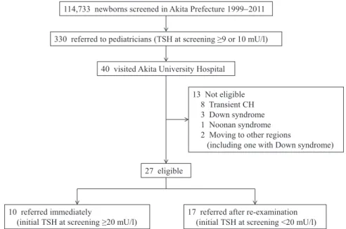
Between April 1999 and March 2011, 114,733 new-
borns were screened for CH in Akita Prefecture, which is a northern area of Japan (Fig. 1). Among them, 330 with TSH upon screening (whole blood) ≥10 mU/l (April 1999 to July 2004) or ≥9 mU/l (August 2004 to March 2011) were suspected of having CH and were referred to pediatricians. We recruited 40 patients who were re- ferred to the Department of Pediatrics, Akita University Hospital. Of them, 13 were excluded because they had transient CH, chromosomal abnormalities, and showed movement to other regions. The remaining 27 patients were diagnosed with or suspected of having permanent primary CH and were enrolled in this study. Moreover, the participants were classified into 2 groups according to the severity of their disease : 10 patients had initial TSH upon screening ≥20 mU/l (i.e., referred immediately) and 17 patients had initial TSH upon screening <20 mU/l (i.e., referred after re
-examination).
We selected the 10 severely affected CH patients with initial TSH upon screening ≥20 mU/l for genetic analysis. Written informed consent to participate in the
Fig. 1. Study subject enrollment. In total, 114,733 newborns were screened for CH in Akita Prefecture between April 1999 and March 2011. Three hundred thirty newborns with TSH upon screening (whole blood) ≥10 mU/l (April 1999 to July 2004) or ≥9 mU/l (August 2004 to March 2011) were referred to pediatricians. Among them, 40 were referred to our institute. Of these patients, 27 were diagnosed with or suspected of having permanent primary CH and were enrolled in this study. Moreover, according to the severity of their disease, the participants were classified into 2 groups : 10 patients with initial TSH upon screening ≥20 mU/l (i.e., referred immediately) and 17 patients with initial TSH upon screening <20 mU/l (i.e., referred after re
-examination).
114,733 newborns screened in Akita Prefecture 1999−2011
13 Not eligible 8 Transient CH 3 Down syndrome 1 Noonan syndrome 2 Moving to other regions
(including one with Down syndrome) 27 eligible
10 referred immediately
(initial TSH at screening ≥20 mU/l) 17 referred after re-examination (initial TSH at screening <20 mU/l) 40 visited Akita University Hospital
330 referred to pediatricians (TSH at screening ≥9 or 10 mU/l)
Fig. 1
study was obtained from the patients or their parents.
This study was approved by the ethical committee of the Akita University Graduate School of Medicine.
Evaluation of patients
During the first visit to our institute, the patients were evaluated for serum levels of TSH, free triiodothyronine (FT3), free thyroxine (FT4), and if possible, thyro- globulin. Thyroid morphology was evaluated by using ultrasonography on all of the patients. For those pa- tients that were 3
-6 years of age, their thyroid function was reevaluated after discontinuation of treatment to dis- tinguish between permanent and transient CH. The pa- tients were evaluated for serum levels of TSH, FT3, FT4, and thyroglobulin. Thyroid morphology was evalu- ated using both ultrasonography and
123I scintigraphy.
The patients who had serum TSH at reevaluation ≥5 mU/l or who had TD were diagnosed with permanent pri- mary CH.
If
123I uptake was increased (≥20%), a perchlorate dis- charge test was performed. A perchlorate discharge rate >90% indicates the presence of a total iodine organi- fication defect, whereas a rate of 10
-90% indicates the presence of a partial iodine organification defect.
Candidate genes
The patients who were diagnosed with or suspected of having permanent primary CH were divided into 3 subgroups : TD (i.e., hypoplasia, aplasia, or ectopia), DH (i.e., iodine organification defect, thyroglobulin synthesis defect, or others), and normal thyroid morphology.
TSHR and PAX8 were sequenced for the TD or normal thyroid morphology patients. TPO and DUOX2 were sequenced for DH patients who had an iodine organifica- tion defect. TG was sequenced for DH patients who had a thyroglobulin synthesis defect and consequently have typically very low levels of thyroglobulin. Other forms of DH were not included in the study.
Genetic analysis
Genomic DNA was extracted from peripheral blood leukocytes by following a standard technique. Amplifi- cation of coding exons and the exon
-intron boundary of the candidate genes was performed by using the poly-
merase chain reaction (PCR) with genomic DNA. The PCR products were purified by using Wizard SV Gel and a PCR Clean
-Up System (Promega, Madison, WI, USA).
They were then sequenced directly with an ABI Prism BigDye Terminator Cycle Sequencing Ready Reaction Kit (Applied Biosystems, Foster City, USA) by using an automated sequencer ABI Prism 310 Genetic Analyzer (Applied Biosystems).
In silico analysis of novel genetic mutations In order to predict the functional effects of the novel genetic mutations that we identified, we performed in silico analysis by using PolyPhen
-2 (http://genetics.bwh.
harvard.edu/pph2/)
23). This online tool can be used to predict the possible impact of an amino acid substitution on the structure and function of a human protein. The genetic mutations were classified as being “benign”,
“possibly damaging”, or “probably damaging”. In addi- tion to the PolyPhen
-2 analysis, we investigated the spe- cies conservation among human and other vertebrates of the mutated amino acids by using Evola (http://www.h
-invitational.jp/evola/)
24).
Results
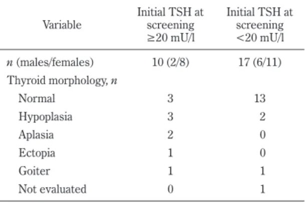
Evaluation of patients and genetic analysis The thyroid morphology of the 2 groups with 27 partic- ipants is shown in Table 1. Among the patients with ini- tial TSH upon screening ≥20 mU/l, 6 of 10 patients (60%) had TD. In contrast, among the patients with ini- tial TSH upon screening <20 mU/l, only 2 of 17 patients
Fig.Table 1
Table 1. Thyroid morphologies of the participants
Variable Initial TSH at screening
≥20 mU/l
Initial TSH at screening
<20 mU/l n (males/females) 10 (2/8) 17 (6/11) Thyroid morphology, n
Normal 3 13
Hypoplasia 3 2
Aplasia 2 0
Ectopia 1 0
Goiter 1 1
Not evaluated 0 1
(12%) had TD.
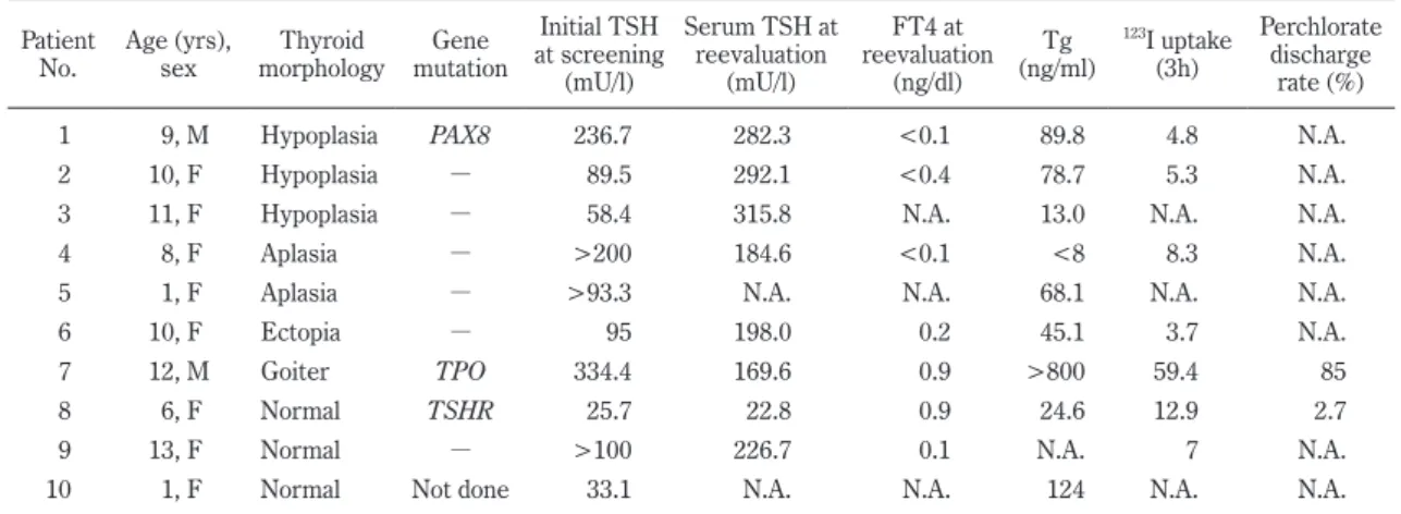
The clinical phenotypes of the patients who had initial TSH upon screening ≥20 mU/l are shown in Table 2.
For patient 1
-6 who had TD and patient 8 and 9 who had normal thyroid morphology, TSHR and PAX8 were sequenced. For patient 7 who had DH that was indica- tive of a partial iodine organification defect, TPO, and DUOX2 were sequenced. For patient 10 who had nor- mal thyroid morphology, genetic analysis could not be conducted because she was an infant at the time of en- rollment and her thyroid function could not be reeva- luated. No patients were suspected to have a thyroglob- ulin synthesis defect.
Three of 9 patients (33%) had mutations in PAX8, TPO, and TSHR (Table 2). The results of their se- quences and family studies of the mutation carriers are shown in Fig. 2 and Fig. 3, respectively. Patient 1 suf- fered from thyroid hypoplasia and had a novel PAX8 mu- tation (heterozygous for p.I34K) (Fig. 2). This missense mutation comprised 2 consecutive point mutations : c.101T > A and c.102C > A. Both of the patient’s par- ents had none of these point mutations (Fig. 3). Patient 7 suffered from a partial iodine organification defect and had a novel TPO mutation (compound heterozygous for p.R540X and p.W732L) (Fig. 2). The nonsense mutation for p.R540X was previously reported
25), but the missense mutation for p.W732L was novel. The mother of this patient was heterozygous for p.R540X and the father was
heterozygous for p.W732L (Fig. 3). Patient 8 exhibited normal thyroid morphology and had a TSHR mutation (homozygous for p.R450H) (Fig. 2), which is common in Japan
26). The genotypes of the parents were not deter-
Fig. 2. Three genetic mutations identified in severe permanent primary CH patients. The identified PAX8 mutation is heterozygous for a novel mutation p.I34K. This missense mutation comprises 2 consec- utive point mutations : c.101T > A and c.102C > A.
The identified TPO mutation is compound heterozy- gous for p.R540X and p.W732L with the latter being a novel mutation. The identified TSHR mutation is homozygous for p.R450H, which is common in Japan.
Fig. 2
PAX8 TSHR
TPO Heterozygous
I34K Homozygous
R450H
Heterozygous
R540X Heterozygous
W732L Table 2. The clinical phenotypes of the patients with initial TSH at screening ≥20 mU/l Patient
No. Age (yrs),
sex Thyroid
morphology Gene mutation
Initial TSH at screening
(mU/l)
Serum TSH at reevaluation
(mU/l)
FT4 at reevaluation
(ng/dl)
(ng/ml) Tg
123
![[原著]Two novel mutations of the FGDl gene in Japanese patients with Aarskog Scott syndrome: 沖縄地域学リポジトリ](data:image/gif;base64,R0lGODlhAQABAIAAAP///wAAACH5BAEAAAAALAAAAAABAAEAAAICRAEAOw==)