Short Report
Mol Syndromol 2018;9:214–218 DOI: 10.1159/000489055
Biallelic WRN Mutations in Newly
Identified Japanese Werner Syndrome Patients
Yoshiro Maezawa
aHisaya Kato
aMinoru Takemoto
a, bAki Watanabe
aMasaya Koshizaka
aTakahiro Ishikawa
aForough Sargolzaeiaval
fMasafumi Kuzuya
cHiroshi Wakabayashi
dTakashi Kusaka
eKoutaro Yokote
aJunko Oshima
a, fa Department of Clinical Cell Biology and Medicine, Graduate School of Medicine, Chiba University, Chiba ,
b Department of Medicine, International University of Health and Welfare, Narita , c Department of Community Healthcare and Geriatrics, Nagoya University Graduate School of Medicine, Nagoya , d Okayama City General Medical Center, Okayama , and e Department of Pediatrics, Kagawa University, Kagawa , Japan; f Department of Pathology, University of Washington, Seattle, WA , USA
classical WS features, further confirmed the pathogenic na- ture of this variant. For the case with c.3233+1G>T, we deter- mined the phase of 2 disease-causing mutations and dem- onstrated that they are on different chromosomes. This as- say would be particularly important for those cases with ambiguous clinical diagnosis.
© 2018 S. Karger AG, BaselSegmental progeroid syndromes are a group of genet- ic disorders in which affected individuals exhibit progres- sive systemic deteriorations characterized by accelerated aging [Hisama et al., 2016]. The best-known example is Werner syndrome (WS; OMIM 277700) caused by bial- lelic pathogenic variants of the WRN gene [Goto et al., 2013; Oshima et al., 2016, 2017]. WS patients develop an aged appearance and age-related disorders such as ocular cataracts, graying and loss of hair, atrophic skin, osteopo- rosis, atherosclerosis, and malignancies at earlier age. The Keywords
Mendelian disorder · Progeroid syndrome · Werner syndrome · WRN
Abstract
Werner syndrome (WS) is a rare autosomal recessive disorder characterized by systemic accelerated aging. It is caused by pathogenic variants of the WRN gene that encodes a nuclear helicase. In this report, we describe 4 newly identified WS cases among those referred to the Japanese Werner Consor- tium, Chiba University, Japan. All 4 cases were compound heterozygotes of the Japanese founder mutation, c.3139–
1G>C, and a novel null pathogenic variant, c.1587G>A, c.2448+1G>A, or c.3233+1G>T, or an amino acid sub- stitution variant, c.1720G>A, p.Gly574Arg. These 3 null pathogenic variants were not previously described. The p.Gly574Arg was previously reported in a European patient, and the identification of the second p.Gly574Arg case, with
Accepted: November 14, 2017 by M. Schmid
Published online: May 15, 2018
Junko Oshima © 2018 S. Karger AG, Basel
most common causes of death are myocardial infarction and cancer at a median age of 54 years [Huang et al., 2006;
Goto et al., 2013]. The Japanese Werner Consortium, Chiba University, in Chiba, Japan, proposed diagnostic criteria consisting of 6 cardinal symptoms: progeroid change of hair, cataracts, changes of skin including in- tractable skin ulcer, soft-tissue calcification, and a bird- like abnormal face [Takemoto et al., 2013].
The WRN gene encodes a nuclear DNA helicase with exonuclease activity which participates in DNA repair during various DNA transactions [Croteau et al., 2014;
Shamanna et al., 2017]. More than 80 different WRN pathogenic variants have been reported from all over the world [Huang et al., 2006; Yokote et al., 2017]. Due to the presence of founder mutations, Japan and the region of Sardinia in Italy are among countries with the highest fre- quencies of WS [Satoh et al., 1999; Masala et al., 2007;
Goto et al., 2013]. Another potential founder mutation has been reported in India/Pakistani patients, although WS may be underdiagnosed due to the relatively low awareness of this disorder [Saha et al., 2013]. A recent re- port showed that the increase of compound heterozy- gotes and decrease of homozygotes among those cases re- ferred to the Japanese Werner patients likely reflects the social trend of a decrease in consanguineous marriages [Yamaga et al., 2017]. Here, we report the results of muta- tion analyses of WRN loci in 4 newly identified WS pa- tients referred to the Japanese Werner Consortium. All 4 patients were compound heterozygotes of the Japanese founder mutation and the other pathological variant, 3 of which have not been described previously.
Materials and Methods
Patient Recruitment
Japanese WS patients were anonymously referred to the Japa- nese Werner Consortium by physicians who requested a molecular confirmation of a
WRN mutation. Consent forms obtained by thephysicians follow local regulations. After enrollment, blood samples collected from the patients were shipped to us for genetic testing.
Genetic Analysis
Blood samples were processed as described previously [Huang et al., 2006]. Genomic DNA was subjected to Sanger sequencing of 36
WRN exons as described before.For the phase study of a compound heterozygote, the region of intron 25–26 of the
WRN gene was amplified from the patient’sDNA. The PCR product was then subcloned into pBlueScriptII-KS linearized with XhoI and HindIII using the Gibson assembly sys- tem according to the manufacturer’s instruction (New England Biolabs, Cata#E2611) [Gibson et al., 2009]. Primers used for the amplification of
WRN exon 26 for Gibson assembly were: I25-F:ACGACTCACTATAGGGCGAATTGGAGCTC GGTAAACAG- TGTAGGAGTCTG ; I26-R: CCTCGAGGTCGACGGTATCGA- T AAGCTT CTTG TGAGAGGCCTATAAACT . The underlines indicate the sequences in the
WRN gene, and the regions withoutunderlines are the sequence of pBlueScript linearized with XhoI and HindIII. The assembled DNA was used for
Escherichia colitransformation, and the plamids isolated from 8 bacterial colonies were analyzed by Sanger sequencing.
Results
The first case is a 62-year-old Japanese woman. Her medical history included bilateral cataract at 42 years of age, diabetes mellitus at age 53, and calcification in the left Achilles tendon at age 55. She subsequently developed a refractory skin ulcer and underwent left leg amputation.
At her first visit to our hospital, she exhibited short statue, a high-pitched voice, a bird-like facial appearance, thin extremities, and refractory skin ulcers on her right foot.
Her height was 1.46 m, weight was 35.2 kg, and her BMI was 14.4 kg/m
2. Her diabetes was controlled by intensive insulin therapy, the HbA1c was maintained less than 7.0%. X-ray examination revealed massive calcification in the right Achilles tendon, which is highly characteristic of WS [Takemoto et al., 2013], and ultrasound demonstrated 1.6 mm atherosclerotic plaque in the left carotid artery ( Fig. 1 ).
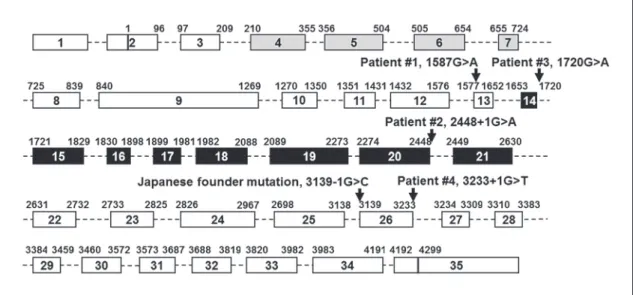
Sanger sequencing of WRN exons in the abovemen- tioned case revealed a novel heterozygous variant, c.1587G>A, p.Trp529 * in exon 13, and a heterozygous Japanese founder mutation, c.3139–1G>C, which results in the deletion of exon 26 (r.3139_3233del95) followed by the premature termination, p.Gly1047Phefs * 14 ( Fig. 2 ).
A B
Fig. 1.
Radiographic findings of a newly identified Werner patient.
A
Calcification in the right Achilles tendon.
BAtherosclerotic
plaque in the left carotid artery.
The second case was referred to us with a clinical diag- nosis of WS, and the sequencing analysis showed a het- erozygous Japanese founder mutation and a novel het- erozygous variant, c.2448+1G>A, which results in the skipping of exon 20 (r.2274_2448del175) followed by the premature termination, p.Ser759Valfs * 3 ( Fig. 2 ).
The third case was referred to us with a clinical diagno- sis of WS, presenting with all 6 cardinal symptoms of the syndrome proposed by the Japanese Werner Consortium.
This patient carried a heterozygous Japanese founder mutation and a compound missense variant, c.1720G>A,
p.Gly574Arg. The p.Gly574Arg was previously reported in a single German patient as a compound heterozygous mu- tation [Tadokoro et al., 2013; Yokote et al., 2017]. Although biochemical studies had already demonstrated the abroga- tion of enzymatic activities in a recombinant WRN protein with p.Gly574Arg [Tadokoro et al., 2013], the identifica- tion of the second p.Gly574Arg case further strengthens the notion of the pathogenicity of this variant and the loss of enzymatic activity as the cause of the WRN mutation.
The fourth case also presented with classical features of WS and was referred to us for genetic testing. This pa-
Fig. 2.
WRN mutations found in newly identified Werner patients. A diagram of the WRN gene is shown with
the locations of mutations described in this study. Boxes indicate 35 exons and dotted lines indicate introns. Gray and black boxes are exonuclease and helicase regions, respectively. Lengths of introns and exons do not show the actual nucleotide lengths.
Fig. 3.
Phase determination of the compound heterozygous mutations, c.3233+1G>T and c.3139–1G>C. Top
lines are amino acid number and sequence of the WRN protein and nucleotide number of coding exon 25 (exon
26). Sequence results of patient DNA and each of 2 alleles are shown with locations of pathogenic variants in red
squares.
tient carried a heterozygous Japanese founder mutation, c.3139–1G>C, and a novel heterozygous variant, c.3233+1G>T ( Fig. 3 ). The c.3139–1G>C is located at 5 ′ of exon 26, and c.3233+1G>T is located at 3 ′ of exon 26, both are expected to cause a deletion of exon 26. Parental samples were unable to be obtained to determine the phase of 2 pathogenic variants. We therefore opted to separate 2 alleles by PCR subcloning using Gibson assem- bly. Following the bacterial transformation and plasmid isolation, 4 out of 8 clones had the c.3139–1G>C change, but not c.3233+1G>T (allele 1 in Fig. 3 ); 3 out of 8 clones had c.3233+1G>T, but not c.3139–1G>C (allele 2 in Fig. 3 ), and 1 did not have the insert, roughly falling into the expected 1: 1 ratio of 2 alleles. This confirms the com- pound heterozygous status of WRN mutations in this pa- tient.
Discussion
We described 3 newly identified null WRN pathogen- ic variants and a previously reported amino acid substi- tution variant found in Japanese WS patients. All were found as one of the compound heterozygous changes in combination with the Japanese founder mutation. It has been noted that the proportion of compound heterozy- gotes increased from 14.2% in 1997 to 31.8% in 2017 in Japanese WS patients. Reciprocally, the homozygotes of the Japanese founder mutation decreased from 73.2 to 63.6% during the same 20-year period [Yamaga et al., 2017]. Considering the trend in Japan to fewer consan- guineous marriages, an increase in compound heterozy- gotes and corresponding decrease in homozygotes is to be expected [Nalls et al., 2009].
Most of the pathogenic variants of the WRN gene were null mutations, either splicing, stop codon, or small in- dels [Yokote et al., 2017]. Amino acid substations within the exonuclease domain found in a German patient causes protein instability [Huang et al., 2006]; thus, they were also functionally null. There have been only 2 likely pathogenic missense variants, namely p.Arg637>Trp and p.Gly574Arg [Uhrhammer et al., 2006; Tadokoro et al., 2013; Yokote et al., 2017], both of which were found in the compound heterozygotes with null mutations. Of those, only p.Gly574Arg was pathogenic [Tadokoro et al., 2013]. Our patient with p.Gly574Arg had all 6 cardinal symptoms, indicating that the combination with a null mutation is sufficient to develop typical WS features.
When a patient presents with classical features of WS, the presence of 2 heterozygous WRN mutations is gener-
ally considered sufficient to make a diagnosis of WS. We, however, felt it is necessary to determine the phase of 2 heterozygous mutations, c.3233+1G>T and c.3139–
1G>C, because both of them result in the skipping of exon 26. Strictly speaking, this should be done for all com- pound heterozygotes when technically feasible. In fact, recommendation of the description of sequence variation (www.hgvs.org/mutnomen/recs-DNA.html#DNA) clearly distinguishes compound heterozygosity with known phases, e.g., c.[3233+1G>T];[3139–1G>C], versus unknown phases, e.g., c.[3233+1G>T(;)3139–1G>C].
Such assay is necessary for the cases with ambiguous clin- ical diagnosis and may become a part of routine proce- dure in the future, as technology progresses.
Acknowledgments
We thank Ms. Julia S. Appelbaum for her editorial assistance.
This work was supported by the grants NIH/NCI R01CA210916 and JSPS KAKENHI 17H04037 to J.O. as well as 17H01558 to K.Y, and the grants from KAKENHI on Innovative Areas “Stem Cell Aging and Disease 26115009,” from AMED 17bm0804016h0001 and 17ek0109126h0003, the Health and Labor Sciences Research 15545420, and from the Project for Elucidating and Controlling Mechanisms of Aging and Longevity, AMED, all to K.Y.
Statement of Ethics
The study was conducted according to the Declaration of Hel- sinki. Written informed consent was obtained prior to clinical pro- cedures. This study was approved by the Internal Review Board of the Chiba University, Japan.
Disclosure Statement
The authors declare no conflicts of interest.
References
Croteau DL, Popuri V, Opresko PL, Bohr VA:Human RecQ helicases in DNA repair, re- combination, and replication. Annu Rev Bio- chem 83: 519–552 (2014).
Gibson DG, Young L, Chuang RY, Venter JC, Hutchison CA 3rd, Smith HO: Enzymatic as- sembly of DNA molecules up to several hun- dred kilobases. Nat Methods 6: 343–345 (2009).
Goto M, Ishikawa Y, Sugimoto M, Furuichi Y:
Werner syndrome: a changing pattern of clin- ical manifestations in Japan (1917 ∼ 2008).
Biosci Trends 7: 13–22 (2013).
Hisama FM, Oshima J, Martin GM: How research on human progeroid and antigeroid syn- dromes can contribute to the longevity divi- dend initiative. Cold Spring Harb Perspect Med 6:a025882 (2016).
Huang S, Lee L, Hanson NB, Lenaerts C, Hoehn H, et al.: The spectrum of WRN mutations in Werner syndrome patients. Hum Mutat 27:
558–567 (2006).
Masala MV, Scapaticci S, Olivieri C, Pirodda C, Montesu MA, et al: Epidemiology and clinical aspects of Werner’s syndrome in North Sar- dinia: description of a cluster. Eur J Dermatol 17: 213–216 (2007).
Nalls MA, Simon-Sanchez J, Gibbs JR, Paisan- Ruiz C, Bras JT, et al: Measures of autozygos- ity in decline: globalization, urbanization, and its implications for medical genetics. PLoS Genet 5:e1000415 (2009).
Oshima J, Martin GM, Hisama FM: Werner Syn- drome, in Pagon RA, Adam MP, Ardinger HH, Wallace SE, Amemiya A, et al (eds):
GeneReviews ® [Internet] (University of Washington, Seattle 1993–2018) Initial post- ing: Dec 2, 2002; last update: Sept 29, 2016.
Oshima J, Sidorova JM, Monnat RJ Jr: Werner syndrome: clinical features, pathogenesis and potential therapeutic interventions. Ageing Res Rev 33: 105–114 (2017).
Saha B, Lessel D, Nampoothiri S, Rao AS, Hisama FM, et al: Ethnic-specific WRN mutations in South Asian Werner syndrome patients: po- tential founder effect in patients with Indian or Pakistani ancestry. Mol Genet Genomic Med 1: 7–14 (2013).
Satoh M, Imai M, Sugimoto M, Goto M, Furuichi Y: Prevalence of Werner’s syndrome hetero- zygotes in Japan. Lancet 353: 1766 (1999).
Shamanna RA, Croteau DL, Lee JH, Bohr VA: Re- cent advances in understanding Werner syn- drome. F1000Res 6: 1779 (2017).
Tadokoro T, Rybanska-Spaeder I, Kulikowicz T, Dawut L, Oshima J, et al: Functional deficit associated with a missense Werner syndrome
mutation. DNA Repair (Amst) 12: 414–421 (2013).
Takemoto M, Mori S, Kuzuya M, Yoshimoto S, Shimamoto A, et al: Diagnostic criteria for Werner syndrome based on Japanese nation- wide epidemiological survey. Geriatr Geron- tol Int 13: 475–481 (2013).
Uhrhammer NA, Lafarge L, Dos Santos L, Domaszewska A, Lange M, et al: Werner syn- drome and mutations of the WRN and LMNA genes in France. Hum Mutat 27: 718–719 (2006).
Yamaga M, Takemoto M, Takada-Watanabe A, Koizumi N, Kitamoto T, et al: Recent trends in WRN gene mutation patterns in individu- als with Werner syndrome. J Am Geriatr Soc 65: 1853–1856 (2017).
Yokote K, Chanprasert S, Lee L, Eirich K, Take- moto M, et al: WRN mutation update: muta- tion spectrum, patient registries, and trans- lational prospects. Hum Mutat 38: 7–15 (2017).