福島県立医科大学 学術機関リポジトリ
This document is downloaded at: 2021-11-08T00:13:29Z
Title Phosphodiesterase 3A1 protects the heart against angiotensin II‒induced cardiac remodeling through regulating transforming growth factor-β expression( 本文 )
Author(s) 岩谷, 章司
Citation
Issue Date 2014-03-25
URL http://ir.fmu.ac.jp/dspace/handle/123456789/599
Rights © 2014 by the International Heart Journal Association
DOI
Text Version ETD
Phosphodiesterase 3A1 protects the heart against angiotensin II-induced cardiac remodeling through regulating transforming growth factor-β expression
Shoji Iwaya
Department of Cardiology and Hematology, Fukushima Medical University, 1 Hikarigaoka, Fukushima, 960-1295, Japan
One table and four figures are contained in this paper.
This manuscript was accepted in International Heart Journal on September
19th, 2013.
Summary
Accumulating evidence suggests that there are direct interactions
between β-adrenergic and angiotensin II signaling pathways, and β-blocker
protects hearts against angiotensin II-induced cardiac remodeling.
Phosphodiesterase 3A (PDE3A) regulates β-adrenergic receptor/protein
kinase A signaling by metabolizing cAMP. Therefore, we hypothesized that
overexpressed PDE3A has cardioprotective effects against angiotensin
II-induced cardiac remodeling by regulating angiotensin II signaling. In the
present study, we used transgenic mice with cardiac-specific overexpressed
PDE3A1. Continuous administration of angiotensin II caused cardiac
hypertrophy in the wild-type mouse heart, but not in the transgenic mouse
heart. Angiotensin II induced cardiac fibrosis in both wild-type and
transgenic mice, but the extent of fibrosis was less in transgenic mice
compared to wild-type mice. Moreover, basal expression levels of
transforming growth factor-β were lower in transgenic mouse hearts, and it
remained lower levels after angiotensin II stimulation. These findings
suggest that PDE3A protects the heart from angiotensin II-induced cardiac
remodeling through modulating the functional connection between
angiotensin II and transforming growth factor-β.
Keywords
Angiotensin II; phosphodiesterase 3A; transforming growth factor-β; cardiac
fibrosis
Introduction
Cardiac remodeling occurs in several clinical conditions, such as
myocardial infarction, cardiomyopathy, and valvular heart diseases, leading
to subsequent heart failure.1) Sympathetic nervous system and
renin-angiotensin system (RAS) are important contributors in the
development of cardiac remodeling. Sympathetic nerve ending secretes
noradrenaline, and it binds to β-adrenergic receptor (β-AR) on cardiac
myocytes to increase in cyclic adenosine 3’ ,5’-monophosphate (cAMP) levels
through activating adenylyl cyclases. Cyclic nucleotide is hydrolyzed by
phosphodiesterases (PDEs), which constitute a superfamily of enzymes
grouped into 11 broad families.2) In cardiac myocytes, there are at least 6
different families of PDEs, including PDE1, 2, 3, 4, 5, and 8.3) Although
functional roles of PDEs are not understood rigorously, several studies using
genetic engineering transgenic mice, PDE gene knock-out mice, or PDE
inhibitors, revealed PDE functions in cardiac myocytes. For instance, PDE2
blunted β-adrenergic cardiac inotropy by affecting cardiac L-type Ca2+
current and it tightly coupled to the pool of adenylyl cyclases activated by
β-AR stimulation.4) PDE4D was related to hyperphosphorylation of
salcolemmal ryanodine receptor, causing a “leaky” receptor.5) PDE5 inhibitor
elicited beneficial effects such as preventing ischemia-reperfusion injury and
chronic pressure overload-induced cardiac remodeling.6,7) PDE8 knockout
hearts showed greater isoproterenol (ISO)-induced increases in Ca2+
transient, L-type Ca2+ currents, and Ca2+ spark activity.8) Among those,
PDE3 has been better understood of its physiological functions. The PDE3
gene family contains two subfamilies, PDE3A and PDE3B.2) PDE3A is highly
expressed in platelets, vascular smooth muscle cells, oocytes, and cardiac
myocytes, whereas PDE3B is a major PDE in adipose tissue, liver, and
pancreas.2) PDE3A regulates β-adrenergic receptor/protein kinase A (PKA)
signaling by metabolizing cAMP, and activated PKA phosphorylates L-type
Ca2+ channels, phospholamban, troponin I, and myosin-binding protein C.
Because these proteins are related to Ca2+ mobilization and Ca2+ sensitivity
of contractile proteins, PDE3A is able to control cardiac inotropic and
lusinotropic effects through regulating PKA activity.9) Moreover, PDE3A is
associated with cardiac remodeling because PKA participates in proliferative
signaling by phosphorylating transcriptional regulators such as cAMP
responsive element binding protein (CREB) and cAMP responsive element
modulator protein, both of which are associated with cardiac remodeling.10,11)
RAS also plays important roles in cardiac remodeling, and
angiotensin II (Ang II), effector molecule of RAS, upregulates expression
level of transforming growth factor β (TGF-β).12) TGF-β exerts potent and
diverse effects on many different cell types and are involved in a wide variety
of biological processes such as embryonic development, cell growth and
differentiation, cell proliferation and survival, fibrosis and inflammatory
responses.13) In the heart, TGF-β is produced by both cardiac myocytes and
cardiac fibroblasts. Although TGF-β1, -β2 and -β3 exhibit distinct patterns of
regulation in infarcted and hypertrophic hearts, the specific role of these
isoforms remains unknown.13) TGF-β signals via binding to TGF-β type II
receptor to activate type I receptor, and subsequently Smad transcription
factors and TGF-β activated-kinase 1, both of which contribute to cardiac
remodeling and dysfunction.13) Several studies have shown that there are
direct interactions between β-AR and RAS signaling, for instance, the Ang II
type 1 receptor blocker effectively blocks downstream signaling of β-AR.14)
Olmesartan inhibits isoproterenol-induced cardiac hypertrophy by
repressing oxidative stress.15)
Given the interactions between β-AR and Ang II signaling, we
hypothesized that PDE3A regulates not only β-AR signaling, but also Ang II
signaling and subsequent cardiac remodeling. To test this hypothesis, we
used transgenic (TG) mice with cardiac-specific overexpressed PDE3A1,
which are characterized by reduced heart rate, reduced left ventricular
ejection fraction, lower response to isoproterenol stimulation, similar
survival rate to wild-type (WT) mice, and high tolerance to
ischemia/reperfusion injury through an anti-apoptotic effect.16) In the
present study, our data revealed that PDE3A1 prevents Ang II-induced
cardiac hypertrophy and fibrosis via regulating Ang II/TGF-β axis.
Methods
Animals
The investigations conformed to the Guide for the Care and Use of
Laboratory Animals 8th edition published by the US National Research
Council. Our research protocol was approved by the institutional review
board, and all animal experiments were conducted in accordance with the
guidelines of Fukushima Medical University Animal Research Committee.
TG mice generated with cardiac-specific overexpression of PDE3A1 have
been described previously.16) The TG mice express PDE3A1 mRNA 10-fold
higher, and protein levels and enzyme activity are also 10-fold increased
compared to WT mice.16) Male PDE3A1 overexpressed TG mice and WT
littermate mice at the age of 10 to 12 weeks were used for experiments.
Study protocol
To induce cardiac remodeling, either Ang II (800 ng/min per kg for
10 days) or vehicle was continuously infused subcutaneously using Alzet
osmotic mini-pumps (model 1002, Durect Corp, Cupertino, CA) in WT and
TG mice. Mouse hearts were excised at 10 days after Ang II infusion. Excised
hearts were washed with saline to remove blood, and whole hearts were
weighed. Hearts were used for histological and immunoblotting analyses.
Measurements of blood pressure
At seven days after subcutaneous infusion of Ang II or saline,
systolic blood pressure was measured by the tail-cuff method using
programmable sphygmomanometer (BP-98A-L, Softron, Tokyo, Japan)
under free from anesthesia as previously reported.17)
Histological analysis
Excised hearts were fixed in 4% buffered paraformaldehyde, and
embedded in paraffin. Hearts were transversely sectioned (5 µm),
deparaffinized and stained with hematoxylin-eosin or Elastica-Masson. The cardiomyocyte cross sectional area was measured in more than 200
cardiomyocytes per section for each animal. The fibrosis fraction was defined
as a ratio of the Elastica-Masson stained blue area to the myocardial area.18)
Echocardiography
Echocardiography was performed in WT and TG mice at 10 days
after Ang II or vehicle administration using Vevo 2100 echocardiography
machine equipped with a 40 MHz frequency probe (VisualSonics, Toronto,
Canada). For anesthesia, 1.5% isoflurane was used. M-mode image
acquisition was performed at the level of cardiac papillary muscle. Anterior
and posterior wall thickness, left ventricular dimensions at end-diastole and
end-systole, and left ventricular ejection fraction were assessed using
analysis software in Vevo 2100 Imaging System.18)
Western blotting
Heart lysates were prepared in a modified RIPA buffer containing
the following: 50 mmol/L Tris-HCl pH 7.4, 1% NP-40, 0.1% SDS, 150 mmol/L
NaCl, 1 mmol/L PMSF, 1 mmol/L sodium orthovanadate, and protease
inhibitor cocktail (Sigma-Aldrich, St. Louis, MO) as previous reports
described.18) Total protein lysates were separated using SDS-PAGE,
transferred to a PVDF membrane and immunodetected with an anti-TGF-β
mouse monoclonal antibody (Cell Signaling, Beverly, MA), anti-α-tubulin
antibody (Santa Cruz, Biotechnology, Dallas, TX). Blots were quantified
using NIH image J software.
Statistics
Data are expressed as mean ± SEM. Comparisons between two
groups were evaluated using student’s t-test. One-way ANOVA followed
Tukey’s post-hoc test was used for multiple comparisons. P-values <0.05
were considered statistically significant.
Results
Overexpressed PDE3A1 attenuated Ang II-induced cardiac remodeling
To assess the effect of PDE3A1 on Ang II-induced cardiac
remodeling, we performed continuous subcutaneous infusion of Ang II using
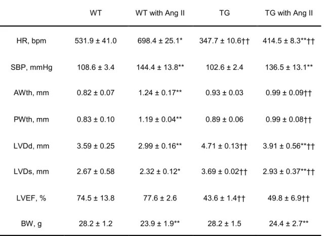
osmotic mini-pumps. Echocardiographic and hemodynamic data at 10 days
after Ang II or vehicle treatment are shown in Table 1. Ang II increased
systolic blood pressure to similar levels in both WT and TG mice. We have
already reported that the TG mice showed reduced cardiac function,
characterized by enlarged left ventricular internal diameter and reduced left
ventricular ejection fraction.16) Consistent with our previous report,
vehicle-TG mice displayed large left ventricular dimensions, lower left
ventricular ejection fraction and slower heart rate compared to vehicle-WT
mice. Interestingly, left ventricular wall thickness was increased in WT mice
after Ang II stimulation, but not in TG mice, suggesting that Ang II-induced
cardiac hypertrophy was attenuated in TG mice. After echocardiography,
mice were sacrificed, and hearts were excised. As shown in Figure 1a, heart
size was enlarged in WT mice after Ang II, but Ang II-TG mice showed
similar size compared to vehicle-TG mice. Although modest cardiac
hypertrophy already occurred in vehicle-TG mouse hearts, Ang II failed to
induce further cardiac hypertrophy in TG mice. Consistent with these data,
heart weight to tibia length ratios (Figure 1b) and cardiomyocyte cross
sectional area (Figure 1c) were increased in WT mice after Ang II, but not in
TG mice.
Ang II-induced cardiac fibrosis was inhibited in TG mouse hearts
Cardiac fibrosis is well known characteristics of Ang II-induced
cardiac remodeling.19) To evaluate the extent of cardiac fibrosis,
Elastica-Masson staining was performed. As shown in Figure 2, Ang II
increased cardiac fibrosis in both WT and TG mice compared to same strain
mice given vehicle. However, the TG mouse heart showed less fibrosis
compared to the WT mouse heart after Ang II infusion.
Overexpressed PDE3A1 inhibited Ang II-induced TGF-β expression
It has been reported that TGF-β is an effector molecule of Ang
II-induced cardiac hypertrophy and fibrosis.12) To investigate the mechanism
by which PDE3A1 attenuated cardiac remodeling and fibrosis, we examined the protein expression levels of TGF-β in the myocardium. As shown in
Figure 3, TGF-β protein expression levels were lower in the vehicle-TG
mouse heart compared with the vehicle-WT mouse heart. Ang II stimulation
increased TGF-β protein levels in WT mice, but it remained lower levels after
Ang II stimulation in TG mice.
Discussion
In the present study, we demonstrated that continuous infusion of
Ang II caused cardiac hypertrophy in WT mice, but not in TG mice. We also
showed that Ang II induced cardiac fibrosis in both WT and TG mice, but the
extent of fibrosis was less in the TG mouse heart compared with the WT
mouse heart. Moreover, basal expression levels of TGF-β, which is implicated
as a downstream effector of Ang II,12) was suppressed in TG hearts, and it
remained lower levels after Ang II stimulation in TG hearts compared with
WT hearts. These findings suggest that PDE3A protects the heart from Ang
II-induced cardiac remodeling and fibrosis through modulating the
functional connection between Ang II and TGF-β.
Pivotal roles of TGF-β in cardiac remodeling are well described in
both experimental and clinical models.20-23) Thus, regulating TGF-β signaling
is expected to be an attractive therapeutic target. Although the association between RAS and TGF-β signaling is reported, β-AR signaling pathways also
regulate TGF-β signaling.12) Several reports have shown that β-AR signaling
is enhanced by TGF-β, which serves as a downstream signaling of Ang
II/TGF-β.24-26) Considering overexpressed PDE3A1 behaves as like β-blocker
by catabolizing cAMP to inhibit β-AR/PKA axis, lower TGF-β level in TG
hearts implies that β-AR signaling may function as an upstream regulator of
TGF-β expression. It has been reported that PDE3A expression levels are
decreased in heart failure.27) Conversely, TGF-β expression levels are
upregulated in the failing heart.22) These findings support the concept that
repressing PDE3A would increase the expression levels of TGF-β, and
subsequently enhance cardiac remodeling in the failing heart. Several
molecules are reported as upstream regulators of TGF-β, such as
nicotinamide adenine dinucleotide phosphate oxidase, protein kinase C, p38
mitogen-activated protein kinase (p38 MAPK), and activator protein-1.28)
There is little evidence of interaction between PDE3A and TGF-β, but a
recent study has shown that A-kinase anchoring protein (AKAP)-Lbc
enhanced p38 MAPK-mediated hypertrophic responses.29) Considering the
fact that AKAP-Lbc is tethered with PKA, PDE3A may affect p38
MAPK-induced TGF expression via PKA regulation (Figure 4). However,
further studies are needed to elucidate more detailed molecular mechanisms
between β-AR/TGF-β signaling.
Recent studies have demonstrated that PDE3A1 has
cardioprotective effects through regulating cardiac apoptosis, which is
regulated by PDE3A/inducible cAMP early repressor feedback loop.30) In the
present study, our findings would provide the novel mechanism of PDE3A1
for cardio-protection by modulating Ang II/TGF-β axis. It would be ideal to
evaluate whether Ang II stimulation exaggerates cardiac remodeling in
PDE3A knock-out mice in the future. Other PDE families might also
contribute to cardiac remodeling. For example, PDE1, which is believed to be
important in the crosstalk of second messenger Ca2+ and cyclic nucleotide
signaling,2) regulates both Ang II and isoproterenol-induced cardiomyocyte
hypertrophy.31) PDE4 also regulated β-AR signaling,32) and PDE4D-/- mice
developed progressive cardiomyopathy and accelerated heart failure after
myocardial infarction.5) Thus, cAMP regulation in the setting of heart failure
might be orchestrated by not only PDE3A but also other PDEs. Further
studies are needed to elucidate roles of other PDEs in the situation of cardiac
remodeling using PDE isoform specific inhibitors and/or genetically
engineered mice.
Clinical perspective
In the clinical situation, a PDE3A inhibitor has been widely used for
the treatment of acute decompensated heart failure, but long-term
administration of it increases in mortality due to excess inotropic effects,
subsequent arrhythmia, and sudden cardiac death. In the present study, we
proposed a novel approach to protect the cardiac remodeling by
overexpressing PDE3A in the heart. Although PDE3A activator is not
available at this time, genetic engineering could express PDE3A in the
cardiac tissues sufficiently.
Conclusions
through modulating the functional connection between Ang II and TGF-β.
Acknowledgements
I thank Ms. Emiko Kaneda for the excellent technical assistance.
This research was supported in part by a grant-in-aid for Scientific Research
from the Japan Society of the Promotion of Science (no. 23790867 to M.O.).
References
1. Jessup M, Brozena S. Heart failure. N Engl J Med 2003; 348: 2007-18.
2. Bender AT, Beavo JA. Cyclic nucleotide Phosphodiesterases: Molecular
regulation to clinical use. Pharmacol Rev 2006; 58: 488-520.
3. Miller CL, Yan C. Targeting cyclic nucleotide phosphodiesterase in the
heart: therapeutic implications. J Cardiovasc Transl Res 2010; 3: 507-15.
4. Mongillo M, Tocchetti CG, Terrin A, et al. Compartmentalized
phosphodiesterase-2 activity blunts β-adrenergic cardiac inotropy via an
NO/cGMP-dependent pathway. Circ res 2006; 98: 226-34.
5. Lehnart SE, Wehrens XH, Reiken S, et al. Phosphodiesterase 4D
deficiency in the ryanodine-receptor complex promotes heart failure and
arrhythmias. Cell 2005; 123: 25-35.
6. Ockaili R, Salloum F, Hawkins J, et al. Sildenafil (Viagra) induces
powerful cardioprotective effect via opening of mitochondrial K(ATP)
channels in rabbits. Am J Physiol Heart Circ Physiol 2002; 283: H1263-9.
7. Takimoto E, Champion HC, Li M, et al. Chronic inhibition of cyclic GMP
phosphodiesterase 5A prevents and reverses cardiac hypertrophy. Nat
Med 2005; 11: 214-22.
8. Patrucco E, Albergine MS, Santana LF, et al. Phosphodiesterase 8A
(PDE8A) regulates excitation-contraction coupling in ventricular
myocytes. J Mol Cell Cardiol 2010; 49: 330-3.
9. Endo M. Amrinone, Forerunner of novel cardiotonic agents, caused
paradigm shift of heart failure pharmacotherapy. Circ res 2013; 113:
358-61.
10. Saucerman JJ, McCulloch AD. Cardiac beta-adrenergic signaling: from
subcellular microdomains to heart failure. Ann N Y Acad Sci 2006; 1080:
348–61.
11. Lewin G, Matus M, Basu A, et al. Critical role of transcription factor
cyclic AMP response element modulator in β1-adrenoceptor-mediated cardiac dysfunction. Circulation 2009; 119: 79-88.
12. Rosenkranz S. TGF-β1 and angiotensin networking in cardiac
remodeling. Cardiovasc Res 2004; 63: 423-32.
13. Dobaczewski M, Chen W, Frangogiannis N. Transforming growth factor
(TGF)-β signaling in cardiac remodeling. J Mol Cell Cardiol 2011; 51:
600-6.
14. Barki-Harrington L, Luttrell LM, Rockman HA. Dual inhibition of
β-adrenergic and angiotensin II receptors by a single antagonist: a
functional role for receptor–receptor interaction in vivo. Circulation 2003;
108: 1611-8.
15. Zhang G, Ohmori K, Nagai Y, et al. Role of AT1 receptor in
isoproterenol-induced cardiac hypertrophy and oxidative stress in mice. J
Mol Cell Cardiol 2007; 42: 804-11.
16. Oikawa M, Wu M, Lim S, et al. Cyclic nucleotide phosphodiesterase 3A1
protects the heart against ischemia-reperfusion injury. J Mol Cell Cardiol
2013; 64: 11-9.
17. Misaka T, Suzuki S, Miyata M, et al. Senescence marker protein 30
inhibits angiotensin II-induced cardiac hypertrophy and diastolic
dysfunction. Biochem Biophys Res Commun 2013; 439: 142-7.
18. Misaka T, Suzuki S, Miyata M, et al. Deficiency of Senescence marker
protein 30 exacerbates angiotensin II-induced cardiac remodeling.
Cardiovasc Res 2013; 99: 461-70.
19. Leask A. Potential therapeutic targets for cardiac fibrosis: TGFβ,
angiotensin, endothelin, CCN2, and PDGF, partners in fibroblast
activation. Circ Res 2010; 106: 1675-80.
20. Deten A, Holzl A, Leicht M, Barth W, Zimmer HG. Changes in
extracellular matrix and in transforming growth factor beta isoforms
after coronary artery ligation in rats. J Mol Cell Cardiol 2001; 33:
1191-207.
21. Li JM, Brooks G. Differential protein expression and subcellular
distribution of TGFβ1, β2 and β3 in cardiomyocytes during pressure
overload-induced hypertrophy. J Mol Cell Cardiol 1997; 29: 2213-24.
22. Li RK, Li G, Mickle DA, et al. Overexpression of transforming growth
factor-β1 and insulin-like growth factor-I in patients with idiopathic
hypertrophic cardiomyopathy. Circulation 1997; 96: 874-81.
23. Felkin LE, Lara-Pezzi E, George R, Yacoub MH, Birks EJ, Barton PJ.
Expression of extracellular matrix genes during myocardial recovery
from heart failure after left ventricular assist device support. J Heart
Lung Transpl 2009; 28: 117-22.
24. Rosenkranz S, Flesch M, Amann K, et al. Alterations of β-adrenergic
signaling and cardiac hypertrophy in transgenic mice overexpressing
TGF-β1. Am J Physiol Heart Circ Physiol 2002; 283: H1253-62.
25. Schlüter KD, Zhou XJ, Piper HM. Induction of hypertrophic
responsiveness to isoproterenol by TGF-β in adult rat cardiomyocytes.
Am J Physiol 1995; 269: C1311-6.
26. Schlüter KD, Frischkopf K, Flesch M, Rosenkranz S, Taimor G, Piper
HM. Central role for ornithine decarboxylase in β-adrenorceptor mediated hypertrophy. Cardiovasc Res 2000; 45: 410-7.
27. Ding B, Abe J, Wei H, et al. Functional role of phosphodiesterase 3 in
cardiomyocyte apoptosis: implication in heart failure. Circulation 2005;
111: 2469-76.
28. Wenzel S, Taimor G, Piper HM, Schlüter KD. Redox-sensitive
intermediates mediate angiotensin II-induced p38 MAP kinase activation,
AP-1 binding activity, and TGF-β expression in adult ventricular
cardiomyocytes. FASEB J 2001; 15: 2291-3.
29. Pérez LI, Cariolato L, Maric D, et al. A-Kinase anchoring protein Lbc
coordinates a p38 activating signaling complex controlling compensatory
cardiac hypertrophy. Mol Cell Biol 2013;33:2903-17.
30. Ding B, Abe J, Wei H, et al. A positive feedback loop of
phosphodiesterase 3 and inducible cAMP early repressor leads to
cardiomyocyte apoptosis. Proc Natl Acad Sci USA 2005; 102: 14771-6.
31. Miller CL, Oikawa M, Cai Y, et al. Role of Ca2+/calmodulin-stimulated
cyclic nucleotide phosphodiesterase 1 in mediating cardiomyocyte
hypertrophy. Circ Res 2009; 105: 956-64.
32. Qvigstad E, Moltzau LR, Aronsen JM, et al. Natriuretic peptides
increase β1-adrenoceptor signalling in failing hearts through
phosphodiesterase 3 inhibition. Cardiovasc Res 2010; 85: 763-72.
Figure legends
Figure 1. Effects of PDE3A on Ang II-induced cardiac hypertrophy. (a)
Representative cross sectional images of the ventricle of WT or TG mice
treated with Ang II or vehicle. Bars, 1 mm. (b) Quantitative data showing
heart weight (HW) to tibia length (TL) ratio after Ang II or vehicle treatment.
(c) Quantitative data showing cardiomyocyte cross sectional area of the left
ventricle from either WT or TG mice treated with Ang II or vehicle. Values
are mean ± SEM (n=5-7 in each group). *P<0.05 compared with same
genotype mice given vehicle. †P<0.05 compared with WT.
Figure 2. Effects of PDE3A on Ang II-induced cardiac fibrosis. (a)
Representative myocardial sections of Elastica-Masson stain of the left
ventricle of WT or TG mice treated with Ang II or vehicle. Bars, 100 µm. (b)
Quantitative data showing fibrosis fraction after Ang II or vehicle treatment.
Values are mean ± SEM (n=4-5 in each group). **P<0.01 compared with
vehicle in the same genotype. †P<0.05 compared with WT.
Figure 3. Protein expression levels of TGF-β after Ang II treatment. (a)
Representative immunoblotting of TGF-β in the left ventricle of WT or TG
mice treated with Ang II or vehicle. (b) Quantitative data showing TGF-β
expression levels normalized to α-tubulin. Values are mean ± SEM (n=6 in
each group). *P<0.05 compared with vehicle in the same genotype. ††P<0.01
compared with WT.
Figure 4. A diagram presenting our hypothesis. PDE3A represses PKA
activity through cAMP metabolism, resulting in inhibited PKA-mediated
cardiac remodeling and reduction of TGF-β expression via
AKAP-Lbc-mediated p38 MAPK activation.
1 2 3 4
0 2 4 6 8 10 12
HW/TL (mg/mm)
WT TG
Vehicle Ang II Vehicle Ang II
1.4 1.2 1.0 0.8 0.6 0.4 0.2
myocytecrosssectionalarea (Foldchange)
0
*
†
c b
*
WT
TG
Vehicle Ang II
a
**
WT
TG
Vehicle Ang II
a
Fi br osi s fract ion (% )
** †
10 8
6 4 2 0
b
TGF-β α-tubulin
WT
Vehicle Ang II
TG
Vehicle Ang II
a
Vehicle Ang II Vehicle Ang II TG F- β/ α- tu bu lin (ar bi trar y uni t)
0 0.2 1.0
0.4 0.6
b *
†† ††
0.8
1.2
1.4
Cardiac function Cardiac remodeling
PDE3A cAMP
PKA β-adrenagic
receptor AngiotensinⅡ
receptor
TGF-β MAPKP38
AKAP PKA
Table 1. Comparisons of heart rate, systolic blood pressure, body weight, and echocardiographic parameters of WT and TG mice.
Values are expressed as mean ± SEM from 6 to 7 mice.
HR, heart rate; SBP, systolic blood pressure; AWth, anterior wall thicknesss; PWth, posterior wall thickness; LVDd, left ventricular end-diastolic dimension; LVDs, left ventricular end- systolic diameter dimension; LVEF, left ventricular ejection fraction; BW, body weight; WT, wild-type mice;TG, PDE3A1 overexpressed mice; AngII, angiotensin II. *P<0.05, **P<0.01 vs. same genotype mice given vehicle, †P<0.05 , ††P<0.01 vs. WT mice.
WT WT with Ang II TG TG with Ang II
HR, bpm 531.9 ± 41.0 698.4 ± 25.1* 347.7 ± 10.6†† 414.5 ± 8.3**††
SBP, mmHg 108.6 ± 3.4 144.4 ± 13.8** 102.6 ± 2.4 136.5 ± 13.1**
AWth, mm 0.82 ± 0.07 1.24 ± 0.17** 0.93 ± 0.03 0.99 ± 0.09††
PWth, mm 0.83 ± 0.10 1.19 ± 0.04** 0.89 ± 0.06 0.99 ± 0.08††
LVDd, mm 3.59 ± 0.25 2.99 ± 0.16** 4.71 ± 0.13†† 3.91 ± 0.56**††
LVDs, mm 2.67 ± 0.58 2.32 ± 0.12* 3.69 ± 0.02†† 2.93 ± 0.37**††
LVEF, % 74.5 ± 13.8 77.6 ± 2.6 43.6 ± 1.4†† 49.8 ± 6.9††
BW, g 28.2 ± 1.2 23.9 ± 1.9** 28.2 ± 1.5 24.4 ± 2.7**