Acta Med. Nagasaki 30 :168-179
Experimental Hypertension by Long-Term Cold Stress and Changes of Cardiovascular System
Masachika ISEKI
Department of Pathology, Atomic Disease Institute, Nagasaki University School of Medicine
Nagasaki, Japan
Received for publication, May 15, 1985
Stress-induced hypertension was produced in rats by exposure to 4±1 °C temperature from one month of age for one hour everyday over several months. Subsequently, signifi- cant changes of the cardiovascular system were observed in cold-stress rats. The present experiment used Sprague-Dawley rats (SD rats) and Wistar rats (W rats). There were a few differences between the two. In SD rats, angionecrosis of the coronary artery, fibro-
muscular sclerosis of the myocardial artery and PN-like lesion in the heart, kidneys and mesentery were observed. In W rats myocardial cell degeneration, fibrosis and mononu- clear cell infiltra ion were detected and when compared to non-stress rats there was sig- nificant medial thickening of the myocardial artery under 100µm in stressed rats. The implications of exposure to cold in the analysis of cardiovascular pathology accompanied by stress-induced hypertension are discussed.
INTRODUCTION
The relationship between stress and hypertension is well known'). Since FARRIS et al.') reported that experimental hypertension was produced in rats by air blasting, reports of experimental hypertension induced by various kinds of stress have been pre- sentedl>-13). Pathological investigation of stress-induced hypertension was reported by HENRY et al. s> , LA WLER et al. 7) , MARWOOD et al."), and so on though the numbers of those reports are few. We have reported that hypertension was produced in rats by using certain long-term stress e.g. "stick-poking harassment" and morphological changes were investigated')-13) . In the present experiment, hypertension was produced in rats by exposure to 4±1 °C temperature daily over several months and the changes of the cardio- vascular system were investigated.
A part of this study was presented at the winter annual meeting of the Japan Atherosclerosis Society, Jan. 18, 1985.
井関充 及
MATERIAL AND METHOD
In a preliminary experiment, 3 male and 3 female Sprague-Dawley rats were used and after one month of age were transferred to a cold room at 4±1 °C for one hour everyday. The cold room was illuminated during the cold stress. During the experimen- tal period, the systolic blood pressure and the weights of the rats were measured every other week. Eleven to twenty months after starting the cold stress, the rats were sacri- ficed by decapitation and sections for light microscopic investigation were prepared by the usual method and examined.
The present experiment was divided into two groups (experiment(1) and experiment (2)].
Experiment(1) : Sprague-Dawley rats were used. After one month of age, the rats were exposed to cold in the same manner as in the preliminary experiment. During the experimental period, the systolic blood pressure and the weights of the rats were measured every other week. The rats in which the systolic blood pressure elevated to 170 to 180 mmHg breeded, and were allowed to breed until the ninth generation using the same methods. The rats which elevated to a maximum blood pressure of 200mmHg and those that seemed unable to survive the cold stress were sacrificed by decapitation.
Experiment(2) : Male Wistar rats were used. They were divided into two groups (the cold stress group (n=4) and the control group (n=5)]. The cold stress group rats were exposed to cold in the same method as in the preliminary experiment after one month o f age. The control group rats were housed at 24--L2 ° C temperature. During the experimental period, the systolic blood pressure and the weights of both groups of rats were measured every other week, and then 10 months later they were sacrificed by decapitation.
Except during exposure to cold, the rats of experiment(1) and (2) were individually housed at 24±2 °C. Food and tap water were provided ad libitum. The systolic blood pressure was measured by the tail-cuff method (RAT automatic blood pressure recorder USM-105-R type). One week before completion of the experiments handling was carried out for 5 minutes everyday and then the rats were sacrificed by decapitation. After dis- sociating plasma from the collected blood, Na, K, Cl, BUN (blood urea nitrogen), cholesterol, free fatty acids, and (3-lipoprotein were measured. The weights of organs were measured and the organ-to-body weight ratio was calculated. The organs were immersed in 10% buffered formaldehyde and embedded in paraffin using a SAKURA automatic tissue processor as usual. For light microscopy sections of the cardiovascular system were stained with Hematoxylin-Eosin (HE) stain, Elastica Van Gieson (EVG) stain, Weigert (W) stain and Mallory Azan (MA) stain.
In experiment(2), when sacrificed, the left adrenal gland was taken out immediately and the catecholamine contents were measured (by the high performance liquid chromato- graphic electrochemical method). Moreover, in experiment(2) the thickness of media was measured in myocardial arteries. The elastica interna of the section was irregularly curved
and so it was stretched to round regularly. The extension of section of myocardial ar- teries with EVG stain was projected and the thickness of media was measured by using image analyzing system (DAIANA-1). The media/radius ratio was calculated. Statis- tical processing was done by the Student's t-test.
RESULT
Preliminary experiment : Hypertension over 160mmHg was produced in all six rats.
The mean value of the maximum systolic blood pressure was 179±4.6 mmHg (means ± S.E.). For pathological findings, subendothelial angionecrosis of the coronary artery was observed in one male rat of six rats (Fig. 1).
Experiment(1) : Hypertension of over 160mmHg was produced one to eight months after starting the exposure to cold. It was expected that the period needed for elevating the blood pressure after starting the cold stress would be shortened by producing suc- ceeding generations, but positive proof could not be obtained. Pathological changes of the cardiovascular system were observed in one male rat of the seventh and ninth genera- tions.
The seventh generation rat which was 13 months of age indicated 164mmHg blood pressure 2 months after starting and then maintained hypertension and the maximum blood pressure was 200mmHg. For macroscopic findings, concentric hypertrophy and fibrosis in the heart (Fig. 2), swelling of the kidneys the surface of which were rough and granular (Fig. 3), and the rosary-like appearance of the mesenteric vessels (Fig. 4) were observed. By light microscopic investigation, a PN-like lesion (fibrinoid necrosis of the arteries and infiltration of neutrophils and lymphocytes and proliferation of fibroblast) was
Fig. 1. Subendothelial angionecrosis (arrowed). (HE stain x300) Fig. 2. Myocardial fibrosis and concentric hypertrophy of the heart.
observed in the heart, mesentery and kidneys (Fig. 5).
The blood pressure of the ninth generation rat which was 10 months of age had risen over 160mmHg, 4 months after starting and then maintained the hypertension. The maximum systolic blood pressure was 170mmHg. Light microscopically, fibromuscular sclerosis was observed in a myocardial artery of the left ventricle (Fig. 6).
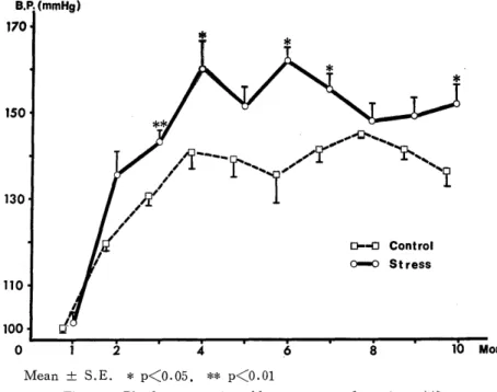
Experiment(2) : There was no significant differences in body weight gain between the two groups (Fig. 7). The blood pressure of the cold stress group had increased 3 months after starting the exposure to cold, and after that it was significantly higher than
Fig. 3. Swellings of kidneys.
Fig. 4. A rosary-like appearance of mesenteric vessels.
Fig. 5. PN-like lesion of the mesenteric artery.
Fig. 6. Fibromuscular sclerosis of the myocardial artery (arrow: elastica interna). (EVG stain x 50)
the control group almost every month (Fig. 8). There were no significant differences between the two groups in organ-to-body weight ratio of each organ and in serum electrolyte and serum lipid (Table 1, 2). Compared to the catecholamine content in the
Mean ± S.E.
Fig. 7. Body weight comparing experimental groups [experiment(2)) .
Mean ± S.E. * p<0.05, ** p<0.01
Fig. 8. Blood pressure in cold stress group [experiment(2)D.
Fig. 9. Myocardial fibrosis. CEVG stain X 50]
Fig. 10. Myocardial cell degeneration (arrowed) and mcnonuclear cell infiltration. [HE stain X275]
Table 1. Comparison of organ-to-body weight ratio
Experimental group Heart* Kidneys* Liver* Lungs* glands Adrenal
Control 0.30±0.01 0.72±0.03 3.41±0.01 0.60±0.03 9.43±0.97
Stress 0.28±0.01 0.67±0.03 3.16±0.01 0.54±0.03 8.30±1.64
mean±S.E.
*(g%) **(mg%)
Table 2. Comparison of serum lipid and serum electrolyte
Experimental Na(mEg/1) K(mEg/1) C1(mEg/1) BUN T.chol FFA jl-lipo
group (mg/dl) (mg/dl) (mg/dl) (mg/dl)
Control 144.3±0.3 7.2±0.1 105.5±1.0 18.3±0.4 99.6±10.8 0.54±0.04 188.4±23.1
Stress 144.4±1.1 6.9±0.5 105.2±1.0 18.9±0.4 107.2±15.9 0.52±0.06 238.0±56.0 mean±S.E.
BUN: blood urea nitrogen, T. chol.: total cholesterol, FFA : free fatty acids, (l-lipo : /3-lipoprotein
Table 3. Catecholamine contents in adrenal norepinephrine epinephrine
(ug) (leg)
control 5.25±0.21 30.74±2.32
stress 4.44±0.41* 29.10±-2.50
mean ±S.E. *p<0.1
Table 4. Pathological findings in heart
Case fibrosis* cell infiltration**
1 + + +
2 + +
3 ++ +
4 + -
*-{- : mild, * : moderate
**- : absence, + : presence
* p<O.05
Fig. 11. Media/radius ratio in myocardial arteries (experiment(2)J.
adrenal gland, norepinephrine was slightly decreased in the cold stress group but there was no significant difference (Table 3). No pathological changes were observed in the control rats. In all the cold stress group rats, myocardial fibrosis (scattered in the myocardium) and myocardial cell degeneration were observed light microscopically in the left ventricle and interventricular septum (Fig. 9, Table 4). Moreover, cell infiltration composed mainly of mononuclear cells was also seen in 3 out of 4 rats (Fig. 10). Comparing the media/radius ratio of myocardial arteries, both groups indicated 0.15-L0.01 (means :L S.E.) in the radius 100 to 200 tern arteries, and no difference was found. However, in the radius to 50pm arteries, the control group indicated 0.26±0.01 and the cold stress group indicated 0.31±0.01. In the radius 50 to 100 pm arteries, the control group indicated 0.16±0.01 and the cold stress group indicated 0.21±0.01. Thus the ratio of the cold stress group was more significantly increased than that of the control group in the radius under 100,um arteries (Fig. 11).
DISCUSSION
There have been many reports that catecholamine is related to the cause of hyper- tension 14>-l6 The system of reactivity to stress is considered to be the sympathetic nerve-adrenal medulla system and the pituitary-adrenal cortex system. KVETNANSKY et al. 17) reported that in rats subjected to repeated immobilization, adrenal medullary activity was increased and urinary excretion of epinephrine was greater than in unstressed or once-immobilized rats. MATSUO et al. 18) reported that there was a significant increase in epinephrine volume of the stress group rats (application of "stick-poking harassment"
stress) by measurement of urine catecholamines than that of the control rats, and they supposed that stress-induced hypertension was caused by acceleration of adrenal medullary activity with acceleration of activity of catecholamine synthesis enzyme. CHIUEH et al. 19) , MCCARTY et al."), and POPPER et al.") reported that by application of stress such as exposure to cold, immobilization and electrical stimulus, activity of the sympathetic nerve- adrenal medulla system was increased and plasma catecholamines were increased. In, the present experiment it was conjectured that activity of the sympathetic nerve-adrenal medulla system increased and also excretion of plasma catecholamine increased and it was assumed that these factors were related to the development of hypertension. It was also supposed that the peripheral resistance was raised by increased plasma level of norepineph- rine and that this affected the development of hypertension.
A combination of factors is said to be more effective in the development of stress- induced hypertension than only one factor'). In the present experiment rats were transferred to a cold room which was illuminated during exposure. Therefore, it was considered that the change of living place and the exposure to cold in an illuminated room could affect the development of hypertension as psychogenic stress in rats which have a nocturnal habit.
LEDUC") reported that adrenal epinephrine and norepinephrine of rats acclimated to cold increased significantly. KVETNANSKY et al. 17) reported that the adrenal epineph- rine of rats subjected to one time-immobilization stress decreases, but that there was no decrease of adrenal epinephrine in rats subjected to repeated immobilizations though urinary excretion of epinephrine was still increased. It was suggested that the "adaptation"
of the adrenal medulla to repeated stress in rats was caused by an enhanced ability to replace the released catecholamine. Although in the present experiment urinary catechola- mines were not measured, from the report of LEDUC"), it was expected that urinary catecholamines would be increased.
In experiment(2), there were no significant differences in adrenal catecholamine contents between the two groups. This result was thought to be caused by "adaptation" to the cold stress.
There have been many reports about the production of experimental hypertension by various kinds of stress, but there are very few reports about stress-induced hypertension pathology6)-13). The vascular changes in the present experiment(1) were as follows : a) angionecrosis of the coronary artery : b) PN-like lesions in the heart, kidneys, and
mesentery ; and c) fibromuscular sclerosis in the myocardial artery. LIMAS et al. 13) supposed that not only the severity of hypertension but also its duration were significant determinants of vascular damage. The blood pressure of all the rats in which a) angio- necrosis, b) PN-like lesion, and c) fibromuscular sclerosis were observed, maintained levels of over 160mmHg for 7 to 11 months and it is suspected that over 160mmHg hypertension and its duration caused the vascular changes. GARDNER et al. 14) supposed that the acceleration of vascular permeability might be the cause of fibrinoid necrosis in experimental hypertension. SHOWA25) reported that fibrinoid material was deposited in the intima and media of the arteries by repeated contraction of the mesenteric artery in the rat. It is considered that aging, hypertension and contraction of the arteries are significant factors
2s> in the PN-like lesion"
It is supposed that the changes of a) angionecrosis and b) PN-like lesion were caused by cold stress-induced hypertension and repeated vascular contraction due to nor- epinephrine.
YAMORI27) supposed that maintained hypertension enhanced not only vascular col- lagen synthesis but also noncollagenous protein metabolism and thereby accelerated arte- riosclerosis. In the rat with vascular change, i.e. fibromuscular sclerosis, hypertension of over 160mmHg was maintained for 7 months, and it was supposed that such lesions may be caused by cold stress-induced hypertension.
LAWLAR et al.7= reported that severe hypertension was produced in borderline hy- pertension rats about 150mmHg by using wheel, noise, and electric stimulus, and that subsequently myocardial cell degeneration, fibrosis and inflammatory cell infiltration were observed. Similar lesions were observed in the present experiment(2). WALDENSTROM et al."') reported that a sudden release of myocardial norepinephrine causes acute myocar- dial damage. UEDA et al. 14) reported that the myocardial norepinephrine increased sig- nificantly by electrical stimulation of the central gray stratum of the midbrain. It is con- jectured that endogenous norepinephrine increases in the heart by exposure to cold and subsequently ischemia by vascular contraction and norepinephrine itself could cause the myocardial cell damage.
In the present experiment the thickness of the myocardial artery under 1001-tin in- creased significantly. LIMAS et al. 23) reported that the thickness of the artery was in proportion to the duration of hypertension. Moreover, MARWOOD et al.') reported that sustained stress-induced hypertension was produced in Wistar rats by sound-withdrawal stimulation and that arterioles from the gastrocremius muscle of rats had thicker walls and hence reduced lumen diameter. SUNAGA29) reported that endothelial damage-permeability acceleration, smooth muscle damage-edematous change and proliferation of collagen were observed by contraction effects of norepinephrine infused into the artery. JOHNSON et al. 30) reported that hypertrophy of the arteries was induced by catecholamine infusion into rats.
In the present experiment, the systolic blood pressure of cold stress group rats was elevated to about 150mmHg, which was not so severe, but compared to the control group rats, the cold stress rats maintained a significantly higher blood pressure for 7 months.
It is suspected that higher blood pressure and endogenous norepinephrine could affect the medial thickening.
There was a few differences in pathology between experiment(1) and experiment(2).
KUHN et al.31) reported that there were endocrine differences between the Wistar and Sprague-Dawley rats exposed to cold. It is supposed that physiological sensitivity to cold stress might be different in different kinds of rats.
CONCLUSION
Changes of the cardiovascular system were observed in rats exposed to cold from one month of age. The results were as follows.
I) In Sprague-Dawley rats ;
i) Over 160mmHg hypertension was produced and maintained by exposure to cold.
ii) Positive proof that the period needed for elevating the blood pressure after starting
the cold stress was shortened by generation could not be obtained.
iii) It was conjectured that stress-induced hypertension causes the pathology in the from of a) angionecrosis of the coronary artery b) fibromuscular sclerosis of the
myocardial artery and c) PN-like lesion in the heart, kidneys and mesentery.
II) In Wistar rats ;
i) The blood pressure of cold stress group rats increased significantly compared to the control group.
ii) Myocardial cell degeneration, fibrosis and mononuclear cell infiltration were ob- served in cold stress rats.
iii) The thickness of the myocardial artery under 100um increased significantly com- pared to the control group. It is suspected that endogenous norepinephrine could
affect the medial thickening.
iv) There were no significant differences in adrenal catecholamine contents between the two groups. It was suggested that the "adaptation" of the adrenal medulla
to repeated cold stress was caused by an enhanced ability to replace the released
catecholamine.
III) It is thought that the physiological sensitivity to cold stress might be different be- tween the Wistar rats and the Sprague-Dawley rats.
ACKNOWLEDGEMENT
The author would like to express his gratitude to Professor I. NISHIMORI, As- sociate Professor I. SEKINE and Associate Professor M. KISHIKAWA for their valuable advices and encouragements. The author also thanks Dr. K. FUJINO for his valuable com- ments and his kind provision of rats, and the cooperative research students and skillful technical assistants in the Department of Pathology, Atomic Disease Institute, Nagasaki University School of Medicine.
REFERENCES
1) FRIEDMAN, R.: Experimental psychogenic hypertension. In: Stress and the Heart, edited by Wheatley, D., 2nd. edition. Raven Press, New York, pp. 209-228, 1981 2) FARRIS, E. J., YEAKEL, E. H., and MEDOFF, H. S.: Development of hypertension
in emotional grey Norway normal rats after air blasting. Am. J. Physiol. 144: 331- 333, 1945
3) HENRY, J. P., MEEHAN, J. P., and STEPHENS, P. M.: The use of psychosocial
stimuli to induce prolonged systolic hypertension in mice. Psychosom. Med. 29: 408- 432, 1967
4) HUDAK, W. J. and BUCKLEY, J. P.: Production of hypertensive rats by experimen- tal stress. J. Phar. Sci. 50: 263-264, 1961
5) ROSECRANS, J. A., WATZMAN, N . , and BUCKLEY, J. P.: The production of hypertension in male albino rats subjected to experimental stress. Biochem. Pharm. 15:
1701 -1718, 1966
6) HENRY, J. P., ERY, D. L., STEPHENS, P. M., RATCLIFFE, H. L., SANTISTE-
BAN, G. A., and SHAPIRO, A. P.: The role of psychosocial factors in the development of arteriosclerosis in CBA mice. Observation on the heart, kidney and
aorta. Atherosclerosis 14: 203-218, 1971
7) LAWLER, J. E., BARKER, B. A., HUBBARD, J. W., and SCHAUB, R. G.:
Effect of stress on blood pressure and cardiac pathology in rats with borderline hyper-
tension. Hypertension 3: 496-505, 1981
8) MARWOOD, J. F., and LOCKETT, M. F.: Stress-Induced hypertension in rats.
In: Stress and the Heart, edited by Wheatley, D., 2nd. edition, Raven Press, New
York, pp. 229-243, 1981
9) NISHIMORI, I., FUJINO, K . , SEKINE, I., KISHIKAWA, M., SHIMIZU, K.
and TSUDA, N.: Experimental hypertension by long-term stress and cardiovascular
change. Presented at the 70th annual meeting of the Japanese Pathological Society, 1981 10) NISHIMORI, I., FUJINO, K . , SEKINE, I., KISHIKAWA, M., TAKAKI, Y., SHIMIZU,
K. and MATSUO, K.: Experimental hypertension induced by long-term psychogenic
stress and cardiovascular changes.
Presented at the 71st annual meeting of the Japanese Pathological Society, 1982 11) SEKINE, I., KISHIKAWA, M., TAKAKI, Y., SHIMIZU, K . , MATSUO, K . , FUJINO,
K., NISHIMORI, I. and NIwA, M.: Experimental hypertension induced by long-term
psychogenic stress and vascular changes. J. Jpn. Atheroscle. Soc. 11: 159-164,
1983 (Japanese)
12) NISHIMORI, I. and SEKINE, I.: Long-term psychogenic stress and vascular changes in rats. J. Jpn. Atheroscle. Soc. 12: 33-39, 1984 (Japanese)
13) ISEKI, M., FUJINO, K. and NISHIMORI, I.: Experimental hypertension induced by long-term cold stress and changes of cardiovascular system in rats. Presented at the
winter annual meeting of the Japan Atherosclerosis Society , 1985
14) UEDA, H., IIZUKA, T., YASUDA, H., TAKABATAKE, Y., IIZUKA, M., IHORI, M.
and YAMAMOTO, M.: Effect of electrical stimulation of central gray stratum of the
midbrain on the blood and myocardial catecholamine. Jpn. Heart J. 7: 277-288, 1966 15) IKOMA, T.: Studies on catechols with reference to hypertension (Report 1) Jpn.
Heart J. 7 : 277-288, 1965
16) REID, J. L., ZIVIN, J. A. and KOPIN, I. J.: Central and peripheral adrenergic mechanisms in the development of deoxycorticosterone-saline hypertension in rats. Circ.
Res. 37: 569-579, 1975
17) KVETNANSKY, R. and MIKULAJ, L.: Adrenal and urinary catecholamines in rats during adaptation to repeated immobilization stress. Endocrinology 87: 738-743,
1970
18) MATSUO, K., SEKINE, I., FUJINo, K., NISHIMORI, I., KOIWA, T., NIWA, M. and OzAKI, M. :,,,Experimental hypertension induced by chronic stress and urinary
catecholamines. Medicine and Biology 106: 123-127, 1983 (Japanese)
19) CHIUEH, C. C. and MCCARTY, R.: Sympatho-adrenal hyperreactivity to footshock stress but not to cold exposure in spontaneously hypertensive rats. Physiol. Behav. 26:
85-89, 1981
20) MCCARTY, R., KVENTNANSKY, R. and KOPIN, I. J.: Plasma catecholamines in rats; daily variations in basal levels and increments in respose to stress. Physiol.
Behav. 26: 27-31, 1981
21) POPPER, C. W., CHIUEH, C. C. and KOPIN, I. J.: Plasma catecholamine con- centrations in unanesthetized rats during sleep, wakefulness, immobilization and after
decapitation. J. Pharmacol. Exp. Ther. 202: 144-148, 1977
22) LEDUC, J.: Catecholamine production and release in exposure and acclimation to cold.
Acta Physiol Scand 53: (Suppl.) : 183, 1961
23) LIMAS, C., WESTRUM, B., and LIMAS, C. J.: The evolution of vascular changes in the spontaneously hypertensive rat. Am. J. Pathol. 98: 357-383, 1980
24) GARDNER, D. L., and MATTHEWS, M. A.: Ultrastructure of the wall of small arteries in early experimental rat hypertension. J. Pathol. 97: 51-62, 1969 25) SHOWA, N .: Pathological study on the arterial lesions by repeated arterial contraction
in rats. J. Jap. Coll. Angiol. 19: 863-876, 1979
26) KYOGOKU, M.: A study on pathogenesis of polyarteritis nodosa. Nippon Rinsho 36: 18-25, 1978 (Japanese)
27) YAMORI, Y.: Vascular protein metabolism in the pathogenesis. Jpn. Circ. J. 40:
879-886, 1976
28) WALDENSTROM, A. P., HJALMARSON, A. C. and THORNELL, L.: A possible role of noradrenaline in development of myocardial infarction. Am. Heart J. 95: 43-51, 1978 29) SUNAGA, T.: The constrictive effects effects of adrenaline or noradrenaline on large
arteries. J. Jpn. Atheroscle. Soc. 12: 3-17, 1984 (Japanese)
30) JONSON, M. D., GRIGNOLO, A., KUHN, C. M. and SCHANBERG, S. M.:
Hypertension and cardiovascular hypertrophy during chronic catecholamine infusion in
rats. Life Sci. 33: 169-180, 1983
31) KUHN, E. R., BELLON, K . , HUYBRECHTS, L. and HEYNS, W.: Endocrine differences between the Wistar and Sprague-Dawley laboratory rat; influence of cold
adaptation. Horm. Metab. Res. 15: 491-498, 1983


![Fig. 9. Myocardial fibrosis. CEVG stain X 50]](https://thumb-ap.123doks.com/thumbv2/123deta/10139655.1973534/6.759.92.658.117.424/fig-myocardial-fibrosis-cevg-stain-x.webp)
