Japancse Joumal of Tropical Medicine and Hygienc
第6巻,第3,4号 昭和53年12月15日
内 容
原 著
沖縄(亜熱帯)生育者と本土(温帯)生育者の発汗反射,能働汗腺数
および体温の比較研究………・………一………・………辻田 純三,堀 東アフリカ・ケニア,タベタ地区におけるヒト住血吸虫症の浸淫状況(英文)
…片峰 大助,Siongok,T.K.A.川島健治郎,中島 野島 尚武,今井 東アフリカ・ケニア,タベタ地区における住血吸虫症の媒介貝類について(英文)
・野島 尚武,片峰 大助,川島健治郎,中島 今井 淳一,坂本 信,嶋田 雅暁,宮原 東アフリカ・ケニア,タベタ地区における住血吸虫症の病原保有宿主としての 野生ネズミ類に関する研究(英文)………∫ll嶋健治郎,片峰 大助,坂本 嶋田 雅暁,野島 尚武,宮原
清記157−165
康雄,
淳一167−180
康雄,
道明181−193
信,
道明195昌203 繕 説・
ラン、ブル鞭毛虫症と栄養失調一〇midazoleでの治療成績一(英文)
・Lasserre,R.205−210
学術記録
日本熱帯医学会九州支部第2回大会講演要旨 会 報
昭和53年度第2回幹事会記録………・…一 昭和53年度評議員会記録一………一・
第20回会務総会記録一……
投稿規定一…
211−221
・・223
−223−224
−224−225
−226−227
日熱医会誌
Japan.」。T.M.H. 日 本熱帯医学会
日本熱帯医学会雑誌 第6巻 第3,4号 1978 157−165頁 157
沖縄(亜熱帯)生育者と本土(温帯)生育者の発汗反射,
能働汗腺数および体温の比較研究
辻 田 純 三・堀 昭和53年4月10日
清 記
受付
緒 目
高温環境に馴化していないヒトに暑熱馴化を行 わせると,同じ暑熱負荷に対して発汗反応が早く 起こり,汗量が増加し,汗による放熱量の増加に より体温上昇度が減少して,高温環境に耐える 能力が獲得されることが知られている(Adolph,
1946;Dil18 磁,1938)。一方,出生時より熱帯に 生活するような長期間高温環境に馴化したヒトは,
中程度の暑熱負荷時には発汗反射の発現が遅延し,
汗量が少なくて体温上昇度も低いといわれている
(Kuno,1956)。発汗潜時の延長と汗量の減少は汗 による放熱量の減少を意味するから,長期間の馴 化によって乾性放熱(対流,伝導,輻射による放 熱)能力の向上があるものと推定される。しかし,
汗量について,熱帯住民は温帯住民や寒帯住民よ り能働汗腺数が多いことから,暑熱負荷量が多く なれば熱帯住民の方が汗量が多くなり,発汗能力 として優れているものと推測されている(久野,
1956)。亜熱帯の沖縄生育者と温帯の本土生育者 の発汗反射については,能働汗腺数および高温環 境下における乾性放熱量の比較を行った研究報告 は少なく,わずかに発汗潜時について発汗性の低 下した冬期における比較報告がなされているにす ぎない(Hori寵α乙,1976)。発汗反射と乾性放熱 は季候の影響をうけること,および乾性放熱量は 平均皮膚温が環境温より高い範囲においては平均 皮膚温の高い方が多いことが知られているので
(Robinson,1949),長期間の高温馴化が発汗反射 兵庫医科大学第一生理学教室
西宮市武庫川町1−1
と高温曝露時の乾性放熱および能働汗腺数にどの ような影響を与えるかを研究することは長期間の 高温馴化の本態を解明するために必要である。そ こで,沖縄生育者および本土生育者で最近沖縄に 移住したヒトについて,夏季に発汗反射と高温環 境下における皮膚温及び能働汗腺数の比較をして,
両者の体温調節のパターンの差異について検討を 加え,長期間の高温馴化と短期間の高温馴化の違 いにづいて考察を加えた。
実 験 方 法
沖縄に生れ育った成人男子7名と,本土に生れ 育って沖縄へ移住してからの期間が3年以内の成 人男子7名について7月に身体計測及び,高温環 境下での体温の測定と発汗反射の測定を行った。
能働汗腺数は生後3年以内にその数が決まるとい われているので,西宮市において沖縄より本土に 最近移住した成人男子30名と本土に生れ育った成 人男子20名について測定した。
皮下脂肪厚は栄研式皮脂厚計を用い,皮膚を 10g/cm2の圧力でつまんでから2秒後に測定し た。測定は5回行って,その平均値をその部位の 皮脂厚とした。皮脂厚の測定部位と平均皮脂厚算
出のための加重平均係数は次の通りである(堀ら,
1974)。
部 位
上腕:右腕背面肩峰と肘頭の中間と 右腕前面中央部の平均値 背:右肩甲骨下角直下
胸:右前腋窩鐡壁と右乳頭の中間 腹:右乳腺上,膀の高さ
係 数
0.141
0.143 0.143 0.139
腰:右肋骨下縁と右腸骨稜の中間 0.139 大腿: 右大腿中央,前面背面の平均値 0.295 体脂肪含有率∫は予知式
∫(彩)ニ
2&9×体表面積(m縫欝脂厚(mm)+a69
により求めた。
体表面積の算出には高比良の式を使用した。高 温環境下での体温の測定は,発汗のない状態での 乾性放熱量の比較が目的であるから体温の日内変 動の影響を避けるため,午後3時に,30C,湿 度70%の室内に水泳パンツだけを着用させて座位 にて30分間安静をとらせ,発汗準備状態としたと きの全身10カ所の皮膚温をパイロメーターを用い て測定した。口内温(舌下温)は婦人体温計を用 いて測定した。皮膚温の測定部位と平均皮膚温測 定のための加重平均係数は次の通りである(Hori
8孟α乙,1977)。
部 位
額:前額中央眉上2cm 胸:乳腺と第4肋骨の交点 腹:乳腺上,膀の高さ 背:肩甲骨下角直下 上腕:三角筋中央点
前腕:前腕内面中央線,下から1/3 手:手背中央線,上から1/2 大腿: 大腿前面中央線,上から1/2 下腿: 下腿後面中央線,上から1/3 足:足背中央線,上から1/2
係数
0.098 0,083 0.162 0.083 0.082 0.061 0.053 0.172 0.134 0.072
発汗反射は右肩甲骨下に内面が12,6cm2の発 汗カプセルを装着しておき,発汗準備状態の被検 者を42Cのよく撹梓された湯に膝から下の両下 肢を湯浴させ(Ohara,1966),2分ごとにカプセ ル内の濾紙をかえて肉眼ではっきり発汗が発現し たことが判るまで濾紙の交換を行って濾紙の重量 の増加より求めた。濾紙の重量の増加が認められ る最初の濾紙からは,発汗時間が不明確で発汗速 度を正確に求めることができないので,便宜的に 次の濾紙の重量増加から初期発汗速度の増加率を 求め,発汗開始後の発汗速度の経時変化を図に画
き,この図形から発汗速度がゼロとなるべき時間 を求め,このときを発汗開始時間とみなして,両 下肢温浴開始時から発汗開始までの時間を発汗の 潜時とした。
能働汗腺数の測定は次のように行った。皮膚表 面にヨードアルコール液を塗布し乾燥したのち,
発汗反射を測定したときと同じ条件で発汗させた 後,ヨードカリ澱粉紙を圧着して,単位面積当た りの汗孔着色点を数えて能働汗腺密度とした(大 原,1977)。下肢の汗腺数は発汗が充分発現した 状態で温浴を中止させて,乾いたタオルでよくぬ ぐった後測定した。能働汗腺の測定部位と平均濃 度汗腺密度算出のための加重平均係数は次の通り である(大原,19771Weiner召緬乙,1969)。
部 位
額: 前額中央眉上2〜3cm 胸:右胸,乳頭上3〜4cm 背:右肩甲骨中央
上腕:左上腕外側肩峰と肘頭の中間 前腕:右前腕内面中央線,上から1/2 大腿:右大腿前面中央,上から1/2 下腿:左下腿外側中央線,上から1/3
結 果
係数 0.095 0.123 0.123 0,120 0.090 0.252 0.197
1.身体計測
沖縄出身者(0群)と本土出身者(M群)の身 体計測の結果を表1に示した。0群の身長および 体重の平均値はそれぞれ,166,6cmと58.6kgで M群のそれぞれの平均値169.7cmおよび61.9kg よりかなり小さかったが,この差は統計的には有 意の差ではなかった。体表面積と体重の比は平均 値で0群の方がM群よりわずかに大きかった。
0群の胸囲,上腕囲および大腿囲の平均値はそれ ぞれ,86石cm,26.1cm,及ぴ49.6cmでM群の それぞれの平均値86.4cm,27.1cm,51.9cmと比 較すると0群はM群より胸囲がわずかに大きく,
上腕囲と大腿囲がわずかに小さく,0群の身体的 特徴はM群と比較して体格は小さいが体型の差
はわずかであることが判る。
Table l Characteristics of suhjects
Gr・ups Age Height Wt
(yr) (cm) (kg)
B.S.A.
(m2) Wt
(cm2/9)
B.S.A. G@ n切 G¢ Am G@ Tm
O
M
21.0 166.6 58.6 土1.9 士5.7 」二5.5 19.4 169.7 61.7
士1.3 土7.5 士6.6
1.67
±0.10 1.73
±0.13
0.285 士0.010 0.281
±0.Oll
86.6
±3.3
86.4
±4.1
26.1
±0.8 27.1
±1.3
49.6
±1.6
51.9
±2.0
0:0kinawa group・M:Main Islands group,Wt:Body weight,B.S.A.:Body sur・
face area, G.C.:Girth ofchest,G.A.:Girth ofupper arm,G.T.:Girth ofthigh.
Mean values are given with their standard deviations.
Table2 Skinfbld thickness and body fゑt of suhjects
Groups Skinfbld thickness(mm)
Chest Abdomen Back Waist Upperarm Mean Body魚t
Thigh (%)
0
M
8.0 11.0 8.6
士3.5 土5.0 ±1.4
8.7 11.3 9、〇 ニヒ3.9 ±6.3 士1.8
10.5 士3.8
11.6
±5.1
4.3 士0.8 5.1
±0.8
9.8 士8.4 11.0 土4.4
9.3
±2.8 9.9
±3.1
11.4
±2.4 11.6 土2.2
0:0kinawa group,M:Main Islands group.
Mean values are given with their standard deviations.
2.皮脂厚
皮脂厚の測定値は表皮と真皮,及び皮下脂肪層 の厚さをそれぞれ二層ずつ含んでいるが,測定値 そのものを皮脂厚として表わした。測定部位別の 皮脂厚と平均皮脂厚を表2に示した。どの測定部 位をみても0群の平均値はM群の平均値より小
さかったが有意差のある部位はなかった。皮脂厚 は部位によって異なるが,0群では胸部が最も厚
く,M群では腰部が最も厚く両群ともに上腕部 が最も薄かった。即ち,両群に多少の部位による 差はあるが,皮下脂肪の部位別分布は類似してい た。皮下脂肪厚と体重,体表面積より算出された 体脂肪含有率の平均値は0群で1L4彩でM群の
11.6%よりわずかに小さかった。
3. 口内温および皮膚温
口内温,全身10カ所の部位別皮膚温,平均皮膚 温,耽。一丁、gradientの平均値及び標準偏差を 表3に示した。0群の口内温の平均値は37.OC でM群の平均値36.7Cよりやや高かった。皮膚 温は測定されたどの部位でも0群の平均値はM
Table3 0ral temperature temperatures
and skin
Groups Okinawa Main Islands Mean S.D. Mean S.D.
T。γ Forehead Chest
Abdomen Back Upper arm Forearm
Hand
Thigh Calf Foot
37.0 35.9 34.4 34.5 35.0 35.2 35.4 35.4 34.8 35.0 34.7
2309097798200101000ααL 36.7
34,9 34.1 34.0 34.4 34.8 35.1 35.0 34.4 34.8 34.4
349568695690LOα00α0ααα
T8
T。γ一丁ε
35.0 0.7 2.02 0.8
34.5 0.4 2.20 0.6
η.:Oral temperature, 73:Mean
skin temperature.
Mean values(in C)are given with
their standard deviations(S.D.).
群の平均値より高く, したがって0群の平均膚 温の平均値は35.OCでM群の平均値34.5Cよ り高かった。しかし,これらの差は統計的には有 意でなかった。皮膚温の最も高い部位は0群で は前額部で359Cで,M群では前腕部の35.1C であった。一方,最も低い部位は0群では胸部 でM群では腹部であった。0群はM群と比較 して前額でLOC躯幹部で0.47C,四肢で0.33C 高く,両群の間で皮膚部位による差がかなりあっ た。T。。一Ts gradientの平均値は0群で2.OCで M群の平均値2.2Cより小さかった。しかし,こ の差は有意差ではなかった。T。。一丁,gradientの 値は体表面積当たりの代謝量が等しい場合は,身 体の熱貫流率と逆比例関係にあるので,O群の熱 貫流率はM群のそれより大きいことが推測され
る。
4. 発汗反射と初期発汗速度
0群とM群の被検者別の発汗の潜時と初期発 汗速度の両群の平均値および標準偏差を表4に示
した。0群の発汗の潜時の平均値は6,1分でM群 の平均値4.0分よりかなり大きく,0群の初期発 汗速度の増加率の平均値は0.24mg/cm2・min/
minでM群の平均値0.33mg/cm2・min/min
よりかなり小さかったが,いずれも有意差ではな かった。以上の結果より,0群の発汗の特徴はM 群と比較して発汗反射の発現が遅く初期発汗速度 が低いことであることが判る。
5.能働汗腺の密度
全身7ヵ所の皮膚の能働汗腺密度と平均能働汗 腺密度の平均値と標準偏差を表5に示した。0群 の能働汗腺密度の平均値は,測定されたどの部位 でもM群の平均値より大きい。しかし,その差 は統計的には有意ではない。両群とも最も密度の 高い部位は前額部であり,以下密度の高い順位は 胸,背,上腕,前腕,大腿,下腿となり一致して いる。平均能働汗腺密度は0群の平均値が147.6 glands/cm2でM群の平均値140.891ands/cm2
よりわずかに大きい。総能働汗腺数は平均能働汗 Table4 Latent period fbr sweating and initial sweat rate
Groups Subjects Onset of sweating (min)
Initial rate of increase in sweat rate
(mg/cm2・min/min)
0
Y.H.
K.Y.
Hl.S.
A.G.
T.M、
T.T.
H.S.
7.2 3.8 5.2 6.0 9.4 5.8 5.5
0.28 0.57 0.17 0.07 0.16 0.20 0.27 M+S.D. 6.1±1.6 0.24±0.15
M
S.T.
M.S.
H.M.
M.K.
Y.M.
H.H.
T.Y.
4.2 5.2 1.0 6.0 4.2 4.0 3.4
0.42 0.36 0.44 0.27 0.05 0.48 0.31 M±S.D. 4.0士L5 0.33±0.13
0:0kinawagroup,M:MainIslandsgroup.
M±S.D.:Meanvaluesandtheirstandarddeviations.
Table5 Numbers ofactive sweat glands
Groups No. Forehead Chest Back Upper arm Forearm Thigh Leg Mean
0
M
30
20
206.O l58.8 156.1
±34.5 ±32.7 ±24.9 197.2 153.l l45.6
」二21.9 ニヒ22.2 ±16.2
141.9 128.4
士17.1 士14.6
129.6 126.3 圭9.8 士13.6
125.5 119.9 138.8
」二16.2 ニヒ13.4 ±19.7 117.3 116.4 131.6
士11.1±10.7 士13.7 0:0kinawagroupラM:Mainlslandsgroup,No.:Numbersofsu切ects。
Mean values(in glands/cm2)are given with their standard deviations・
腺密度と体表面積の積で求められるが,0群の体 表面積はM群のそれより小さい傾向にあるため,
総能働汗腺数はほとんど差がないものと推定され
る。
考 察
季候はヒトの体温調節機能に影響を及ぼし,異 なる季候下に長期間生活した場合には,発育状態,
身体構成や体型にも差異が生じることはよく知ら れている(Lewisα磁,1960)。沖縄は亜熱帯に 属し,沖縄の季候は温帯の日本本土の季候と比較
して,夏の高温期間が長く,冬は温暖で, した がって季節による気温の変化が少なく,また,
一年を通じて気温の日内変動巾が少ない。日本 本土に生育したヒトが季候の異なる沖縄に移住し た場合に,季候の人体生理機能に及ぼす影響は 比較的短期間に現われ(Williamsθ孟磁,1967;
Whyndham,1967),本土生育者の体温調節機能と,
沖縄に生育したヒトとの体温調節機能の差は,主 として生育期における長期間の季候の差異によっ てもたらされたものとみなしうる。
身体計測の結果より,沖縄生育者の体格,体型 上の特徴は本土生育者と比較して,身長が低く,
体重が軽く,身体幹部に比して四肢の囲径が小さ いことで,この体格,体型の特徴は低温季候下で 生息する哺乳動物が体格が大きくなるという Bergmanの法則およぴ高温季候下では躯幹部に 比し四肢が細長くなるAllenの法則と一致するも のである(堀ら,1976;Coon8雄乙,1950)。
体表面積と体重の比は沖縄生育者の方が本土生 育者より大きい。高温環境下での歩行やランニン
グあるいは階段を昇るような体重の移動を伴なう 身体運動を行うときは,エネルギー消費量及び代 謝量(産熱量)は体重に比例するが,放熱量は皮 膚温が環境温より高い場合には体表面積に比例す るので,体表面積と体重との比は大きい方が高温 環境下での体温調節に有利である。したがって沖 縄生育者の体型は本土生育者に比べて高温環境下 で有利な体型を有する傾向があると言える。
脂肪の熱伝導率は非脂肪組織の熱伝導率より低 く,そのため皮下脂肪層が厚い場合は体熱の放散 に不利である(Keysθ孟磁,1953)。表2に示され たように,沖縄生育者の皮下脂肪厚は測定された どの部位をみても本土生育者より薄く,ことに放 熱に有効な四肢に於て薄い傾向を示し,皮下脂肪 厚の差からも,沖縄生育者の体熱放散は本土生育 者より有利であると推定される。高温季候下に生 活すると摂取養価量が減少すること(Johnson8孟 磁,1947;Welchα磁,1958)及び摂取養価量の減 少は体脂肪量の減少をきたすことが知られている ので,沖縄生育者の皮下脂肪厚の減少は長期間の 高温環境下での生活による摂取養価の減少が原因 になっているものと推定される。皮膚よりの伝導,
対流および輻射の機転による放熱(乾性放熱)量 は平均皮膚温と環境気温の差に比例するので,発 汗のない状態での放熱量は平均皮膚温が高いほど 大きい。表3に示された皮膚温は環境温30Cで の安静時の皮膚温で,この状態は発汗準備状態と いわれ,皮膚血管の拡張による皮膚温の上昇によ り放熱を増加させて体温を調節できる極限状態で の両群の皮膚温を示している。沖縄生育者の皮膚 温は測定されたどの部位でも本土生育者より高く,
従って平均皮膚温も高い。このことはおそらく沖
縄生育者の皮膚血管の拡張度が本土生育者のそれ より強く,皮膚循環血液量が多くなって皮膚温が 高く維持されたものと推定される。
皮膚温の部位差をみると,沖縄生育者は本土生 育者と比べて躯幹部の皮膚温に比して四肢の皮膚 温が低くなっている。環境温度が中和温域より上 昇すると,躯幹部より四肢の皮膚温の上昇度が大 きくなって躯幹部と四肢の皮膚温の差が小さくな るので,沖縄住民の四肢の皮膚温は,環境温が 30Cより少し高くなるとさらに上昇する可能性
を有するものと推察される。表3に示されたよ うに,沖縄生育者のTち。一丁、gradientの平均値 2.02Cは本土生育者の平均値2.2Cよりかなり小
さい。代謝量に大きな差がないときはT。.一鶉 gradientと身体の熱貫流率とは逆比例の関係に ある。したがって高温環境下では沖縄生育者は本 土生育者と比較して高い熱貫流率を有し,体熱の 体表への運搬能力が秀れていることが推察される。
以上のように両群の皮膚温と身体の熱貫流率の差 異より,沖縄住民の乾性放熱能力が本土生育者よ
り秀れていることを推定させる。
高温環境に馴化していないヒトに短期間の高温 馴化を行わせると発汗反射の発現が早くなり汗量 が増加することはよく知られているが(Adolph,
1946;Dillθ彦孤,1938),熱帯に生下時より生活す るような長期間の高温馴化者は,少なくとも中程 度の高温負荷に対しては発汗の潜時が長く,汗量 も少ないといわれている(Kuno,1956)。本研究 においては表4より明らかなように,沖縄生育者 は本土生育者と比較して発汗の潜時が長い。汗腺 細胞より分泌される汗のprecursor fluidは汗腺 の導管部で吸収されるものとされているので,真 の発汗反射の発現の潜時は皮膚に汗が分泌される 時間よりも早いものであろう。しかし,汗腺細胞 での分泌反射の発現時間と皮膚への汗の分泌が現 れる時間とは相関が強いと思われるので,皮膚へ の汗の分泌の潜時は真の発汗反射の発現の潜時の 長短を示すものと考えてよい。沖縄生育者は本土 生育者より初期における発汗速度の増加の割合も 少ないので,同一高温負荷に対して汗量の少ない こ.とは明らかである。しかし,我々はすでに沖縄
生育者は汗量が少ないにもかかわらず,体温の上 昇度は変らないことを報告している(Horiθ∫磁,
1976)。その理由は,体温中枢の馴れ Habitu・
ation (Glaser寵α乙,1953)によって,かえって 発汗反射を生じる中枢の感受性が低下するためと 言われている。本研究においても,下肢をつける ことによって発汗準備状態より,下肢より体内に 熱の入る量,従って体温上昇度が両群においてほ ぼ等しいと思われるので,発汗速度の増加度は体 温上昇に対する発汗中枢の感受性を表わすとも解 釈できるが,この値は表4より,沖縄住民の平均 値0.24mg/cm2・min/minは明らかに本土生育 者の平均値0.33mg/cm2・min/minより小さく,
沖縄生育者の発汗中枢の感受性が本土住民のそれ より低いことがうかがわれる。しかし,発汗速度 は中枢機序の他,末梢の機序も関与していること が知られているので,末梢での発汗に関与する生 理反応,例えば汗腺の反応性等に差があれば,必 ずしも中枢での感受性の変化がなくても発汗速度 の差異を説明することは可能である。
高温環境下での体熱の平衡を考えてみると発汗 反射の発現がおくれ,汗量が少ないことは汗によ
る放熱量が少ないことを示す。それにもかかわら ず体温上昇度がむしろ低い(Horiθ孟謡,1976)の は,沖縄生育者が放熱に有益な体型をもち,皮下 脂肪厚が少ない放熱に有利な体構成を有し,その うえ,おそらく皮膚循環が良好であることによっ て皮膚温を高く維持する能力に秀でており,乾性 放熱能力が高いことによって補っているものと思 われる。
能働汗腺数密度の平均値は表5に示されたよう に,測定されたすべての部位で沖縄生育者の方が 本土生育者より大きいがその差は少ない。能働汗 腺密度に関してはfunctioningactivesweat gland
(実際に発汗を行う汗腺)は温度負荷により変わ るとする学者もあるが,久野(1956)によれば,
温熱性発汗は全身のすべての能働汗腺に同時に起 こるとして,これを発汗の普現法則と呼んでいる。
本研究での能働汗腺密度を発汗の普現性によって 真の能働汗腺数を示しているものとすれば,沖縄 生育者は能働汗腺密度が高いので,もし汗腺の発
汗能力が等しいと仮定すると皮膚表面積当たりの 発汗能力が秀れており,皮膚面積当たりの汗量 が等しい場合,沖縄生育者は皮膚の濡れ面積
(wetted area)の割合が大きくなり,発汗による 放熱効率がよい傾向を示すものと思われる。総能 働汗腺数は,平均能働汗腺数と体表面積の積に比 例する。体表面積は沖縄生育者の方が本土生育者 より小さい傾向があるので,総能働汗腺数は両群 の差はほとんどないものと推定される。しかし,
上述のように発汗による放熱能力の優劣は,総能 働汗腺数より能働汗腺密度の方がより関連が強い と考えられるので,能働汗腺密度からも沖縄生育 者の方が本土生育者より高温環境下での体熱放散 能力が秀れているものと推論される。
要 約
沖縄に生れ育った成人男子7名,及び本土に生 れ育って沖縄へ移住してからの期間が3年以内の 成人男子について,7月に身体計測および30C,
湿度70%の室内に安静を保たせたときの口内温,
及ぴ全身10カ所の皮膚温を測定し,その後で42C の水に両下肢を温浴させ,背部の局所発汗を濾紙 法で測定することにより,発汗反射の観察を行っ て次のような結果を得た。
1. 沖縄生育者は本土生育者より身長は低く,
体重は軽く,四肢の囲径が小さく皮下脂肪厚が薄 かった。これらの身体的特徴は一般に高温環境下 に生活するヒトの身体的特徴と一致し,高温環境 下での体熱放散に有利な体型,体格を有すること
が判った。
2,沖縄生育者の皮膚温は本土生育者の皮膚温 より高く,乾性放熱能力が秀れており,また口内 温と平均皮膚温の差が少ないことから,身体の熱 貫流率が高いものと推定される。沖縄生育者の秀 れた乾性放熱能力が,高温環境下での発汗の発現 が遅くて発汗量が少量であるにもかかわらず,体 温上昇度がむしろ低い理由の一つと考えられる。
3.沖縄生育者の発汗反射発現の潜時の平均値 は6.1分で本土生育者の4.0分よりかなり長く,従 来報告されている熱帯住民の発汗反射の特徴と一 致した。沖縄生育者の発汗反射の発現時に起こる 発汗速度の増加率は,本土生育者のそれより低く,
体温の上昇に対する発汗の増加量が少ないことが 示唆された。
4. 沖縄生育者の能働汗腺密度は,測定された すべての皮膚部位に於て本土生育者より高く,発 汗による放熱能力が秀れていることが推定された。
5. 沖縄生育者の高温環境下における体温調節 能力が,本土生育者で沖縄へ移住した者より秀れ ていることは,生下時より高温環境に曝露される 機会が多く,長期間の高温馴化により獲得された
ものと思われる。
謝 辞
稿を終わるにあたり,研究に際し多大の御協力 を頂きました琉球大学保健学部,赤松 隆教授に 心から感謝の意を表します。なお,本研究は文部 省科学研究費の援助によって行われた。
文 献
1) Adolph E.F.(1946):The initiation of sweating in response to heat,Am・J・PhysioL,145ン 710−715
2)Coon,C.S,Gam,S.M.and Birdsell,J.B.(1950):Races:A study of the problems of race formation in man,Charles C.Thomas,111inois
3) Dill D.B.,Ha11,F.G,and Edwards,H.T.(1938):Changes in composition of sweat during acclimation to heat,Am.J.PhysioL,123,412−419
4)Glaser,EM.and Whittow,G,C.(1953):Evidence for a non−speci丘c mechanism of habitu−
ation, J.Physio1.(Lond),122,43
5)Hori,S,,Ihzuka,H.andNakamura,M.(1976):Studies on physiological responses of resi・
dents in Okinawa to a hot environment,Jap.J.PhysioL,26ン235−244
6) 堀 清記,飯塚平吉郎,中村 正(1974):沖縄住民と本土住民の皮下脂肪厚および体脂肪含 有率の比較,栄養と食糧,27(7),335−339
7)Hori・S・Ohnaka,M・,Shiraki,K・,Tsulita,J.,Yoshimura,H.,Saito,N.and Pamta,M.(1977):
Comparison ofphysical characteristics,body temperature and basal metabolism between Thai and Japanese in a neutral temperature zone,Jap,J.Physiol.,27,525−538
8)
9)
10)
11)
12)
13)
14)
15)
16)
17)
18)
堀 清記,斉藤 昇,吉村寿人(1976):熱帯住民の高温環境への適応に関する試論,東南ア ジア研究,14(1),123−131
Johnson・R・K and KarkナR・M・(1947):Environment and food intake in man,Science,
105,378−379
Keys・A・and Broゑek,J・(1953):Body fat in adult man,Physiological reviews,33,245−325 Kuno,Y.(1956): Human perspiration, Charles C.Thomas,Springfleld
Lewis7H・E・Masterson,J.P・and Posenbaum,S.(1960):Body weight and skinfold thick−
ness of men on a polar expedition, Clin.Sci.,19,551−561
0hara,K(1966):Chloride concentration in sweat:Its individual,regiona1,seasonal and some other variations and interrelations between them,Jap.J.PhysioL,16,274−290
大原孝吉(1977):発汗の観察と測定法,130−131,生理学実習書,日本生理学会編,南江堂,
東京
Weiner・J・S・and Lourie,J・A・(1969):IBP Handbook No.9.Human Biology,A guide to field methods, Oxford and Edinburgh,London
WelchンB.K,Buskirk,E.R.and Iampietro(1958):Relationofclimate and temperature to food and water intake in man,Metal.Clin.Exp,7,141−148
Whyndham,C.H.(1967):E任ect ofacclimatization on the sweat rate/rectal temperature re−
lationship, J.ApPL PhysioL,22,27−30
Williams,C・G,Whyndham,C.H.and Morrison,J.F.(1967):Rate ofloss ofacclimatization in summer and winter,J.ApPL Physio1.,22,21−26
COMPARATIVE STUDIES ON SWEATING REFLEX
,
NUMBER.S OF ACTIVE SWEAT GLAND AND BODY TEMPER.ATURE OF SUBTROPICAL NATIVES AND TEMPERATE NATIVES
JuNzo TsuJITA AND sEIKI HoRI
Received fbr publication lO April1978
Anthr・P・metricmcasurementsandmeasurements・fb・dytemperatureandlatentperi・df・r。n−
set・fsweatingweremadeinthesummerinokinawa・n7y・ungmaleJapanesewh。wereb。mand
ThelstDepartment・fPhysi・1・gy,Hy・9・C・llege・fMedicine,Nishin・miya663,Japan.
reared in Okinawa, subtropical zone (Group O), and 7 young male Japanese who were born and reared in the Main Islands ofJapan, temperate zone, but moved to Okinawa in less than three years (Group M). Measurement of numbers of active sweat glands were made on 30 subjects in Group O and 20 subjects in Group M in Nishinomiya.
After staying at rest for 30 min in a room with a temperature of 30 C and 70 per cent R. H., oral temperature and skin temperatures at 10 sites were measured. Sweating was induced by immersing the legs just below the knees and the time of the onset of sweating was determined by measuring the weight of the filter paper mounted on the back which was changed at 2 min intervals. Counting of numbers of the active sweat glands at seven sites was made by the Minor's calorimetric methods after sweating was induced by the foot bath method.
Group O showed a little shorter height, Iighter body weight and higher ratio of body surface to body weight than Group M. The skinfold thickness for Croup O was thinner than that for Group M. The mean values of oral temperature and skin temperatures for Group O were slightly greater than those for Group M. The mean value of To' T gradient for G・roup O was slightly smaller than that for Group M. The mean value of the density of active sweat gland for Group O was greater than that for Group M.
These results suggest that the capacity of Group O for non‑evaporative heat dissipation is superior to that of Group M, Group O has higher conductive‑convective heat transfer coefficient from core to skin than Group M, and the efficiency and capacity of evaporative heat dissipation for Group O are superior to those for Group M.
The anthropometrical characteristics, higher skin temperature, higher conductive‑convective heat transfer coefficient, Ionger latent period of sweating refiex, and greater density of the active sweat gland for Group O might be explained as due to a result of long‑term heat acclimatization to hot climate.
Japan . J. Trop. Med. Hyg., Vol. 6, No. 3, 4, 1978, pp. 167 180
PREVALENCE TAVETA
OF HUMAN SCHISTOSOMIASIS IN THE AREA OF KENYA, EAST AFRICA
DAISUKE KATAMlNE2, T. K. ARAP SIONGOK3, KENJIRO KAWASHIMA4 YASUO NAKAJIMA2, HISATAKE NOJIMA2 AND JUN‑ICHI IMA12
Received for publication I August 1978
Abstract: A total of 963 individuals in three villages were examined for schistosomiasis by both skin test and schistosome ova detection in stool and urine in 1974.
The antigen used for skin test was VBS adult S. japonicum antigen ( I : 10,000 dilution) . Stool and urine samples were examined through the concentration methods. Egg‑positive rate was 62.2 per cent in Jipe, 68.0 per cent in Eldoro, 69.6 per cent in Kivalwa. Jipe was infested mostly by S. mansoni, Kivalwa by S. haematobium and Eldoro by both two schistosomes. The egg‑positive rate was higher in females than in males in Eldoro. In Jipe and Kivalwa, however, the differences in the rate between males and females were not statistically significant. The rate increased with age in children, reached a peak between the ages of 5 and 14 years and then decreased gradually. The positive rate ofskin test was 76.4 per cent in total, higher than that of stool and urine examinations. The skin reaction was weak or absent among many egg‑positive children. The skin‑test‑
positive rate increased as the age advanced and reached 95 per cent in inhabitants from 40 years up. The positive rate of skin test was higher among males than females in Jipe. No significant difference in the rate between males and females was found in Eldoro and Kivalwa. Among the egg‑positive subjects there was no significant difference in skin reaction between S. mansoni infection and S. haematobium infection. In 1975 stool
and urine samples from Jipe, Kivalwa, Kuwahoma and Chala were examined.
Kuwahoma proved to be infested by S. haematobium. In Chala schistosome infection was rare. There exist villages infested by S. mansoni and/or S. haematobium in the small area.
It seems that VBS adult S. japonicum antigen for skin test and the concentration methods for stool and urine examinations are of use in the epidemiological survey in the areas where S. mansoni and!or S. haematobium infections are prevailing.
INTRODUCTION
It was revealed by the previous investigations made by the Division of Vector Borne Diseases, National Public Health Laboratory Services, Kenya and some others that schistosomiasis mansoni and haematobia are prevailing in villages around
l Studies on Schistosomiasis in Kenya, East Africa (Report 1) conducted by Schistosomiasis Research Team (Leader: D. Katamine), Institute for Tropical Medicine, Nagasaki University and Kenyan Counterparts, supported by a Scientific Research Grant from the Ministry of Education, Japan.
2 Department of Parasitology, Institute for Tropical Medicine, Nagasaki University, Nagasaki, Japan. 3 Division of Vector Borne Diseases, National Public Health Laboratory Services, Ministry of Health, Nairobi, Kenya. 4 Laboratory of Medical Zoology, School of Health Sciences, Kyushu University, Fukuoka, Japan.
Taveta Town located at the base of Mt. Kilimanjaro, near Kenya‑Tanzania border, within the boundaries of Kenya (Highton, 1974). Some villages surrounding this town (i.e. Jipe, Eldoro, Kivalwa, Kuwahoma and Chala) were selected for our three years investigations into schistosomiasis in Kenya beginning in 1974. Jipe is a fishing village of Luo people totally depending on fishing in Lake Jipe. Eldoro, Kivalwa and Kuwahoma are situated along the irrigation canals from the Lumi River and Chala on the Lumi River. The inhabitants of these villages are engaged In cotton, maize and banana production or stock farming except some working at
sisal estates.
The present paper reports the data collected on stool and urine examinations and skin test for schistosomiasis in the area during the first and second years of the investigations.
MATERIALS AND METHODS
In the three villages, Jipe, Kivalwa and Eldoro, l, 1 70 individuals were subjected to stool and urine examinations and skin test using VBS S. japonicum antigen during the survey period of the first year (December, 1974). However, the statistics of three combined examinations were compiled from 963 inhabitants, because some of them did not provide enough quantities of stool andjor urine samples (Figure 1).
Stool examination : The fecal sample, at least I g, usually 3 to 5 g, was processed according to the MIFC method (Blagg et al., 1955) which was modified by Ota and Sato (1957) for schistosome ova detection. Ten ml of MF solution and one drop of Tween 80 were added to each I g of stool sample and mixed well. The suspension was strained through gauze into a centrifuge tube. Approximately 2 to 4 ml of ether was poured into the tube for each I g of initial sample. The centrifuge tube was stoppered, shaken vigorously for 30 sec, Ieft standing for 2 min and centrifuged at 1,600 rpm for I min. The plug under the ether was loosen and the supernatant was discarded. The deposit in the bottom of the tube was examined under micro‑
scope for schistosome ova. The MF solution consisted of 200 ml of stock solution, 250 ml of water, 25 ml of formalin and 5 ml of glycerine. To make I ,OOO ml of stock solution, water was added to the mixture of sodium merthiolate I g, 500 ml of ethyl alcohol and 100 ml of acetone.
Urine examination : The urine sample, usually 100 to 200 ml, was provided at noon or in the early afternoon. Immediately, 4 ml of formalin was pipetted to each 100 ml of sample. The sample was then placed in the conical flask and sedimented for more than 30 min. The upper layer was discarded and the bottom laver was centrifuged at I ,OOO rpm for 2 min. The sediment was examined microscopically.
Skin test: The antigen solution (1 : 10,000 dilution of VBS extract of adult S.
japonicum) was prepared after the method of Chaffee et al. (1954) with slight modifi‑
cation. Intradermal in.jection of 0.02 ml of VBS antigen solution was given to the volar surface of the forearm of each individual. At 15 min after injection the edge
/ 'l / " 4 OSb
' /
I C ¥
: )Gi
/ / IfldP
i eQ J: co ' c:
‑ :
y
av
ToWn
, l' dd‑
/'/
Eldoro
..tf ,b ll"
' i<Ivalvva
o
/
a t
((c
,
lb I 'd' ll.
,
llb 'b'l‑'1 'll"I
t・plP' ¥ ¥ lb "P" '/ / "P,¥' 7 'P .b l /"b' '
i
!
¥ {
' .̲.
'
/
' Mata
¥.
:t! c
, i
Lak t Jipe .
,
t i
:,.
t
Voi
.‑ .
:・t.
¥ L¥
, f / /
.
KENYA
¥. ¥.. .
" .
l.
¥.
¥.
C5 ;
A, : ;
Figure l
Jl pe ViHage
Km
Map indicating three villages examined during the survey Hatched areas indicate the same villages; solid lines, the irrigation cana]s; parallel lines, main roads in the area.
period of the first Lumi River, streams
year.
and
170
of wheal was outlined with a ball‑point pen. The tracing was transferred to the paper with individual's name and other information by moistening with alcohol and pressing over the inked area. Two diameters of the wheal were measured at right angles. An average of diameters was designated as the wheal size. The wheals with sizes smaller than 8 mm in average diameter were classified as negative reaction, and the wheals with sizes 8 mm or more as positive reaction.
During the survey period of the second year (November, 1975) 1 73 individuals of the two villages (i.e. Jipe and Kivalwa) who were examined in the previous year, and 1 42 inhabitants of Kuwahoma as well as 302 inhabitants of Chala provided urine and stool samples. The samples were examined by the same methods as in the previous year's survey.
As shown in Tables I and by stool and urine examinations
RESULTS
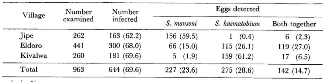
3 the prevalence rates of schistosomiasis determined in 1974 were 62.2 per cent in Jipe, 68.0 per cent in
Table 1 Results of stool and of the Taveta area in
urine 1974
examinations for schistosome eggs in three villages
Village Number
examined Number
infected
Eggs detected
S. mansoni S. haematobium Both together Jipe
Eldoro Kivalwa
2 62 44 l 2 60
163 (62.2) 300 (68.0) 181 (69.6)
156 (59.5) 66 (15.0) 5 (1.9)
1 (0.4) 115 (26.1) 159 (61.2)
6 (2.3)
1 19 (27.0) 17 (6.5) Total 963 644 (69.6) 227 (23.6) 275 (28.6) 142 (14.7) ( ):o/'
Table 2 Positive rates for schistosome egg examinations and skin test by sex in three villages
Village S ex Number
examined
Egg‑positive
(o/o)
Skin‑test‑positive
(o/o)
Jipe
Male Female Total
l 48 l 14
262
64.2 59.6 62.2
85. l 71.1 79.0
Eldoro
Male Female Total
l 83
258
44 l
61.2 72.9 68.0
73.2 72.9 73.0
Kivalwa
Male Female Total
121 1 39
2 60
71.l
68 . 3
69.6
78.5 80.6 79.6
Totals Male
Female
452 511
64.8 68.7
78.5 74.6
Total 963 66.9 76.4
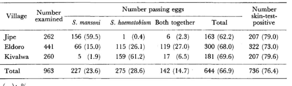
Table 3 Results of stool and urine examinations for schistosome eggs and skin test in three villages
Number Number passing eggs
Village examined S mansom
' S, haematobium Both together Total
Number
skin‑test‑
positive
Jipe 262 156 (59.5) 207 (79.0) l (0.4) 6 (2.3) 163 (62.2) l 19 (27.0) 300 (68.0)
Eldoro 441 66 (15.0) 322 (73.0) ll5 (26.1) 181 (69.6)
Kivalwa 260 5 (1.9) 207 (79.6) 159 (61.2) 17 (6.5) Total 963 227 (23 6) 275 (28 6) 142 (14.7) 644 (66 9) 736 (76 4)
( ):"/o
Eldoro and 69.6 per cent in Kivalwa. In Jipe S. mansoni eggs were detected in 156 individuals (59.5 /o)' S. haematobium eggs in one and both together in six (2.30/0)' In Eldoro S. mansoni eggs were found in 66 individuals (15.00/0)' S. haematobium eggs in l 15 (26.10/0) and both together in 1 19 (27.00/0)' In Kivalwa S. mansoni eggs were
detected in five individuals (1.90/0)' S. haematobium eggs in 159 (61.20/0) and both together in 1 7 (6.50/0)' The results indicated the predominance of S. mansoni infec‑
tion in Jipe, the predominance of S. haematobium infection in Kivalwa and nearly even occurrence of two species in Eldoro.
In Eldoro 112 males out of 183 (61.20/0) and 188 females out of 258 (72.90 ) were found positive f'or schistosome eggs (Table 2). Using the X2 test the difference in the egg‑positive rate between males and females was statistically significant (p<
0.025) in this village. In Jipe and Kivalwa the egg‑positive rates seemed to be some‑
what higher among males than females (Table 2), but the computed values of x2 were not statistically signiflcant. As shown in Tables 2 and 3, the positive rates of skin test with VBS S. japonicum antigen were 79.0 per cent in Jipe, 73.0 per cent in Eldoro, 79.6 per cent in Kivalwa and 76.4 per cent in total, higher than those of egg examinations. The positive rate of skin test was higher among males than females in Jipe. The difference was statistically significant (p<0.01) in the x2 test. In Eldoro and Kivalwa, however, the data did not indicate any statistically signiflcant difference in the positive rate of skin test between males and females.
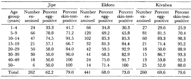
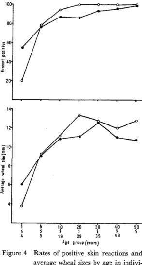
The age distribution of egg‑positive rate and that of skin‑test‑positive rate are shown in Table 4 and Figure 2. The youngest age of egg‑positive individuals was two years. The egg‑positive rate was 33 per cent in the age group of I to 4 years, 75 per cent in that of 5 to 9 years, and 80 per cent among teen‑agers. In the older age groups the rate declined to 60 per cent. On the other hand, skin‑test‑positive rate increased as the age advanced, reaching 95 per cent in the age groups from 40 years up. Children of 9 years and downward have a higher egg‑positive rate than the skin‑test‑positive rate (Figure 2). At every village the peak of the egg‑positive rate was found either in the age group of5 to 9, or in that of 10 to 14 years, and then gradually declined at the older age, remarkably in the cases of S. haematobium (Table 4, Figure 3).
There were found 541 skin‑test‑positive individuals among 644 egg‑positive cases, or in 84 per cent. The rate was higher than the incidence of 61 per cent among the