第34回日本熱帯医学会総会御案内
内 容 原 著
雄ヒトスジシマカの吸血源への誘引飛来についての室内試験(英文)
・栗原 毅125−128 アフリカ風土病型カポシ肉腫
一9例における病理組織学的検討ならびにフローサイトメトリー法 による核DNA量解析一(英文)
・・江藤 秀顕,鳥山 寛,板倉 英喜,田川 泰,Noah O.Kamidigo l29−134 ジャワ島産ナンヨウブユ亜属の1新種について(英文)
・高岡 宏行,Singgih H.Sigit l35−142 ガーナ,アクラにおける乳幼児の栄養状態が寄生虫感染率に及ぼす影響(英文)
・・水上 惟文,Reginald K.Anteson,Maxwell A.Appawu 143−147 ラオス国ビエンチャンにおけるデング熱および日本脳炎ウイルス感染症の血清疫学的研究(英文)
一Khanthong Bomlu,只野 昌之,牧野 芳大,新垣 栄,加根村和美,福永 利彦 149−156 プラジカンテルによる治療後短期間に起こったビルハルツ住血吸虫の再感染
一行天 淳一,木村 英作,N.D.Muhoho,勝又 達哉,
D.K、Migwi,W.R.Mutua,後藤牧人,宇賀 昭二,
塚本 増久,嶋田 雅暁,青木 克己 157−164 短 報
インドネシア,北スマトラにおける条虫Bθ漉召伽s伽4副の人体感染の2症例(英文)
一影井 昇,Ymita Purba,坂本 修 沖縄県における糞線虫感染の現状(英文)
…安里 龍二,仲宗根民男,吉田 朝啓,新垣 民樹,
池城 毅,村上 秀親,崎山 八郎 寒天平板法による糞線虫の検出
寒天平板上に見られる様々な遊走痕について (英文)
一新垣 民樹,田港 朝弘,仲宗根 勇,浦崎 裕子,
中村 哲,岩永 正明 (裏面に続く)
165−168
169−173
175−178
日本熱帯医学会
IS THE MALE AEDES ALBOPICTUS ATTRACTED TO THE BLOOD SOURCE?
TAKESHI KURIHARA
Received January 29 1992/Accepted March 4 1992
Abstract: The responses of male Aedes albopictus to a bait mouse were examined under laboratory conditions using the cage‑test method. In most of the tests fewer males were recovered in the bait cage than the control cage. There was no evidence that male mosquitoes were fond of the mouse or attracted to the animal.
During daytime mosquito collections we often experience male Aedes (Stegomyia) mosquitoes landing on the human body. This phenomenon has been noted by several researchers in different locations. During their human bait collection in Bangkok, Yasuno and Tonn (1970) observed that as many Ae. aeg pti males as females are attracted to the human bait. Following this, Hartberg (1971) in Tanzania, and Gubler and Bhattacharya (1972) in Calcutta also reported, respectively, on the responses of male Ae. aeg)pti and Ae.
albopictus mosquitoes to the human body. Does the male really perceive a blood source and approach the body?
The cage‑test method is useful in examining the behavioral responses of blood sucking fiies in the laboratory (Kurihara et al., 1991) . With some modification of this method, male behaviour, particularly in its response to a blood source was studied.
MATERIALS AND METHOD
Three wire cages were used (30 X 30 x 30 cm) , each with two round holes 15 cm in diameter on opposite walls, and a piece of 6 mm mesh net. In the first series, the three cages were set side by side with the holes aligned, and the net was stretched between the holes of two adjoining cages as a physical barrier. The mosquitoes were, however, able to pass through the netting easily if they wished. In the second series the two holes of the middle cage were covered with the net, and each hole was then connected to the hole of the neighbouring cage by a transparent plastic pipe, 15 cm in diameter and 30 cm in length. Where it contacts the hole of the neighbouring cage the end of the pipe was covered with a fiap to prevent mosquitoes from passing through.
The first series of seven tests utilized different combinations of males and females as shown in Table l. In the second series, three tests were carried out with different age males.
In both series, mosquitoes were released at the middle cage. One of the connecting two cages was a bait cage containing a mouse, and the other was an empty control cage, the only
Department of Medical Zoology, Teikyo University School of Medicine, Kaga, Itabashi, Tokyo 173, Japan
exceptiQn being t st No. 7 in which a small net container (15 x 15 x 15 cm) holding 20 virgin females was placed ip< the cage.
In the first series, mosquitQb vyere released at 3 p.m. and recovered at 10 a.m. in the following morning. In the second series, m0 qY ,toes were lightly anesthetized with ether and released at around 2 p.m. One hour later, the flaps were opened. Observation was made for 15 min, the observer himself also acting as an attractant, and watching the m0Squitoes, that flew into the bait cage.
These cage tests were carried out in a room at 25'C, 80% RH, without circulation of air, and on a constant condition of surroundings such as illumination and colour of the back‑
ground. Each cage was supplied with cotton soaked with 5 ml of a 10% sucrose solution. The Ae. albopictus used in the experiments were a laboratory colony, maintained in an insectar‑
ium at 23‑25'C, 80% RH, and with 16 : 8 (hr. L:D) photoperiod. In the pupal stage or just after emergence the sexes were separated for each test. The number of mosquitoes was checked before releasing them. After recovery, the number of each sex from each cage was also recorded.
RESULTS AND DISCUSSION
The number of mosquitoes examined is shown in Table I together with the results of each test. Each test was replicated 5 times, and the results of recovery are shown as mean number with standard deviation. In all tests in which females were released together with males, more females were recovered in the bait cage than the control cage. In most of the tests fewer males were recovered in the bait cage than the control cage. Significant difference in the number of males recovered from the bait cage was found in two tests only: in Test No.
3 the number was unexplaindly increased in control cage, and in Test No. 7 the number was Table 1 Number of mosquitoes released in the cage test, and mean number recovered
and standard deviation in each test No.
released
No. recovered
Series Test Bait cage Control cage
C71 c7t Cfl
1.
2.
No. 1
3
5 6 7 No. 8 9 10
50 70 70 5 Om
70m 70m
50t 50t 50S 50
O 25
25m
O 25
25m
O O O O
6.8i2.7 9.7i2.4 7.2 3.8*
9.1:!:4.1
7.7i3.8 7.0il.7
7 . 6d: I . 8,*
3.6 1.2 4.3 1.2 4.6 1.6
14 10
.2d:4
.8i2
5.
14 .
.5*
.8
7+1.
0 3.
2*
O*
9 13 21 8 9 9 ll 4.
3.
4.
.4+5 .7 5 .0+2
.6d:1 .5:1:1 .O:i:4 .O:!:2 Od:2.
Od:O.
3 2.
.7 .9 .3 .6 .6
1 8 6
3.
6.
3.
5.
8 1 5+2
.4 .6
0+2.1 6d:4.4
m *
t
$
Mated mosquitoes; others were unmated.
Significant difference with number in control cage.
20 virgin females were held in control cage.
Age at days 3 after emergence, and S : day 4. All others were day 5.
also greater when virgin females were held in the control cage.
In the second series, visually checked was the number of mosquitoes that fiew into the bait cage. However, during a 15 min period, there were almost no males actively approaching the bait cage and hovering above the bait. No significant difference was found within a particular age group.
There was no evidence that male mosquitoes were fond of the mouse or attracted to the animal. On the contrary, more unmated males were in the control cage than in the bait cage in series 1; they appeated to avoid entering the bait cage.
Gubler and Bhattacharya (1972) found that during periods of low density in the study area, males were seldom attracted to the observers' bodies. Thus, it seems that the phenome‑
non of "males landing on a human body" is an accidental happening due to their high density as they rest in vegetation. They may not have been attracted to the body but have had to move from blades of grass or plant leaves to another object which had entered the area.
Unquestionably, many males were also attracted by female mosquitoes which came to bite. We often overlook the presence of females and notice only males which are slower in movement. The males were usually active in approaching the females as shown in Test No.
7.
In a field experiment, however. Mclver et al. (1980) observed that the Mansonia males were significantly more numerous at a distance 3 m from the bait than at 7 m‑15 m.
Therefore, it is still early to conclude that males are not attracted to a mouse. It is possible that the size of the present test apparatus was somewhat small and the males could not orientate to the mouse from the release cage. It is also possible that the bait mouse was not appropriate for attracting the males. Grant (1969) found that certain chemicals attracted male Ae. aeg)pti, and Fay (1968) observed the swarming of male Ae. aeg)pti to objects of certain colours and shapes. The mouse may not have had the appropriate attractancy in colour and/or shape.
REFEREN CES
1 ) Fay, R.W. (1968) : A trap baSed on visual responses of adult mosquitoes, Mosquito News, 28, l‑7
2 ) Grant, G.G. (1969): Dioxane and Dioxaspiro derivatives as attractants for male yellow‑fever mosquitoes, J. Econ. Ent., 62, 786‑789
3 ) Gubler, D.J. and Bhattacharya, N.C. (1972): Swarming and mating of Aedes (S.) albopictus in nature, Mosquito News, 32, 219‑223
4 ) Hartberg, W.K. (1971) : Observations on the mating behaviour of Aedes aeg)pti in nature, Bull.
Wld Hlth Org., 45, 847‑850
5 ) Kurihara, T., Kikuchi, T. and lchimori. K. (1991): Effects of malaria infection in Anopheles stephensi mosquitoes on passage through a wide‑mesh net, Jpn. J. Sanit. Zool., 42, 141‑146 6 ) Mclver, S.B., Wilkes T.J. and Gillies, M.T. (1980): Attraction to mammals of male Mansonia
(Mansonioides). Bull. Ent. Res., 70, Il‑16
7 ) Yasuno, M. and Tonn, R.J. (1970): Study of biting habits of Aedes aeg)pti in Bangkok, Thailand, Bull. Wld Hlth Org., 43, 319‑325
雄ヒトスジシマカの吸血源への誘引飛来についての室内試験 栗原 毅
昼間屋外で蚊の人おとり採集法を実施していると,ヒトスジシマカの雄が人体へ飛来接近する 現象を観察することがある。こうした経験を共有するためか,既に雄蚊の人体誘引現象について の野外観察成績が,2,3報告されている。この現象を室内試験で再現するべく実験を行った。
供したケージテスト法は,30cm角の二面に開孔部をもつ金網ケージを3つ,相互に蚊の往来を可 能なように一線に並べ,中央ケージに蚊を放ち,1つはベイトケージでマウスをいれ,他にはコ ントロールケージとして空のままで,どちらに選択飛来をするか観察するものである。午後3時 から翌朝10時までのどの試験でも,雌蚊はベイトケージに多数集まるが,雄のヒトスジシマカが 特にマウスのいるケージを選択して集まるような現象は観察できなかった。ただコントロール ケージに20匹の未交尾雌を容した場合は,有意に多数集まった。次にケージ間を口径15cmのパイ プで連ねて,観察者自身もマウスのいる側におとりとなりながら雄蚊の飛翔を観察しても,やは り雄蚊がマウスのいるケージに近づく様子が見られなかった。今回の成績から考え,従来の野外 での観察成績は,本種の著しい高密度に由来する機械的な飛来現象か,あるいは雌蚊に伴っての 雄の飛来を注目したものではないかと思わせる。
帝京大学医学部医動物学教室(〒173東京都板橋区加賀2−11−1)
AFRICAN ENDEMIC‑TYPE KAPOSI'S SARCOMA A HISTOPATHOLOGIC STUDY AND FLOW CYTOMETRIC DNA ANALYSIS OF NlNE CASES
HIDEAKI ETOl,*, KAN TORlYAMA*, HIDEYO ITAKURAl YUTAKA TAGAWA2 AND NoAH O. KAMIDIG03
Received February 5 1992/Accepted March 14 1992
Abstract: Flow cytometric DNA analysis using formalin‑fixed, parafnn‑embedded tissues were performed on nine cases of African endemic‑type Kaposi's sarcoma (KS).
Histologically, these cases were classified into the following three types; granulation tissue‑
like (one case) , angioma/angiosarcoma‑like (four cases) and spindle cell (four cases) . Although these three types showed a variety of cellularity and cellular atypism, there were no fundamental differences in the view point of proliferation of spindle cell. Mitotic figures were not prominent. All cases were exclusively diploid and lacked an aneuploid population by flow cytometric measurement of DNA content. These results suggest that African endemic‑type KS is a less aggressive disease rather than a malignant neoplasm.
INTRODUCTION
Kaposi's sarcoma (KS) was first described by Kaposi (1872) as an "Idiopathic multiple pigmented sarcoma of the skin". Since then, many cases of KS have been reported in European, American and African countries. Recently, it has been observed frequently in patients with acquired immune deficiency syndrome (AIDS).
KS is broadly classified into four categories; classical (European) type, African endemic‑
type, AIDS‑related type and the type associated with immunosuppressive therapy. The clinical manifestations of these types are not necessarily same (Safai, 1985; Itakura et al..
1986) . Furthermore, African endemic‑type KS is divided into cutaneous form and lymph node form by the affected site (Toriyama et al., 1987a, b) . Generally, cutaneous KS is long standing, spontaneously regressive, and not fatal. On the other hand, Iymph node KS mainly occurs in children, and shows an aggressive clinical course similar to that of adult AIDS patients (Olweny et al.. 1976; Bayley, 1983). However, it has been reported that twenty‑
seven years was the longest duration of lymph node KS following initial diagnosis in childhood (Dutz and Stout, 1960) . The true nature of KS including histogenesis has been widely disscussed, but still remains obscure.
Flow cytometric measurement of DNA content has been increasingly used as an objec‑
1
2
3
Department of Pathology, Institute of Tropical Medicine, Nagasaki University, 12‑4 Sakamoto‑machi, Nagasaki 852, Japan ( * correspondent author)
First Department of Surgery, Nagasaki University School of Medicine, 7‑1 Sakamoto‑machi, Nagasaki 852, Japhn
Histology Department, Rift Valley Provincial Hospital, P.O.Box 71, Nakuru, Kenya
tive determinant of biological behavior and prognosis in certain solid neoplasms. Abnormal DNA ploidy appears to indicate poor prognosis in various malignant tumors (Hedley et al..
1985; Merkel et al., 1987; Kiyabu et al., 1988; Stanley et al., 1988) . However, there have been few flow cytometric studies of KS. We have performed flow cytometric DNA analysis of parafnn‑embedded tissues of African endemic‑type KS, and discussed the relationship between histological findings and DNA ploidy.
MATERIALS AND METHODS
Materials:
Nine cases of KS obtained from 1986 to 1989 in Provincial General Hospitals in Nakuru and Kisumu, the Republic of Kenya, were examined. Clinical data and relevant information were recorded as accurately as possible.
Histopathologic studies:
For light microscopic examination, each specimen was prepared with hematoxylin‑eosin stain (H.E.) , periodic acid‑Schiff reaction, Azan‑Mallory's stain and silver impregnation for reticulin fibers. Histological growth pattern, cellularity, cellular atypism ahd mitotic rate
(per 10 high power fields; x 400) were determined.
Flow Cytometry:
The technique of Schutte (1985) for DNA analysis was employed using the formalin‑
fixed, paraffin‑embedded tissues. In brief, single cell suspension was obtained by mechanical and enzymatic treatment of three or four 50 pm paraffln sections of each specimen, and stained with propidium iodide. Cellular DNA content was measured by a FACScan equipped with an argon laser. The excitation wavelength was 488 nm. The number of cells in each measurement was at least 2 x 10⁴. The coefflcient of variation (CV) of the diploid peak ranged from 4.0 to 8.7. To confirm the presence of the lesion in sections used for DNA analysis, we cut further 4 pm sections adjacent to the analyzed sections from each specimen.
They were stained with hematoxylin‑eosin and examined to reconfirm the histological diagnosis and features.
RESULTS
The clinical manifestation, histologic features and DNA ploidy in the nine cases were summarized in Table 1. The age of these patients ranged from 18 to 56 years (mean 40 years) . All patients were male. Eight of the nine cases occurred in the skin of the lower extremities. One case occurred in the lymph nodes of the upper arm.
According to the predominant histological features, three main types of growth were recognized. One case was classified as the granulation tissue‑like type. This was character‑
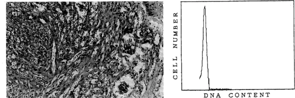
ized by an angioproliferative process with an infiammatory cell infiltration, but was less cellular than the other two types (Fig. 1) . Four cases were angioma/angiosarcoma‑1ike type.
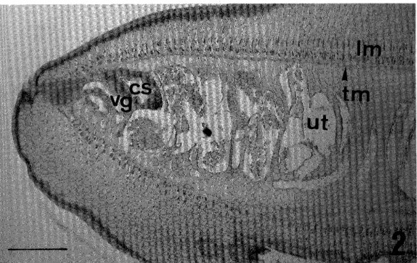
The lesions were composed predominantly of well‑formed vascular spaces, with slit‑like or anastomosing vasculature similar to that seen in angiosarcoma, but with minimal cellular atypism (Fig. 2). Four cases were spindle cell type. This type showed a high cellularity, but its cellular atypism and mitotic figures were not prominent (Fig. 3) . Transitional zones and intermingling of these three types were also frequently observed in the same case. Mitotic
Table l Clinical manifestation, histologic features and DNA ploidy of African endemic‑type Kaposi's sarcoma
Case Age(yr) Sex Site of lecation Histological ty pe
Cellular
Cellulanty atypism Mitotie rate
(per 10 HPFs) DNA ploidy
1 55 M Skin (leg) S +++ ++ 4 D
2 32 M Sk in (limb) S +++ + 2 D
3 U M Skin (leg) A ++ + 1 D
4 39 M Skin ( Ieg) A ++ + 3 D
5 53 M Skin (limb) s +++ + 5 D
6 56 M Skin ( f oot) A ++ + l D
7 18 M Skin (knee) A ++ ++ o D
8 38 M Skin (leg) G + + o D
9 30 M Lymph node
(upper arm) s +++ ++ 8 D
U:unknown; G:granulation tissue‑like HPF:high power field; D:diploid
type; A:angioma/angiosarcoma‑like ty pe; S:spindle cell type;
rate ranged from O to 8 per 10 high‑power fields ( x 400) .
All cases were diploid by fiow cytometric measurement of DNA content .
DlscUssloN
Many studies have been performed about the histogenesis of KS, such as vascular endothelial cell (Hashimoto et al., 1987; Scully et al., 1988) , Iymphatic endothelial cell (Beckstead et al.. 1985; Dictor and Anderson, 1988) and mesenchymal c̲ell (Komuro and Toriyama, 1991) . Some immune factors with angiogenic activity, such as thymosin, interfer‑
on, Iymphokine8 and prostaglandin, are thought to be crucial to the development of KS (Levy
Figure 1
p(
eq FO
i{
:D :
,1 I (,
(left) Case 8 (granulation tissue‑1ike type) ized by an angioproliferative proeess infiltration (H.E., x 100). (right)
(CV 8.2) .
DNA CONTENT
The・ Iesion is character‑
with an inflammatory cell DNA histogram showing diploidy
Figure 2
e ,
;
・i
l
O
(left) Case 6(angioma/angiosarcomalike composed predominantly of anastomosing well‑formed vessels (H.E., x 100) ,
diploidy (CV 7.7) .
DNA CONTENT
type). The lesion is vascular channels and (right) DNA histogram showing
Figure 3
4
eo :
;D
・
l
M U
DNA CONTENT
(1eft) Case 9 (spindle cell type) . The lesion ls formed by interlacing bundle8 of spindle shaped cells (H.E., x 100). (right) DNA histo‑
gram showing diploidy (CV 4.0),
and Ziegler, 1983) . However, the true nature of KS, whether hyperplasia or neoplasia, remains unknown (Costa and Rabcon, 1983; Brooks, 1986; Mirra, 1986) .
African endemic‑type KS is usually divided into cutaneous form and lymph node form by the affected site (Tariyama et al.. 1987a, b) . Generally, cutaneous KS usually occurs in lower extremities af adults, and is long standing, growing slowly and occasionally regressive Itakura et al., 1986) . On the other hand, Iymph node KS mainly occurs in children without cutaneous lesions, and shows an aggressive clinical course with generalized lesions often involving the visceral organs similar to that of adult AIDS patients (Olweny et al,, 1976;
Bayley, 1984). However, it has been reported that twenty‑seven years was the longest duration of lymph node KS following initial diagnosis in childhood (Dutz and Stout, 1960) . In this study, African endemic‑type KS was histalogically classified into the following three types; granulation‑tissue like type, angioma/angiosarcoma like type and spindle cell type. Transitional zoncs and intermingling of these three types were also frequently observed
in the same case. Although thes three types showed a variety of cellularity and cellular atypism, there were no fundamental differences in the view point of proliferation of spindle cell. Mitotic figures were not prominent.
Recently, DNA ploidy analysis by flow cytometry has shown that DNA aneuploidy is common in several malignant tumors and is a useful prognostic indicator (Hedley et al.. 1985;
Merkel et al.. 1987; Kiyabu et al.. 1988; Stanly et al., 1988) . However, there has been no flow cytometric studies of African endemic‑type Kaposi's sarcoma. In our study, all cases of African endemic‑type KS were exclusively diploid and lacked an aneuploid population by flow cytometric measurement of DNA content. Histological appearance and DNA ploidy of lymph node KS were identical with cutaneous KS. These results are consistent with the reported findings in AIDS‑related type KS cases (Fukunaga and Silverberg, 1990) . Although there are no sufiicient follow‑up studies, our results suggest that African endemic‑type KS is a less aggressive disease rather than a malignant neoplasm.
REFERENCES
1 ) Bayley, A.C. (1984): Aggressive Kaposi's sarcoma in Zambia, 1983, Lancet, 1, 1318‑1320 2 ) Beckstead, J.H., Wood, G.S. and Fletcher, F. (1985): Evidence for the origin of Kaposi's
sarcoma from lymphatic endothelium, Am. J. Pathol., I19, 294‑300
3 ) Brooks, J.J. (1986): Kaposi's sarcoma: A reversible hyperplasia, Lancet, 2, 1309‑1 10 4 ) Costa, J. and Rabson, A.S. (1983): Generalized Kaposi's sarcoma is not a neoplasm, Lancet,
l, 58
5 ) Dictor, M. and Anderson, C. (1988) : Lymphaticovenous differentiation in Kaposi's sarcoma:
Cellular phenotypes by stage, Am. J. Pathol., 130, 411‑417
6 ) Dutz, W, and Stout, A.P. (1960): Kaposi's sarcoma in infants and children, Cancer, 13, 684‑694 7 ) Fukunaga, M. and Silververg, S.G. (1990) : Kaposi's sarcoma in patients with acquired immune deficiency syndrome: A flow cytometric DNA analysis of 26 Iesions in 21 patients, Cancer, 66, 74‑77
8 ) Hashimoto, H., Muller, H., Falk, S. and Stutte, H.J. (1987): Histogenesis of Kaposi's sarcoma associated with AIDS: A histologic, immunohistochemical and enzyme histochemical study, Pathol. Res. Pract., 182, 658‑668
9 ) Hedley, D.W., Freidlander, M.L. and Taylor, I.W. (1985): Application of DNA flow cytometry of paraffin‑embedded archival material for the study of aneuploidy and its clinical significance, Cytometry, 6, 26‑30
lO) Itakura, H., Toriyama, K. and Uzuta, F. (1986): Kaposi's sarcoma, pathology and local epidemiology in Kenya, Trop. Med., 28 (suppl.), 3‑8
ll) Kaposi, M. (1872): Idiopathisches multiples Pigmentsarkom der Haut, Arch. Derm. Syph., 4, 265‑273
12) Kiyabu, M.T., Bishop, P.C., Parker, J.W., Turner, R.R. and Fitzgibbon, P.L. (1988): Smooth muscle tumors of the gastrointestinal tract: flow cytometric quantitation of DNA and nuclear antigen content and correlation with histologic grade, Am. J. Surg. Pathol., 12, 954‑960 13) Komuro, S. and Toriyama, K. (1991) : Geopathology of endemic pediatric lymph node Kaposi's
sarcoma in western Kenya, Jpn. J. Trop. Med. Hyg., 19, 251‑263
14) Levy, J.A. and Ziegler, J.L. (1983): Acquired immunodeficiency syndrome is an opportunistic infection and Kaposi's sarcoma results from secondary immune stimulation, Lancet, 1, 78‑80 15) Merkel, D.E., Dressler, L.G. and Mcguire, W.L. (1987): Flow cytometry, cellular DNA content,
and prognosis in human malignancy, J. Clin. Oncol., 5, 1690‑1703
16)Mirra,」.M.(1986):Kaposi s Sarcoma :Is it a sarcoma−at al1P,Trop.Med.,28(suppl.),49−62 17)Olweny,C.L.M.,Kaddumukasa,A.,Atine,1.,Ower,R.,Magrath,L and Ziegler,J.L(1976):
Childhood Kaposi s sarcoma:Clinical features and therapy,BrJ.Cance二,33,555−560 18)Safai,B.(1985): Kaposi s sarcoma and other neoplasms in Acquired Immunodeficiency
Syndrome:Advances in Host Defense Mechanisms,5,59−73,Ravin Press,New York 19)Schutte,B.,Reynder,M.M.J.,Bosman,F、T.and Blijam,G.H.(1985):Flow cytometric
dete㎜ination of DNA ploidy level in nuclei isolated from paraf五n−embedded tissue,
Cytometry,6,26−30
20)Scully,P.A.,Steinman,H.K.,Kennedy,C.,Trueblood,K,Frisman,DM.and Voland,J.R (1988):AIDS−related Kaposi s sarcoma displays differential expression of endothelial surface antigens,Am.J.Pathol.,130,244−251
21)Stanley,J.R.,Wooldridge,T.N.and Linder,」.(1988):Flow cytometric DNA analysis of malignant fibrous histiocytoma and related fibrohistiocytic tumors,Hum.PathoL,19,74−77 22)Toriyama,K.,Uzuta,F.and ltakura,H.(1987a):Geopathological study on endemic Kaposi s sarcoma in westem Kenya,Trop.Med.,29(2),87−100
23)Toriyama,K.,Uzuta,F.and ltakura,H.(1987b):Kaposi s sarcoma of lymph node(s)in westem Kenya,Trop.Med.,29(2),10HO6
アフリカ風土病型カポシ肉腫
一9例における病理組織学的検討ならびにフローサイトメトリー法 による核DNA量解析一
江藤 秀顕1・鳥山 寛1・板倉 英喜1・
田川 泰2・Noah O.KAMIDIGO3
カポシ肉腫(Kaposi ssarcoma,KS)は一般には,欧米古典型,アフリカ風土病型,AIDS関連
型,その他の免疫不全型の4型に大別され,さらにアフリカ風土病型KSは,発生部位により皮膚型 とリンパ節型に分けられている。それぞれの型において若干の病態的な差異が認められているが,
その組織発生を含め本態は未だ不明のことが多い。最近ではフローサイトメトリー(FCM)法に よる核DNA量解析が腫瘍,あるいは腫瘍様病変の悪性度評価に,広く用いられている。今回9例 のアフリカ風土病型KSにおいて,病理組織学的検討,ならびにホルマリン固定,パラフィン包埋 ブロックを用いたFCM法による,核DNA量解析を行った。組織学的には次の3型に分けられた。
すなわち肉芽組織類似型(1例),血管腫あるいは血管肉腫類似型(4例)と紡錘形細胞型(4例)
である。これら3型においては,細胞密度や細胞異型に程度の差はあるものの,紡錘形細胞の増 殖という点からは,基本的な差は見られなかった。核分裂像も目立たなかった。FCM法による核 DNA量では,全例がdiploidを示しており,aneuploid例は見られなかった。これらの結果より,
アフリカ風土病型KSは悪性腫瘍というより,むしろ1ess aggressiveな病変であると思われた。
長崎大学熱帯医学研究所病理学部門(〒852長崎市坂本町12−4)
長崎大学医学部第一外科学教室 リフトバレー州立病院,ケニア
A NEW BLACKFLY SPECIES OF SIMULIUM (GOMPHOSTILBIA) FROM JAVA, INDONESIA
(DIPTERA= SIMULIIDAE)
HIROYUKI TAKAOKA* AND SINGGIH H. SIGIT2 Received February 7 1992/Accepted March 16 1992
Abstract: A new blackfly species, Simulium (Gomphostilbia) parahiyangum sp. nov.
is described based on the female, male, pupal and larval specimens collected from Java.
This new species is very distinctive among the Gomphostilbia species in possessing promi‑
nent dorsal protuberances on abdominal segments 1‑5 and a deep postgenal cleft reaching the hypostomium in the larval stage. By the number of antennal segments (11 vs. 10) the male of this species is easily distinguished from that of S. (G.) varicorne Edwards, 1927 reported from Sumatra, to which the larva and pupa of this species had been once thought to be conspecific.
INTRODUCTION
The Simuliidae of the Sunda Islands has not been studied since Edwards (1934) described 11 new species from Sumatra, Java and Bali, making a total of 19 taxa (including two subspecies) for this archipelago.
Our recent preliminary survey on the blackfiies in East and West Java yielded a total of 16 species of Simulum Latreille s. l. including several new species, of which two belonging to the subgenus Simulium Latreille s. str. have already been described (Takaoka and Hadi, 1991) . This paper describes one new blackfly species of the subgenus Gomphostilbia Ender‑
lein.
The classification follows that of Crosskey (1969). Collecting and rearing methods, as well as dissection of anatomical parts for description, were mentioned in Takaoka (1983) .
DESCRIPTION
Simulium (Gomphostilbia) parahiyangum sp. nov.
Female. Body length 1.8 mm. Head slightly narrower than width of thorax. Frons brownish, whitish grey pruinose, densely covered with whitish yellow recumbent pubescence except middle longitudinal portion narrowly bare; frontal ratio (i.e., ratio of the greatest width at vertex, the narrowest near antennal base, and the height of the frons) 1.7:1.0:1.8.
l Division of Medical Zoology, Oita Medical University, Hasama, Oita, 879‑55, Japan (correspondent author)
2 Entomology Laboratory, Department of Parasitology and Pathology. Faculty of Veterinary Medicine, Bogor Agricultural University, Taman Kencana 3, Bogor 16151, Indonesia
Frons‑head ratio (i.e., ratio of the greatest width of the frons against that of head) 1.0:3.9.
Clypeus brownish black, whitish grey pruinose, and densely covered with whitish yellow pubescence interspersed with several dark hairs. Antenna composed of 2 + 9 segrnents, brownish black except scape and pedicel (in one female, base of Ist flagellar segment too) yellow. Maxillary palp composed of 5 segments with proportional length of 3rd, 4th and 5th segnlents being 1.0:1.1:2.3; 3rd segnent not so enlarged, but sensory vesicle (Fig. 1) enlarged, 0.6x as long as 3rd segment and with its opening on distal 1/3. Maxilla with 14 inner and 15 outer teeth. Mandible with ca. 30 small inner teeth and devoid of outer ones. Cibarium with heavily sclerotized arms but without any denticles medially. Thorax. Scutum brownish black in ground colour, thinly whitish grey pruinose, with three dark longitudinal lines which are distinct when viewed anterodorsally; scutum densely covered with whitish yellow recum‑
bent pubescence. Scutellum brown with whitish yellow pubescence as well as long upstanding dark hairs along posterior margin. Postscutellum brownish black, whitish grey pruinose and bare. Pleural membrane bare. Katepisternum brownish black, whitish grey pruinose, with numerous pale and dark hairs, and longer than deep; sulcus distinct. Legs. All coxae and trochanters yellow except mid and hind coxae brown. All femora yellow, somewhat dar‑
kened distally and brown on distal cap. All tibiae yellow to dark yellow on basal 2/3, brown on distal 1/3, and with subbasal dark spot. Fore tarsi brownish black. Mid and hind tarsi brown except basal 1/3 of mid basitarsus, basal 2/3 of hind basitarsus and basal 1/2 of hind 2nd tarsal segment yellowish. Fore basitarsus slightly dilated, ca. 5.9 x as long as its greatest width. Hind basitarsus (Fig. 3) slender, parallel‑sided. Calcipala (Fig. 3a) moderately developed, nearly as long as wide, and ca. 0.6x width of basitarsal tip. Pedisulcus also distinct at basal 1/3 of 2nd tarsal segment. Claws (Fig. 3b) each with large basal tooth which is l/2 x as long as claw. Wing. Length 1.7 mm. Costa with spinules as well as hairs.
Subcosta haired. Tuft hairs at base of stem vein dark brown. Basal portion of radius fully haired. Abdomen. Basal scale yellow with a fringe of pale yellow hairs. Dorsal surface of abdomen brown to brownish black except that of 2nd segment entirely pale yellow; tergite of 2nd segment whitish pruinose; tergites of 6th, 7th and 8th segments wide and shiny. Geniialia.
Sternite 8 (Fig. 5a) bare medially, and with ca. 18 dark macrosetae on each side. Anterior gonapophyses (Fig. 5b) thin, membraneous, rounded posteriorly, covered densely with microsetae except posterointernal margins narrowly bare, and with a few short setae near anterior border; inner margins well sclerotized. Genital fork (Fig. 5c) of usual reversed‑Y form, with arms each produced inwards to some extent but lacking any projection directed forwards. Paraproct (Figs. 6 and 7) of usual form, and with ca. 12 macrosetae ventrally and laterally. Cercus (Fig. 7) short, 1/2 x as long as wide, rounded posteriorly, when viewed laterally, and covered with ca. 14 macrosetae on outside surface. Spermatheca (Fig. 5d) oblong, well sclerotized except small adjacent area near tubal juncture unsclerotized.
Male. Body length 1.8 mm. Head slightly wider than width of thorax. Upper eye consisting of 17 vertical colums and 15 horizontal rows of large facets. Clypeus brownish black, whitish pruinose, and covered densely with yellow pubescence interspersed with dark hairs. Antenna composed of 2 + 9 segments, dark brown except scape and pedicel yellow; Ist flagellar segment elongated, 2 x as long as 2nd flagellomere. Maxillary palp with 5 segments;
proportional length of 3rd, 4th and 5th segment 1.0:1.1:2.3; sensory vesicle (Fig. 2) small, ca.
0.17 x as long as 3rd segment and with very small opening distally. Thorax. Scutum brownish
,i*:L.'.
,・: i:i: .'
. }: '・
i: ・; ::
1
e
,:* =
: '
:i*F *: '
**'+
. ,, : ; * s
・ !.*・*
r・: .
t.. ' '‑
5
c‑‑
a ,,..'. "r! :i':i' l
¥̲ ,i i;;; ;. s';;:*E
"s:'; :;; L'f':{.: ::
' ::""'f:*
si:i ::t'i":i' : ': "': : s:
':":: 't:" l " ""'
"* ':':; ""': '*i': :t' : "i
": ::!;; ;st: "'t:t ' " ;r: :' " 't{; ; {;J !'jt'tlitl
f; j:.,' 1 "s " ':' t
"h": ':is ':'!d!;' I ' "::'i' Itt
:' :! ' b
,:‑
, t:
1
If* ‑
/ ‑‑‑
Fh l̲r ‑ ‑‑‑r
(/
/4 ;‑'.
¥! ¥'///
/
/
d
3
Figs.
̲ b
1‑14
4
l' a
,,
¥e
9
,, '
10 '
., . ,
,
/
a It
'̲̲̲ ""Il'It
' l:: "
Illl'IttthLI I u i' 'littE lha ;;i : 'l I!{7!LlfllUit l]lt L t'
t. rtr.i,tLV L
, ,t
' I'Is I
¥b
8
t ,I LL' t,1 Il
: It
12
C ' k*..
11
tl
, .,,
13
l{':. {s'̲('F
i ^ = E :.
'14
Adult of S. (G.) rahiyangum sp. nov.: I and 2, 3rd segments of maxillary palp showing sensory vesicle in front view (1, female; 2, male) ; 3 and 4, hind legs (3, female; 4, male‑a, calcipala; b, claw); 5‑7, female genitalia {5, 8th sternite (a), anterior gonapophysis (b) , genital fork (c) and spermatheca (d) in ventral view; 6 and 7, paraprocts and cerci in ventral view (6) and in lateral view (7) }; 8‑14, male genitalia {8, coxite (a), style (b) and ventral plate (c) in ventral view; 9 and 10, styles viewed mediolaterally (9) and ventrolaterally (lO); 11 and 12, ventral plates viewed laterally (11) and posteriorly (12); 13, paramere with 3 distinct parameral hooks; 14, median
sclerite } .
black, entirely whitish pruinose in certain angle of light and densely covered with bright yellow recumbent pubescence. Scutellum brownish black, with bright yellow pubescence and several long marginal hairs. Postscutellum brownish black, whitish pruinose, and bare.
Pleural membrane and katepisternum as in female. Legs. Mostly brown to brownish black except hind trochanter, base of hind femur and tibia, basal 2/3 of hind basitarsus and basal 1/2 of hind 2nd tarsal segment whitish yellow. Fore basitarsus slender, ca. 6.9 x as long as its greatest width. Hind tibia (Fig. 4) subequal to hind femur in greatest width. Hind basitarsus (Fig. 4) slender, parallel‑sided. Calcipala (Fig. 4a) well developed, 1.2 x as long as wide, and 0.65 x as wide as basitarsal tip. Pedisulcus well developed. Wing. Length 1.6 mm. Other features as in female except subcosta bare. Abdomen. Basal scale dark brown and its hair fringe dark basally and pale distally. Dorsal surface of abdominal segments brownish black except that of 2nd segment brow and covered with short hairs; 'a pair of dorsolateral whitish pruinose patches on segments 2, 6 and 7. Genitalia. Coxite (Fig. 8a) nearly rectangular in ventral view, ca. 1.6x as long as wide. Style (Figs. 8b, 9 and 10) much shorter than coxite, gently curved inwards and without apical spine. Ventral plate (Fig. 8c, ll and 12) fiat, with microsetae almost entirely on ventral surface but only medially on posterior surface. Paramere (Fig. 13) with 3 Iong parameral hooks and a few small, indistinct ones. Median sclerite (Fig, 14) plate‑like, with widened tip.
Pupa. Body length (excluding gill filaments) 2.0 mm. Head and thorax. Integument yellowish brown, covered densely with cone‑shaped tubercles. Antennal sheath (Fig. 15) along its length with 9 well marked ridges on outer margin, each covered with several cone‑
shaped tubercles. Head with I facial and 3 frontal pairs of simple IQng trichomes. Thorax with 5 pairs of simple long trichomes on anterior 1/2. Gill (Fig. 16) with 8 slender greyish brown filaments arranged in 3 groups, i.e., upper and middle triplets and I Iower pair; all filaments very short (ca. 0.7 mm) , subequal in length and thickness to one another, with numerous transverse furrows becoming indistinct towards apex, and covered with minute tubercles. Abdomen. Terga I and 2 slightly darkened, and without tubercles; tergum I with a long simple seta on each side, and tergum 2 on each side with 6 simple minute setae, of which I seta is longer than others. Terga 3 and 4 each with 4 hooked spines directed forwards along posterior margin, and a short seta medially on each side. Tergum 5 with 5 very minute setae but devoid of spine combs. Terga 6‑8 each with spine combs in transverse row, and comb‑like groups of very minute spines on each side; terga 6‑8 each also with a pair of minute setae on each side; tergum 9 with comb‑like groups of minute spines and a pair of simple terminal hooks (Fig. 17) . Sterna 4‑8 each with comb‑like groups of minute spines scattered all over. Sternum 4 with I simple slender hook on each side. Sternum 5 with a pair of bifid or trifid hooks situated close together on each side. Sterna 6 and 7 each with a pair of inner bifid or trifid and outer simple hooks widely spaced on each side. Sternum 9 with 3 grapnel‑
shaped hooks on each side. Cocoon simple, slipper‑shaped, moderately woven, extending ventrolaterally, and with thick anterior margin which has no anterior projection.
Mature larva. Body length 4.0 mm. Body colour greyish yellow to yellowish brown. Head moderately covered with minute setae; cephalic apotome (Fig. 19) pale on anterior 1/2, somewhat darkened on posterior 1/2 and with negative head spots. Antenna longer than cephalic fan stem, with 4 segments, proportional length of Ist, 2nd and 3rd segrnents 1.0:0.9:
'II " "b 'tl' ,," e ',,fe.JI 'fL c r . , llL J' .,
"I I ・1 .,1 ". ) ;
'l
15
I '.S‑
f,.kf:,:: ;;:*:r{
16
: i, .・t;,.
,
)
i ; ;J:?Sd ' :: t;: ,:;,
. t.'
' "lj・,.・.:.
17
*::i : "
::i:.:r. :.
::;:;:. ;:;: ; :":
?t' '.iil"?.
̲ ' 'S e. ;;"'
"fi; :. '*.
' t : 'i :i 'i::
';j ;':": : ::'*L.1,i : ' " ,i ;;;*ilii :'.,,:
L':td i. "
. ' "r':'
,,
18
,1$.・l
1‑
'ii':
*.
,f. ' a・il : ,・.:
, ::
,:V:・ t ‑1 :,P' ;S. 't
'.? *s. :; ::: : :;. ii"i.,
, ,V ・ .$. ";.1'
:・ ;S$"' , , 1 ':,t ・, f: ;.;{:,S ':tl"i̲・!i:1'.‑
Sl"' ' ' "'1 : t'll b' : b I"
.: ,' .t '::
I: "" ' 1 :1'/"!.,t
.
, ! ,
20
21
'==e='
L
le
15‑24
' 'j"=== '=;==;/
23
22
,
)
)
l :,fl'/
Figs. Pupa and larva of S. (G.) parahiyangum sp. nov. 15‑17, pupa (15, antennal sheath showing tuberculate projections; 16, respiratory filaments; 17, termi‑
nal hooks in dorsal view) ; 18‑24, Iarva (18, apical tip of mandible; 19, cephalic apotome showing negative head spots; 20, ventral surface of head capsule showing hypostomium and deep cleft; 21, dorsal view of larva showing the distribution of spinules and spines; 22, Iateral view of larva showing dorsal and dorsolateral protuberances on abdominal segments 1‑5 (spinules and spines omitted) ; 23, flat spinules of various sizes on thorax and abdomen‑2 spinules from left are seen laterally; 24, black spines on abdomi‑
nal segments 6‑8) . Scale bars indicate 0.2 mm for 16 and 0.02 mm for 23 and 24.
0.9. Cephalic fan with ca. 30 main rays. Mandible comb teeth decreasing in length and thickness from
(Fig. 18) with 2 mandibular serrations;
Ist to 3rd tooth. Hypostomium with a
row of 9 apical teeth, of which corner and median teeth are moderately developed; Iateral serration absent; 4 hypostomial setae lying parallel to lateral margin on each side. Postgenal cleft (Fig. 20) deep, widely reaching posterior margin of hypostomium. Histoblast of pupal gill with 8 slender filaments, arranged in 3 + 3 + 2. Thoracic segments densely covered, on dorsal and lateral' surfaces, with small dark fiat spinules which have several transparent slender branches apically (Fig. 23). Abdominal segments 1‑5 each with a dorsal pair of conical protuberances and a dorsolateral pair of rather smaller protuberances (that of segment 5 not well defined) (Figs. 21 and 22) which are covered with a few colorless setae and numerous small dark spinules similar to those on thoracic cuticle; abdominal cuticle, besides on protuberances, covered densely with similar spinules dorsally and laterally except on intersegment spaces; there are also 12‑20 black distinct spines scattered dorsally on segments 6‑8 (Figs. 21 an 24); colorless setae alsQ present on dorsal and lateral surface of last segment. Rectal gill lobes compound, each lobe with 8‑10 finger‑like lobules. Anal sclerite of usual X‑form, posterior arms a little longer than anterior ones; basal portion of arms widely sclerotized. Ventral papillae well developed. Posterior circlet with ca. 74 rows of up to 13 hooks per row.
Type specimens. Holotype slide‑mounted together with its pupal skin and cocoon on glass slide, reared from pupa, taken at foot of Mt. Tangkubanperahu, 6 km north of Bandung, West Java. Indonesia, 30.XII.1990, H. Takaoka. Allotype crl, slide‑mounted, same data as holotype.
Paratypes, 3 , I cf' , reared from pupae, and 3 mature larvae (all preserved in alcohol except I Iarva slide‑mounted), same data as holotype. Holotype , allotype cfl , I paratype and I Iarval paratype will be deposited at the Department of Parasitology and Pathology, Faculty of Veterinary Medicine, Bogor Agricultural University, and I and I Iarval paratype at the Department of Entomology, Bogor Museum of Zoology, Bogor, and other paratypes (l , I c; and I Iarva) at the Natural History Museum, London.
Di,stribution. Java.
Ecological note. The pupae and larvae of this species were found on banana leaves and on slender plant roots trailing in the water of a small shaded stream (ca. I m wide; ca. 1,000 m in altitude) running down through the sloped field cultivated for tea plantation not far from the northern exit of National Park of Mt. Tangkubanperahu, along the road to Segalaherang.
Collected together with this species were S. (G.) batoense Edwards, 1934 and S. (S.) sigiti Takaoka and Hadi, 1991.
DrscUSsloN
This new species was named after the Sundanese "parahiyanga" ( = remarkable) because its larva is very distinct in possessing prominent dorsal protuberances (Fig. 22) on the abdomen and the deep postgenal cleft (Fig. 20) reaching the hypostomium, a combination of these characters being unusual among the known species of the subgenus Gomphostilbia.
There has been no report of any Gomphostilbia species with these peculiar characters except S. ( G.) nr, varicorne (only larval stage known) which was collected from Assam (Datta, 1975) . The larva of S. ( G.) parahiyangum resembles that of S. ( G.) nr. varicorne, from which
it is differentiated by the branching method of respiratory filaments as seen in the histoblast (i.e., two triplets plus one pair vs. four pairs) . The pupa of S. (G.) parahiyangum is characterised by the short slender filaments. In this aspect, this species is similar to S. ( G. ) montiblense Takaoka, 1983 from Palawan Island of the Philippines (Takaoka, 1983) , and S.
( G. ) darjeelingense Datta, 1973 from India (Datta, 1973) . However, there is a clear difference in the branching method of the respiratory filaments.
The larva and pupa of S. ( G.) parahiyangum are probably conspecific to those thought as the immature stages of S. ( G. ) varicorne Edwards, 1925 which was known only from the type male specimen collected from Sumatra (Edwards, 1934). Our study showed that Edwards' tentative association was incorrect since the reared adults of S. ( G. ) parahiyangum had a normal antenna composed of ll segments {not 10 as seen in S. ( G. ) varicorne}. The immature stages of S. (G.) varicorne may be of diffeyent form. In this connection, it should be noted that two Malayan specimens of male adults labelled as S. varicorne at the Natural History Museum, in London, had their pupal exuvia with long‑stalked respiratory filaments which were apparently different from those of S. ( G. ) parahiyangum (Takaoka, unpublished
data) .
The male of this new species is similar to S. (G. ) friederichsi Edwards, 1934 known only from male type specimen taken from Java in having the dark leg coloration and the slender hind basitarsus. The male of S. ( G.) parahiyangum is easily distinguished from the latter by lacking a distinct terminal spine on the style as well as any pruinose scutal pattern.
ACKNOWLEDGEMENTS
We thank Drh. Upik K. Hadi, Entomolygy Laboratory of Department of Parasitology and Pathology, Bogor Agricultural University for arranging the collection trip. Our apprecia‑
tion goes to Miss Siti Zaenab and Mr. Pupun Purnama for their cooperation in the field survey. The senior author would like to thank Dr. D.M. Davies, Professor Emeritus, McMaster University, Ontario, and Dr. H. Takahashi, Tokyo for their encouragement.
The research was supported by a grant‑in‑aid from Ministry of Education, Science and Culture, Japan to H.T.
REFEREN CES
1 ) Crosskey, R.W. (1969): A re‑classification of Simuliidae (Diptera) of Africa and its islands, Bull. Br. Mus. Nat. Hist. (Entomol.) Suppl., 14, 195
2 ) Datta, M. (1973): New species of black fiies (Diptera: Simuliidae) of the subgenera Eusimulium Roubaud and Gomphostilbia Enderlein from the Darjeeling area, India, Oriental Insects, 7, 363‑402
3 ) Datta, M. (1975): Simuliidae (Diptera) from Assam foot‑hills, India, Jpn. J. Sanit. Zool., 26, 31‑40
4 ) Edwards, F.W. (1934): Deutsche Limnologische Sunda‑Expedition. The Simuliidae (Diptera) of Java and Sumatra, Arch. F. Hydrobiol., Suppl., 13, 92‑138
5 ) Takaoka, H. (1983): The blackflies (Diptera: Simuliidae) of the Philippines, pp. 212, Japan Society For The Promotion of Science, Tokyo
6 ) Takaoka, H. and Hadi, U.K. (1991): Two new blackfiy species of Simulium (Simulium) from Java, Indonesia (Diptera: Simuliidae), Jpn. J. Trop. Med. Hyg., 19, 357‑370
ジャワ島産ナンヨウブユ亜属の1新種について
高岡 宏行1・Singgih H.Sigit2
1990年12月から1991年1月にかけて,ジャワ島において吸血性昆虫ブユの採集調査を行った。
得られた標本を検討した結果,数種の新種が含まれていることが分かった。本論文では,ブユ属 ナンヨウブユ亜属に属する,1新種の記載を行った。本種は,幼虫の頭部腹面のpostgenal cleft が深く,腹部第1節から5節の背面に顕著な瘤様の突起を持つなど,これまでこの亜属のなかで は見られない珍しい形態的特徴を有する。
大分医科大学医動物学教室(別冊請求先:〒879−55大分県大分郡挾間町医大ケ丘1−1)
インドネシア国ボゴール農科大学獣医学部寄生虫・病理学部門昆虫学研究室