Illuminating the Onco-GPCRome: Novel G protein
coupled receptor-driven oncocrine networks and
targets for cancer immunotherapy
著者
Victoria Wu, Huwate Yeerna, Nijiro Nohata,
Joshua Chiou, Olivier Harismendy, Francesco
Raimondi, Asuka Inoue, Russell Robert B.,
Pablo Tamayo, J. Silvio Gutkind
journal or
publication title
Journal of biological chemistry
volume
294
number
29
page range
11062-11086
year
2019-07-19
URL
http://hdl.handle.net/10097/00128411
doi: 10.1074/jbc.REV119.005601Illuminating the Onco-GPCRome: Novel G protein– coupled
receptor-driven oncocrine networks and targets for cancer
immunotherapy
Published, Papers in Press, June 5, 2019, DOI 10.1074/jbc.REV119.005601
Victoria Wu‡, Huwate Yeerna§,X Nijiro Nohata‡1,X Joshua Chiou¶,X Olivier Harismendy§ §§,
Francesco Raimondi储**2,3, Asuka Inoue‡‡4, Robert B. Russell储**3,5, Pablo Tamayo§, and J. Silvio Gutkind‡6
From the Departments of‡Pharmacology and§Medicine, UCSD Moores Cancer Center, La Jolla, California 92093, the¶Department of Pediatrics and§§Division of Biomedical Informatics, University of California San Diego School of Medicine, La Jolla, California 92093, the储CellNetworks, Bioquant, Heidelberg University, Im Neuenheimer Feld 267, 69120 Heidelberg, Germany, the **Biochemie Zentrum Heidelberg (BZH), Heidelberg University, Im Neuenheimer Feld 328, 69120 Heidelberg, Germany, and the‡‡Graduate School of Pharmaceutical Science, Tohoku University, Sendai, Miyagi 980-8578, Japan
Edited by Henrik G. Dohlman
G protein– coupled receptors (GPCRs) are the largest gene family of cell membrane–associated molecules mediating signal transmission, and their involvement in key physiological func-tions is well-established. The ability of GPCRs to regulate a vast array of fundamental biological processes, such as cardiovascu-lar functions, immune responses, hormone and enzyme release from endocrine and exocrine glands, neurotransmission, and sensory perception (e.g. vision, odor, and taste), is largely due to the diversity of these receptors and the layers of their down-stream signaling circuits. Dysregulated expression and aberrant functions of GPCRs have been linked to some of the most prev-alent human diseases, which renders GPCRs one of the top tar-gets for pharmaceutical drug development. However, the study of the role of GPCRs in tumor biology has only just begun to make headway. Recent studies have shown that GPCRs can con-tribute to the many facets of tumorigenesis, including prolifer-ation, survival, angiogenesis, invasion, metastasis, therapy resis-tance, and immune evasion. Indeed, GPCRs are widely dysregulated in cancer and yet are underexploited in oncology. We present here a comprehensive analysis of GPCR gene expression, copy number variation, and mutational signatures
in 33 cancer types. We also highlight the emerging role of GPCRs as part of oncocrine networks promoting tumor growth, dissemination, and immune evasion, and we stress the potential benefits of targeting GPCRs and their signaling circuits in the new era of precision medicine and cancer immunotherapies.
The G protein– coupled receptor (GPCR)7family of proteins
includes over 800 members and comprises⬃4% of the encoded
human genome, making it the largest gene family involved in
signal transduction (1, 2). Common to all GPCRs is the
7-trans-membrane domain structure, which has an extracellular N ter-minus and an intracellular C terter-minus. The importance of the multiple biological roles GPCRs is reflected in the range of key physiological processes that they regulate, including vision, olfaction, neurotransmission, hormone and enzyme release, immune response, hemostasis, cardiac response and blood pressure regulation, epithelial cell renewal, stem cell fate deci-sions, tissue development, and homeostasis. In fact, dysfunc-tion of GPCRs contributes to some of the most prevalent
This work was supported in part by National Institutes of Health Grants R33CA225291 and U54CA209891 (to S. G. and V. W.) and U01CA196406 (to O. H.) from NCI, U01DE028227 from NIDCR (to S. G. and V. W.), and National Institutes of Health Grants U01-CA217885 and P30-CA023100 (to P. T. and H. Y.), and R01-HG009285, R01-GM074024, R01-CA172513, U24-CA194107, and U24-CA220341 (to P. T.). J. S. G. is a member of the Scientific Advisory Board of Oncoceutics Inc. and Domain Therapeutics. The content is solely the responsibility of the authors and does not necessarily repre-sent the official views of the National Institutes of Health.
This article containsTables S1–S6 and Fig. S1.
1Present address: Oncology Science Unit, MSD K.K., Tokyo 102-8667, Japan.
2Supported by an Alexander Von Humboldt post-doctoral fellowship.
3Supported by the Cell Networks Excellence Initiative of the Germany
Research Foundation (DFG) and a Michael J. Fox Foundation Research Grant.
4Supported by JSPS KAKENHI Grant 17K08264, the PRIME JP17gm5910013,
and the LEAP JP17gm0010004 from the Japan Agency for Medical Research and Development (AMED).
5Part of the Germany Research Foundation SFB/TPR186 Molecular Switches
in the Spatio-Temporal Control of Cellular Signal Transmission and the BMBF German Network for Bioinformatics (de.NBI).
6To whom correspondence should be addressed. Tel.: 858-534-5980; E-mail:
7The abbreviations used are: GPCR, G protein– coupled receptor; 7TM, 7
transmembrane; CNV, copy number variation; COAD, colon adenocarci-noma; COX, cyclooxygenase; DC, dendritic cell; DLBC, diffuse large B-cell lymphoma; GBM, glioblastoma multiforme; GEF, guanine nucleotide exchange factor; GISTIC, Genomic Identification of Significant Targets in Cancer; GOF, gain– of–function; KSHV, Kaposi’s sarcoma herpesvirus; LOF, loss– of–function; MDSC, myeloid-derived suppressor cell; NGF, nerve growth factor; NK, natural killer cell; NSAID, nonsteroidal anti-inflamma-tory drug; OV, ovarian serous cystadenocarcinoma; PAAD, pancreatic ade-nocarcinoma; PD-1, programmed cell death protein 1; PGE2, prostaglan-din E2; PKA, protein kinase A; SHH, Sonic hedgehog; SKCM, skin cutaneous melanoma; SMO, Smoothened; STAD, stomach adenocarcinoma; TAM, tumor-associated macrophage; TCGA, The Cancer Genome Atlas; Treg, regulatory T cell; UVM, uveal melanoma; VEGF, vascular endothelial growth factor; FDA, Food and Drug Administration; mAChR, muscarinic acetylcholine receptor; FZD, Frizzled; TSHR, thyroid-stimulating hormone receptor; PAR, protease-activated receptor; GRP, gastrin-releasing pep-tide; NMB, neuromedin B; SCLC, small cell lung cancer; CTCL, cutaneous T-cell lymphoma; FAK, focal adhesion kinase; MAPK, mitogen-activated protein kinase; DAG, diacylglycerol; mTOR, mammalian target of rapa-mycin; LPA, lysophosphatidic acid; S1P, sphingosine 1-phosphate; BCC, basal cell carcinoma; YAP, Yes-associated protein; ERK, for extracellular sig-nal-regulated kinase; PI3K, phosphatidylinositol 3-kinase; GI, gastrointesti-nal; IL, interleukin; MHC, major histocompatibility complex; EGFR, epider-mal growth factor receptor.
REVIEWS
11062
J. Biol. Chem. (2019) 294(29) 11062–11086at TOHOKU UNIVERSITY on May 19, 2020
http://www.jbc.org/
human diseases, which is reflected by the 475 currently approved drugs that target 108 unique GPCRs and represent
34% of all FDA-approved drugs (https://www.centerwatch.
com/drug-information/fda-approved-drugs8(3, 4). Although
drugs for GPCRs represent⬃34% of the global therapeutic drug
market (3, 4), only a handful of these are drugs for oncology; of
the current FDA-approved anti-cancer drugs, only eight of these target GPCRs, as described in detail below. GPCRs have been a longstanding topic of interest in the Journal of Biological Chemistry, and here we will expand on the impact of GPCRs in cancer biology. This review will summarize the current knowl-edge of how GPCRs are altered in cancer and how these aber-rations can contribute to cancer initiation and progression. We also bring forth an emerging role of GPCRs as part of autocrine and paracrine signaling processes, which we refer to collec-tively as oncocrine networks that drive tumor formation, growth, and immune evasion. We also highlight the poten-tial benefits of targeting GPCRs in the new era of precision cancer immunotherapies.
Historical perspective
The first evidence demonstrating a role for GPCRs in tumor-igenesis came over 30 years ago in 1986 when studies illustrated that the GPCR encoded by the Mas1 gene (MAS1) produced
tumors in nude mice (5). This finding was largely
underappre-ciated, likely because in contrast to most oncogenes discovered at the time, these receptors did not harbor activating mutations, similarly to the behavior of WT 5HT1c receptors (HTR1C) that
resulted in NIH3T3 cell transformation (6). Further work,
how-ever, revealed that WT GPCRs can become tumorigenic in a ligand-dependent fashion. This was best demonstrated in 1991 in studies depicting the oncogenic transforming ability of mAChRs in NIH3T3 cells only in combination with the agonist,
carbachol, and exclusively for G␣q-coupled mAChR subtypes
(M1, M3, and M5, gene names CHRM1, CHRM3, and CHRM5,
respectively) (7). With this, these studies brought to light the
possibility of G protein– dependent oncogenic roles for GPCRs when activated by locally produced or circulating ligands and raised the possibility that activating mutations in key conserved GPCR residues could result in transforming potential even without agonist stimulation.
These early studies introduced GPCRs as a new class of receptors capable of oncogenic transformation. Aligned with
this possibility, mutational alteration of␣1B adrenergic
recep-tor (ADRA1B) can lead to transformation, providing an
enhanced ability for tumor generation in nude mice (8). The
identification of activating mutations in the thyrotropin recep-tor gene (TSHR) in hyperfunctioning thyroid adenomas pro-vided the first evidence that mutant GPCRs can initiate a
neo-plastic disease (9). Downstream of the receptor, somatic
mutations that impair the GTPase activity of G␣s conferred
constitutive activation of adenylyl cyclase, leading to develop-ment of hyperfunctioning thyroid adenomas and pituitary
tumors (10 –12). Although these lesions are benign in nature,
and hence often neglected in cancer biology, recent studies
demonstrated similar activating mutations in the G␣s
-encod-ing gene (GNAS oncogene) in multiple cancer types, includ-encod-ing
pancreatic and colorectal cancer (13–15). In addition, our
sys-tematic analysis of the transforming potential of G proteins
revealed that the genes encoding the G␣q/11 (GNAQ and
GNA11) and G␣12/13(GNA12 and GNA13) G protein␣
sub-units harbor transforming potential (16 –18), thus contributing
to the more recent discovery of multiple G protein– driven can-cers (see below).
Remarkably, many human cancer-associated viruses utilize GPCR signaling for their life cycle. These include Kaposi
sarco-ma-associated herpesvirus (KSHV/HHV8) (19, 20), human
cytomegalovirus (21, 22), and Epstein-Barr virus (23), which
encode receptors in their genomes that resemble human chemokine receptors, and deploy them to recruit immune cells and exploit the immune system for viral dissemination
(reviewed in Ref.24). Specifically, the discovery that the GPCR
encoded by KSHV/HHV8, often referred to as vGPCR or ORF74, initiates Kaposi’s sarcomagenesis provided the first link between GPCRs and virally-associated human malignancies (20, 25). Viral GPCRs can signal through G␣ proteins indepen-dent of ligand activation, and they take advantage of this “con-stitutive activation” to promote tumorigenesis and aid in tumor
survival, growth, and metastasis (24, 26). The dawn of these
studies opened a new door to establish the link between GPCRs and cancers.
Despite this large body of information, GPCRs were gener-ally not thought to represent traditional “genetic drivers” in cancer, thus pursuing GPCRs in oncology was neglected for some time. In the past decade, however, studies bloomed link-ing GPCRs to many cancers and mechanisms of tumorigenesis, metastasis, and immune evasion. The goal of the comprehen-sive expression, mutation, and copy number alteration omics information presented in this review is to shed light on under-studied GPCRs and G proteins in different cancers and, for leading experts in studying particular cancers, to direct more attention in considering GPCRs as potential therapeutic targets.
Canonical and noncanonical G protein and GPCR signaling
As a result of the use of alignment tools and gene ontology,
342 functional nonolfactory human GPCRs (1) have been
doc-umented and further classified into major receptor families
based on sequence similarity and function (27, 28) (http://
gpcrdb.org/). To date, over 600 inactivating and almost 100 activating mutations in GPCRs have been identified, which are
responsible for more than 30 different human diseases (29).
Consequently, GPCRs have remained a long-standing interest as pharmacological targets.
GPCRs bind a wide variety of agonists, including ions, amines, purines, lipids, peptides, and proteins. Upon agonist binding, a conformational change is induced in the extracellu-lar loops of the transmembrane region for ligand binding and in the intracellular loops (primarily in the second, third, and fourth loops), which promotes receptor activation and G
pro-tein coupling (30 –32). The basic signaling unit of a GPCR
sys-tem includes five main components: the receptor; the trimeric
8Please note that the JBC is not responsible for the long-term archiving and
maintenance of this site or any other third party hosted site.
at TOHOKU UNIVERSITY on May 19, 2020
http://www.jbc.org/
␣␥ G protein; an effector; RGSs (regulators of G protein sig-naling) that accelerate GTP hydrolysis and inactivate G pro-teins; and arrestins that control receptor fate and signal
modu-lation (2). Once activated, the receptor binds the heterotrimeric
G proteins, which promotes the release of GDP from the G␣
subunit and the exchange for GTP and the functional
dissocia-tion of the GTP-bound␣ subunit from ␥ dimers (2, 31). Both
parts remain attached to the plasma membrane but free from the GPCR to interact with downstream signaling proteins.
A defining feature of GPCRs is the ability to activate one or
multiple G␣ proteins, which can be subdivided into four major
families based on sequence similarity: G␣s, G␣i, G␣q/11, and
G␣12/13(Fig. 1). As reviewed previously (33, 34), G␣sactivates adenylyl cyclases to catalyze the conversion of ATP to cAMP, which is produced as a second messenger and activates protein kinase A (PKA) and in some cells guanine nucleotide exchange
factors (GEFs) for the small GTPase RAP1. Members of the G␣i
family primarily inhibit cAMP production, activate a variety of
phospholipases and phosphodiesterases, and promote the
opening of several ion channels. The G␣q/11family converts
phosphatidylinositol 4,5-bisphosphate to DAG and inositol 1,4,5-trisphosphate to activate PKC and elevates intracellular
Ca2⫹levels. In a noncanonical fashion, G␣q/11also stimulates
Rho GEFs thereby stimulating Rho GTPases (35, 36), whereas
DAG activates Ras-GEFs (37). G␣12/13signaling involves a
fam-ily of RhoGEFs harboring an RGS domain by which they
asso-ciate with active G␣12/13and stimulate Rho GTPase (reviewed
in Ref.38). In turn, as depicted inFig. 1, the coordinated
acti-vation of second messenger systems and Rho and Ras GTPases will result in the stimulation of multiple kinase cascades regu-lating key cellular functions. These include one or more mem-bers of the mitogen-activated protein kinases (MAPK) (e.g.
ERK1 and ERK2, JNK1–3, p38␣-␦, and ERK5, AKT, and
mTOR), second messenger–regulated kinases (e.g. PKA, PKC, PKD, PKG, and CAMKs) and phosphatases (e.g. calcineurin), and multiple kinases regulated by Rho (e.g. ROCK, LIMK, PKN,
Figure 1. GPCR signaling. Agonist-activated GPCRs promote the dissociation of GDP bound to the␣ subunit of heterotrimeric G proteins and its replacement
by GTP. G␣ and G␥ subunits can then activate numerous downstream effectors. The 16 human G protein ␣ subunits can be divided into the four subfamilies, and a single GPCR can couple to one or more families of G␣ subunits. Downstream effectors regulated by their targets include a variety of second messenger systems (red), GEFs (yellow), and Rho and Ras GTPases (green), which will result in the stimulation of multiple kinase cascades (blue) regulating key cellular functions. These include members of the MAPK, AKT, and mTOR, second messenger regulated kinases and phosphatases, and multiple kinases regulated by
Rho and Ras GTPases. In addition, G␣s-coupled receptors inhibit and G␣12/13-, G␣i-, and G␣q/11-coupled receptors activate the transcription coactivator YAP
and its related protein TAZ, the most downstream targets of the Hippo kinase cascade, as well as-catenin and the Shh pathway, among others. Ultimately,
these large numbers of effector molecules can have multiple effects in the cytosol and nucleus to regulate gene expression, cell metabolism, migration, proliferation, and survival by GPCRs, which can contribute to normal and malignant cell growth. See text for details.
at TOHOKU UNIVERSITY on May 19, 2020
http://www.jbc.org/
Citron kinase, PAKs, and MLKs) and Ras (e.g. BRAF, ARAF, and CRAF) GTPases, which in turn regulate nuclear events
contrib-uting to normal and malignant cell growth (reviewed in Refs.33,
34). In addition, G␣s-coupled receptors activate and G␣12/13-,
G␣i-, and G␣q/11-coupled receptors inhibit LATS1/2 kinases,
which are key components of the recently described Hippo
kinase cascade (39). LATS kinases phosphorylate and inhibit
the transcription coactivator Yes-associated protein (YAP) and its related protein, TAZ, thereby causing their cytoplasmic
retention and degradation (40). By inhibiting LATS1/2, G␣q
-and G␣12/13-coupled GPCRs stimulate the ability of YAP/TAZ
to promote the expression growth and anti-apoptotic genes
(39). See below for exciting new information on how oncogenic
G␣qproteins regulate the Hippo pathway and its therapeutic
potential for G␣q-driven malignancies.
Once functionally dissociated from the G␣ protein, G␥
dimers also play a central signaling role, first described in the
context of ion channel regulation. For example, G␥ can inhibit
some voltage-activated Ca2⫹channels and activate G protein–
activated inwardly rectifying K channels (GIRKs) (41). In the
context of cancer signaling, G␥ dimers were initially shown to
mediate the activation of ERK downstream from GPCRs linked
to G␣i(42). We now know that G␥ stimulates PLC, adenylyl
cyclases, PI3Ks (primarily PI3K␥ and PI3K in cells lacking
PI3K␥ expression) and GEFs stimulating the small GTPase Rac,
such as PREX1 (33). By doing so, G␥ signaling contributes to
the prosurvival and migratory activity of many GPCRs, with emphasis on chemokine receptors involved in cancer
metasta-sis, such as CXCR4 (see below and Ref.43).
Ultimately, the signaling pathways stimulated by each GPCR depends on its G-protein– coupling specificity, which can be distinct for each ligand (often referred to as “biased agonism”), the intensity and duration of receptor activation, and the level of expression of each G protein subunit and the repertoire of signaling molecules expressed in each cell type. The most prox-imal signaling pathways stimulated by each G protein subunit
are summarized inFig. 1.
In addition to canonical signaling through heterotrimeric G proteins, some classes of GPCRs can initiate G protein–inde-pendent signal transduction. For example, some GPCRs also initiate intracellular signaling by engaging the scaffolding
activ-ity of-arrestins, particularly for the activation of ERK and
JNK3 (44). However, it is possible that G proteins may be
required to initiate signal transduction, with-arrestins
play-ing a more important modulatory role in signal transmission,
by shaping and fine-tuning dynamic GPCR responses (45).
G protein–independent signaling is well-exemplified by the Frizzled (FZD) family of receptors. In this case, the FZD ligand, WNT, stimulates a signal transduction cascade that results in -catenin activation through the protein disheveled (DVl), which plays a key role in embryonic development and cancer. WNT proteins bind FZD and a single-pass transmembrane molecule, low-density lipoprotein receptor–related proteins 5 and 6 (LRP5/6), leading to the dimerization of the two receptors
(46). The resulting conformational changes cause the
phosphor-ylation of the cytoplasmic tail of LRP at multiple residues and
the recruitment of GSK3 bound to scaffold protein Axin,
whereas FZD associates with Dishevelled. This complex
forma-tion prevents the persistent phosphorylaforma-tion and consequent
degradation of -catenin bound to its degradation complex,
which includes Axin, the tumor suppressor APC, the kinases
GSK-3␣/ and CK1, and the E3-ubiquitin ligase -TrCP,
thereby stabilizing-catenin and promoting its
nuclear-signal-ing activity (46). The WNT/-catenin pathway is frequently
dysregulated in cancer, with particularly high incidence in
colo-rectal cancer (47).
An interesting aspect of WNT signaling is that FZDs are persistently ubiquitinated and down-regulated by the trans-membrane proteins ZNRF3 and RNF43, and that this negative effect can be circumvented by secreted proteins of the R-spon-din family that bind ZNRF3/RNF43 together with the GPCRs LRG4 and LGR5, suppressing ZNRF3/RNF43 function and
leading to enhanced WNT signaling indirectly (46). However,
as recently reviewed, FZD can also activate G proteins of the
G␣i, G␣q, and G␣13families, mediating many of the responses
initiated by WNT exposure (48).
Another GPCR involved in development and cancer, partic-ularly in basal cell carcinoma, is smoothened (SMO), which acts in the sonic hedgehog (SHH) pathway primarily by regulating the activity of the GLI transcription factor by a not fully
under-stood mechanism in mammalian cells (49). Traditionally, this
effect was considered to be G protein–independent, but GLI activation requires the inhibition of PKA, and growing evidence
suggests that this aspect may require the activation of G␣i
pro-teins or the inhibition of G␣sor its coupled receptors (reviewed
in Ref.47). How G protein signaling by FZD is coordinated in
space and time with canonical-catenin signaling, and how
SMO regulates G protein–independent and G protein– regulated pathways to activate GLI and other signaling events in the context of cancer stemness and metastasis is an active
area of current investigation (48 –50). Its full elucidation may
have important implications for the design of new pharmaco-logical interventions in cancers that involve persistent G protein–independent and/or -dependent WNT and SHH signaling.
Mutational landscape of G proteins and GPCRs in cancer The Cancer Genome Atlas (TCGA) is a comprehensive, pub-licly available database launched by the National Institutes of Health, which includes large-scale genome sequencing analyses through multiple omics platforms for a variety of cancer types
(51). In addition to this, the TCGA database also includes
array-based DNA methylation sequencing for methylation pro-filing and reverse-phase protein array for large-scale protein expression profiling. These platforms can add a multidimen-sional view to the landscape of GPCRs and G proteins in cancer.
Here, we built on our prior cancer genome-wide study (13),
performing an in-depth omics analysis of the mutational land-scape of 33 cohorts of cancer patients in TCGA by new
bioin-formatics approaches (Table S1B).
The power of this analysis revealed that 20% of all human tumors sequenced contained mutations in genes encoding GPCRs. In particular, we used MutSig2CV, a now widely used computational biology tool that takes mutations discovered by DNA sequencing to illuminate genes that are statistically more frequently mutated relative to the background mutation rate of
at TOHOKU UNIVERSITY on May 19, 2020
http://www.jbc.org/
individual lesions (52). Many G proteins and GPCRs were found to be mutated. For visualizing the data, we used a very
stringent criterion (MutSig2CV q-value⬍0.25) to identify the
most statistically significant mutated genes in each cancer type. An unexpected observation was that among all cancer cohorts, cancers arising in the gastrointestinal (GI) tract, including colon adenocarcinoma (COAD), stomach adenocarcinoma (STAD), and pancreatic adenocarcinoma (PAAD) displayed the highest number of significantly mutated GPCRs and G
pro-teins (Fig. 2andTables S2, A and B). This may be independent
of the mutational burden of these tumors, which are lower than that of other typical highly-mutated cancers such as melanoma
and lung cancer, for example (53). However, the phenotypic
and biological outcome of these mutations remains largely unknown, and thus these findings provide a wealth of
informa-tion for the development of hypothesis-driven approaches to investigate their cancer relevance.
In addition to our analysis of the most statistically significant mutated and genomically altered G proteins and GPCRs in
can-cer (q⬍ 0.25), we have compiled the frequency of mutations of
all G proteins and GPCR genes for each cancer type
investi-gated in TCGA (Table S6). We expect that this color-coded
table will provide easy access and visualization of the cancers in which G proteins and GPCRs of interest are most frequently mutated. We generated this table using the more recent and robust Multi-Center Mutation Calling in Multiple Cancers
(MC3) Project TCGA PanCancer 2018 dataset (54). This
data-base includes mutation-calling algorithms that account for variance and batch effects to enable more precise cross-tumor–
type analyses (54). We have also provided a direct link for each
Figure 2. Top significant mutations of GPCRs and G proteins in cancer. From MutSig2CV analysis, the proportion of TCGA cohorts (sample number) with
highly-significant (MutSig2CV q-value⬍0.25) mutations in genes encoding GPCRs (black) and G proteins (red) are shown. The statistically significant mutated
genes for each cohort are plotted outside of the pie; cohorts are colored based on number of significant genes.
at TOHOKU UNIVERSITY on May 19, 2020
http://www.jbc.org/
gene to their corresponding page in cBioPortal Cancer
Genom-ics portal (http://www.cbioportal.org/)8(55, 240) for the
visu-alization, analysis, and download of mutational information. The cBioPortal for Cancer Genomics is a web resource for dis-secting and visualizing multidimensional cancer genomics data. These data include information about somatic mutations, copy number alterations, mRNA expression, DNA methyla-tion, and transcript and protein abundance from multiple
can-cer omics studies (55). Please note that the percentage of
mutated samples may vary with our analysis, as cBioPortal anal-ysis uses different instances of the TCGA PanCancer dataset
from 2013–2018 (56). We encourage our colleagues to follow
the corresponding links to gain easy access to the following: (a) “Cancer Types Summary,” in which all genomics alterations are displayed for all cancer types; (b) “Mutations,” which provide a visual representation of the most frequently mutated and altered residues and a downloadable list of samples that includes their corresponding protein change mutations, muta-tion type, and CNV type; (c) “Survival,” which shows the overall survival (length of time that the patients are alive) of cancer patients harboring genomic alterations versus those without (although, we recommend to perform this analysis for each par-ticular cancer type of interest); and (d) “Expression,” which pro-vides a graphical representation of the mRNA expression level of each sample in every cancer type, together with their muta-tional status.
Significantly mutated G proteins in cancer
Whereas the contribution of each GPCR mutation in cancer is still under evaluation, the recent discovery of hot spot muta-tions in G proteins as oncogenic drivers in multiple highly prev-alent cancer types has accelerated tremendously the research in this field. Indeed, many G protein genes (GNAS, GNA11,
GNAQ, and GNA13) are part of the current⬃400 gene panels
of cancer-associated genes sequenced routinely by clinical oncology services in many cancer centers and by all large cancer genomic testing providers and institutional genomics cores. Among them, the summary of our MutSig2CV analysis revealed that GNAS is the most highly mutated G protein in
human cancer (Table S2B). From this analysis, GNAS is
signif-icantly mutated in COAD (6.19%), PAAD (5.09%), and STAD (7.52%). As described above, GNAS is a known oncogene that was first described in growth hormone–secreting pituitary ade-nomas and has since been found to be mutated in a number of
neoplasms, predominantly at the codon 201 hotspot (13, 57).
Mutations occurring at arginine 201 of GNAS activate adeny-late cyclase and lead to constitutive cAMP signaling by
reduc-ing the rate of GTP hydrolysis of the active GTP-bound G␣s, as
well as by adopting an active-like conformation even when
bound to GDP (13, 58). In COAD, a synergistic effect with the
MAPK pathway is likely, as GNAS is co-mutated with KRAS in a large portion of adenomas and carcinomas. Similarly, GNAS
mutations are found in⬃50% of low-grade appendiceal
muci-nous neoplasms (59) and are highly prevalent in a subset of
pancreatic tumors, including intraductal papillary mucinous
neoplasms and adenocarcinomas (14). In this regard, recent
mouse models revealed that GNAS and KRAS mutations are
necessary and sufficient to initiate this particular subtype of
pancreatic adenocarcinomas (60, 61).
Emerging studies have begun to explain the functional impact of GNAS mutations. In 1991, GNAS mutations were discovered in McCune-Albright syndrome and pituitary
tumors (62). In cancer, GNAS has been linked to
pro-inflam-matory functions, which could mimic the impact of chronic
inflammation on tumor development. G␣sis well-documented
to mediate the effects of inflammatory mediators like cyclooxy-genase (COX) 2-derived prostaglandins. Its inflammatory role in cancer is best shown in colon neoplasia where COX2-derived prostaglandin E2 (PGE2) enhances colon cancer progression via activation of PI3K and AKT and relieving the inhibitory
phosphorylation of-catenin as part of G␣soncogenic
signal-ing (63). Activating mutations in GNAS have also been found in
gastric adenocarcinomas, leading to activation of the Wnt/
-catenin signaling pathway (64).
Mutations in GNAQ and GNA11 are most relevant in uveal melanoma (UVM) incidence, as 93% of patients harbor
muta-tions in these genes encoding constitutively active G␣qfamily
members (65, 66). All cancer mutations in G␣qor G␣11occur at
either glutamine 209 or, in a smaller proportion, arginine 183 (Gln-209 and Arg-183, respectively; Arg-183 is the identical
position to Arg-201 in G␣s) (65, 66). Mutations affecting
Gln-209 in GNAQ or GNA11 are present in most primary UVM
lesions and their metastases (66). Mutated residues impair
GTPase activity (diminish GTP hydrolysis), which ultimately leads to prolonged signaling. Although initial studies supported a role of ERK signaling in UVM development, targeting this pathway did not improve the survival of UVM metastatic
patients (67). Instead, our genome-wide RNAi screens revealed
that the noncanonical activation of RhoGEFs, specifically
TRIO, by G␣qmediates UVM progression (68). Furthermore,
we discovered that the activation of YAP, the most downstream target of the Hippo pathway, by the novel TRIO–RHO signaling arm is essential for UVM, thus identifying a druggable target
downstream from mutated G␣q(68).
GNAQmutations are also associated with a smaller
propor-tion of skin cutaneous melanoma (SKCM) and have been recently described in vascular tumors, such as hemangiomas
and angiosarcomas (15, 69). GNAQ R183Q mutations are also
specifically responsible for a frequent congenital neurocutane-ous disorder characterized by port wine skin lesions that are vascularly-derived, which is known as Sturge-Weber syndrome
(70). Thus, mutations in GNAQ appear to be responsible for
numerous disease conditions for which there are no current targeted therapeutic options.
Mutations in GNA13 have been characterized in both liquid and solid tumors and are present at high frequency in bladder carcinoma. In addition, recent genome-wide sequencing efforts have unveiled the presence of frequent mutations in GNA13 in lymphomas, specifically Burkitt’s lymphoma and diffuse large
B-cell lymphoma (DLBCL) (71–73). These mutations in
GNA13as well as in RhoA, a downstream target of G␣13, have been shown to be inhibitory in nature, suggesting a
tumor-suppressive role for G␣13and RhoA in Burkitt’s lymphoma and
DLBCL (71). In this case, loss– of–function (LOF) mutations
rather than gain– of–function (GOF) mutations underlie the
at TOHOKU UNIVERSITY on May 19, 2020
http://www.jbc.org/
oncogenic activity of GNA13, likely by disrupting the normal
differentiation program of B cells (71). In contrast, WT GNA13
overexpression has been implicated in many solid tumors, such
as in gastric cancer (74), nasopharyngeal carcinoma (75),
pros-tate cancer (76), and breast cancer (77). Furthermore, GNA13
levels modulate drug resistance and tumor-initiation pheno-types in patient-derived head and neck squamous cell
carci-noma cells in vitro and in vivo (78). In this case, GNA13 or
GNA12overexpression may enhance the proliferative and pro-migratory function of multiple GPCRs that converge to activate
these G protein ␣ subunits. A causal role of excessive G␣12
signaling may be elucidated by a use of a recently developed
G␣12-coupled chemogenetic designer GPCR (Designer
Recep-tors Exclusively Activated by Designer Drugs (DREADD)) (79).
Mutations in G subunits are infrequent, and yet activating
mutations in G1 and G2 (GNB1 and GNB2, respectively) has
been identified in myeloid and B-cell neoplasms, which act as an oncogenic driver and confer resistance to kinase inhibitors targeting typically mutated kinases in these malignancies,
including BCR–ABL, BRAF, and JAK2 (80). Certainly, this
information suggests that other G subunit mutations may also
harbor tumorigenic potential. Mutated oncoGPCRome
The most frequently mutated GPCRs in each cancer type are
depicted inFig. 2and are listed inTable S2Awith the
corre-sponding statistical significance (q-value) and frequency. As mentioned above, the high frequency of GPCR mutations specifically in tumors arising from the gastrointestinal tract is intriguing as it likely reflects their ability to stimulate
organ-specific growth-promoting pathways in these cancers.
Although a discussion of each specific GPCR is beyond the goals of our review, we will discuss new emerging concepts and specific cases that may exemplify the challenges and opportu-nities for future exploration in this area and its potential for drug discovery.
Whether mutations in GPCRs result in GOF or LOF, or rep-resent passenger mutations with little impact on cancer pro-gression, in most cases is still unknown. A complicating factor is that most GPCRs do not harbor hotspot mutations, meaning that mutations in each GPCR do not occur with high frequency in a single or limited numbers of codons, and in addition, each tumor exhibits a different repertoire of mutated GPCRs. To address this daunting question, we have recently developed new bioinformatics approaches analyzing GPCR mutations in the context of multiple sequence alignments (MSA) defining the conserved seven-transmembrane (7TM) domain, as well as
considering 3D structures and interaction partners (241). We
have used this approach to model the most significantly
mutated GPCRs (Table S2A). Remarkably, visualization of the
most mutated 7TM positions on a representative GPCR 3D structure revealed that most mutations occur in “hotspot
struc-tural motifs” rather than being randomly distributed (Fig. 3and
Table S3). This includes frequent mutations in the DRY argi-nine motif, which is as important for class A GPCR activation as it is responsible for the intramolecular polar contacts that keep
the receptor inactive until ligand binding (81). Other structural
mutation hotspots are found at or nearby highly-conserved
GPCR regions, including the ligand and G protein– binding sites, as well as the NPXXY and other conserved motifs that
regulate in an allosteric way receptor’s activation (82).
Collec-tively, this supports that most cancer-associated mutations in GPCRs occur in “structural hotspots,” similar to other onco-genes and tumor suppressor onco-genes, a property that could have not been predicted from the analysis of individual GPCRs.
Although the functional impact of these alterations may need to be investigated for each GPCR, our recent computational
analysis of cancer genomes indicates that most G␣i-linked
GPCRs exhibit DRY mutations that are inhibitory in nature (inhibit function), which typically occur mutually exclusively
with GNAS9-activating mutations (241). This suggests the
exciting possibility that mutations in G␣i–GPCRs may mimic
GNASmutants leading to higher cAMP activity to drive
tumor-igenesis (241).
A particular challenge when analyzing the potential impact of cancer mutations is that longer genes exhibit a higher num-ber of mutations, which would achieve statistical significance (MutSig2CV analysis) only when higher than the background mutation rate of individual lesions. This is well-exemplified by GPR98, which is the most frequently mutated GPCR across all cancer types and, concomitantly, is the GPCR with the highest number of amino acids. GPR98 is an adhesion receptor, and its ligand and physiological functions are currently poorly under-stood. GPR98 mutations are known to cause febrile seizures and one form of Usher syndrome, the most common genetic
cause of combined blindness and deafness (83, 84). GPR98 has
Figure 3. Significantly mutated genes in 7TM positions. 3D “putty”
draw-ing of most mutated 7TM positions in significantly mutated genes from the TCGA database is shown. A prototypical GPCR structure (i.e. ADRB2, Protein Data Bank code 3NYA) is used for representation. Cartoon diameter and col-oring (blue to red) are directly proportional to the number of unique samples carrying mutations at given 7TM positions. To identify these, mutated recep-tor sequences were aligned (using PFAM 7tm_1 Hidden Markov Model), and
Ballesteros/Weinstein numberings were assigned (seeTable S3). Conserved
functional motives are highlighted and labeled.
at TOHOKU UNIVERSITY on May 19, 2020
http://www.jbc.org/
been shown to have significant association with glioblastoma
(GBM) (85) and lymphoblastic leukemia (86), and the
evalua-tion of the impact of GPR98 mutaevalua-tions in cancer warrants fur-ther investigation. The family of metabotropic glutamate GPCRs, GRM1– 8, are also frequently mutated in many cancer cohorts. Mutations of GRM1, GRM5, and GRM3 have been
shown in breast cancer and melanoma (87–89). In addition,
their transforming potential and increased secretion of their ligand, glutamate, by the tumor microenvironment makes the GRM receptor family an intriguing area of study.
The analysis of the mutational landscape of GPCRs suggest that COAD harbors the highest incidence of significantly mutated receptors. Among them, thyroid-stimulating hor-mone receptor (TSHR) was the most frequently mutated
GPCR, involving⬃14% of COAD patients. Mutations in the
P2Y purinoceptor 13 (P2RY13) gene were the most statistically
significant in this cancer type and occurred in⬃5% of COAD
patients. P2RY13 encodes for a purine receptor and has been shown to be overexpressed in acute myeloid leukemia samples
but not involved in other nonhematologic malignancies (90).
On a related note, mucosal biopsies from the colon of Crohn’s disease and ulcerative colitis patients have shown abnormalities in P2RY13, which may suggest a role for the receptor in GI
inflammatory diseases (91). The importance of
TSHR-activat-ing mutations in human neoplasia was first demonstrated in
thyroid adenomas (9) and are also found in some thyroid
carci-nomas. However, the roles of both TSHR and P2Y13in COAD
remain largely unexplored.
Recently, analysis of hotspot mutations in oncogenes uncov-ered a mutation in cysteinyl leukotriene receptor 2 (CYSLTR2) in a UVM cohort. This GOF mutation results in an L129Q
substitution and leads to the G␣q-coupled receptor to be
con-stitutively active (92). This mutant protein is insensitive to
leuk-otriene stimulation, constitutively activates G␣q, and can
pro-mote tumorigenesis in melanocytes in vivo (92). According to
MutSig2CV analysis, CysLT2is the most frequently mutated
GPCR (3.75%) in UVM. While representing a small fraction of all UVM cases, these mutations in CYSLTR2 are mutually exclusive with known drivers in UVM (GNA11 and GNAQ)
(92). Therefore, CYSLTR2 mutations promote persistent G␣q
activation substituting for GNA11 and GNAQ mutations to
drive aberrant G␣q signaling in UVM. This receptor is also
mutated in COAD at a distinct amino acid, and hence its con-sequences (GOF or LOF) are still unknown. Recently, small molecules have been discovered and utilized against WT
CysLT2, but development of higher-affinity molecules or
anti-bodies that can stabilize the mutated receptor in its inactive state will be required to explore the therapeutic benefit of
tar-geting CysLT2in UVM.
Our current analysis also identified many adhesion receptors and class A GPCRs that are mutated with high frequency in cancer. The former includes GPR98, BAI3, ADGRL1, CELSR1, GPR125, GPR110, GPR112, and GPR126, which can now be prioritized for their individual analysis. A recent comprehen-sive mutagenesis screen in ADGRL1 revealed that many can-cer-associated mutations result in GOF alterations and
persis-tent activity (93).
Among the typical class A GPCRs, some of the more fre-quently mutated genes are muscarinic receptors M2 and M3 (CHRM2 and CHRM3), multiple P2Y receptors, serotonin receptors (HTR1E, HTR1F, HTR2A, and HTR7), and adenosine receptors (ADORA3), among others, all of which could be acti-vated by locally produced ligands as well. Notable mutated GPCRs also include the PAR2 receptor (F2RL1), which is often amplified and will be discussed below, as well as multiple orphan GPCRs whose coupling specificity and biological activ-ity is still largely unknown. Given the emerging studies support-ing the notion that aberrant GPCR activity leads to tumor initiation and progression, we expect that the emerging muta-tional information will guide new cancer-relevant studies addressing each of these frequently mutated GPCRs. Given that many ligands of GPCRs may be produced in significantly higher amounts in the hypoxic, metabolic, and acidic tumor microen-vironment, the tumorigenic synergism between ligand avail-ability and activating mutations in receptors should also be explored.
Gene copy number alterations and G protein and GPCR expression in cancer
In addition to mutations, alterations in gene expression and copy number of G protein and GPCR genes have been detected. Determining the contribution of such alterations to cancer initiation and progression remains a significant challenge, yet it may be critical both for the discovery of driver oncogenic processes and for the development of tar-geted therapeutics. Indeed, aberrant expression of many WT G proteins and GPCRs can contribute to cancer growth even if not mutated, often as part of oncocrine signaling networks (see below).
Somatic alterations are acquired at random during cell divi-sion, and some of these participate in tumorigenesis or tumor growth. Here, we used GISTIC (Genomic Identification of Sig-nificant Targets in Cancer), an algorithm that identifies genes targeted by somatic CNVs that may contribute to tumorigene-sis by evaluating the frequency and amplitude of observed
events (94). To illuminate the most relevant GPCR candidates
in tumorigenesis, we also filtered the large list of CNVs for those that correlated with mRNA expression. Our analysis revealed that 28 out of 33 TCGA cancer cohorts included alter-ations of GPCR and G protein that are significantly correlated
with mRNA expression of the corresponding genes (R⬎ 0.33)
(Tables S4, A and B, and S5, andFig. 4).
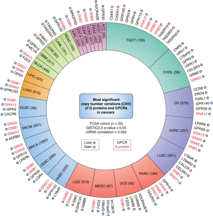
Among the G proteins, copy number gain in GNA12 is remarkably significant in ovarian cancer (OV). This cancer type is characterized by few driver mutations and by the accumula-tion of high concentraaccumula-tions of LPA in ascites fluids, which
may work through G␣12 to promote growth and metastasis
(reviewed in Ref.95). Similarly, GNAI1 (encoding G␣i1) is
sig-nificantly amplified in breast-invasive carcinoma (BRCA), a
cancer type in which many G␣i-coupled GPCRs, including
CXCR4, are well-established as metastatic drivers (see below). The significance of other genomic alterations in G proteins,
including copy number gains in G subunits (GNAB1, GNAB2,
GNAB3, and GNAB5) and G␥ (GNG4, GNG5, GNG7, GNG12,
at TOHOKU UNIVERSITY on May 19, 2020
http://www.jbc.org/
and GNGT1) in multiple cancers likely reflect the broad
signal-ing capacity of G␥ dimers (seeFig. 1).
Testicular germ cell tumor displayed the most genomic alter-ations in genes encoding GPCRs, which included mostly orphan, taste, and adhesion receptors. In contrast, F2RL1, the gene encoding -activated receptor (PAR) 2, was the most sig-nificantly altered gene in OV. PAR2 is a protease-activated receptor and is expressed in many organs. The ability of pro-teases to degrade extracellular matrices and to activate PARs render them important in the facilitation of tumor growth and
metastasis (96, 97). Overexpression of F2RL1 has been linked to
some of the most diagnosed cancers, including lung, breast,
colon, and pancreatic cancers (96, 98, 99). Functionally, PAR2
has also been linked to cancer cell migration and stimulates vascular endothelial growth factor (VEGF) production for
angiogenesis (100, 101). Another unexpected observation was
that most kidney cancers (KIPAN) exhibit highly-significant copy number gains in genes for multiple chemokine receptors (CCR2, CCR5, CCR6, CCR9, CX3CR1, and CXCR6) and hista-mine receptors (HRH2), among others. The frizzled family of GPCRs and LPA receptors (in particular LPAR6) were also genetically altered in multiple cancer types. Overall, although gene copy gains and losses may reflect cancer-associated genomic instability, most cancers exhibit a very specific pattern of copy number variations in G protein and GPCR genes, whose biological relevance can now be examined.
Figure 4. Top significant CNVs of GPCRs and G proteins in cancer. From GISTIC analysis, the proportion of TCGA cohorts (sample number) with highly
significant (GISTIC q-value⬍0.05 and mRNA correlation ⬎0.333) CNVs in genes encoding GPCRs (black) and G proteins (red) are shown. The significant genes
for each cohort are plotted outside of the cohort pie; cohorts are colored based on the number of significant genes, and amplification is denoted by red highlighting, and deletion is denoted by a blue highlighting.
at TOHOKU UNIVERSITY on May 19, 2020
http://www.jbc.org/
G proteins and GPCRs as tumor suppressor genes? An interesting observation of the pattern of genomic altera-tions is that many cancers lose one or both copies of specific G protein and GPCR genes. This raises the possibility that certain G protein/GPCRs may act as tumor suppressors rather than oncogenes. Indeed, as described above, GNA13 is significantly mutated in diffuse B-cell lymphoma and Burkitt’s lymphoma, and detailed experimental analysis revealed that in all cases this involves LOF mutations, resulting in the inability of B cells to undergo terminal differentiation and hence increasing their
uncontrolled growth (71). Analysis of candidate G␣13-coupled
GPCRs identified inactivating mutations in P2RY8 and LOF
mutations in RHOA (71, 102). These mutations appear to be
mutually exclusive, supporting the notion that in these B-cell malignancies P2RY8 –GNA13–RHOA are part of a tumor-sup-pressive axis.
Surprisingly, while conducting conditional gene knockout
studies of G␣sin the skin, we observed that mice develop
mas-sive basal cell carcinomas (BCC) in only 2 weeks after Gnas
excision (103). This involved a widespread activation of the
SHH pathway in Gnas⫺/⫺ tumor lesions (103), thus
pheno-copying the effects of LOF mutations in PATCHED (PTCH) or GOF mutations in SMO, which are the best known BCC tumor suppressor and oncogenes, respectively. Activation of the SHH pathway is also typical of a subset of medulloblastomas, a
child-hood malignancy (104). Remarkably, homozygous GNAS gene
loss was identified in a group of SHH subtype
medulloblasto-mas that does not harbor mutations in PTCH or SMO (104). In
these particular cancers that express SHH pathway
compo-nents, G␣sand its downstream target PKA act as tumor
sup-pressors by preventing the activation of GLI transcription
fac-tors (103), whereas in most GI tissues, GNAS and PKA signaling
act as tumor promoters. The former implies that certain yet to
be identified G␣s-coupled receptors may exert a
tumor-sup-pressive function in BCC. The latter raises the possibility that in
GI cancers, G␣i-coupled GPCRs may act as tumor suppressors
and hence that their LOF mutations might be pro-tumorigenic.
This is aligned with the large number of G␣i-coupled GPCRs
that are mutated in GI tumors; however, whether they exhibit GOF or LOF mutations has not yet been tested formally. These particular predictions are of high clinical relevance, as overac-tive cAMP/PKA activity in many GI tumors could be
counter-acted therapeutically by stimulating locally expressed G␣i–
GPCRs, whereas BCCs and SHH-subtype medulloblastomas may be treated by raising cAMP using phosphodiesterase
inhibitors (as proposed in Ref.104) or by stimulating locally (or
systemically) G␣s–GPCRs expressed in these tissues. These
exciting possibilities will likely be explored in the near future. pan-Cancer GPCRs expression
In addition to mutations, normal GPCRs can play a key role in cancer progression, and they can be targeted pharmacologi-cally for therapeutic purposes. A typical problem when analyz-ing gene expression changes in cancer is that often both normal and cancerous tissues are heterogeneous, including multiple cell types. Hence, relative changes (fold changes and over- and underexpression) may reflect cellular heterogeneity more than
the progression from a normal cell to its distinct cancer states. For example, comparison of GPCRs expressed in cutaneous melanoma with normal skin may grossly overestimate the rel-ative changes in expression between normal and cancerous melanocytes, as the normal skin includes a very limited number of melanocytes. Moreover, although fold changes can provide useful information, this takes attention away from GPCRs that may exert important functions for cancer transformation through increased local ligand secretion or aberrant down-stream signaling activity. A recent study has documented
rela-tive changes in GPCR expression in cancer (105). Instead, we
focus here on illuminating absolute expression levels of each GPCR and provide visual representations to gauge absolute GPCR levels. Certainly, a limitation of this analysis is that the precise cells that express each GPCR within the tumors, such as cancer and tumor stromal cells (e.g. cancer associated fibro-blasts, blood vessels, and immune infiltrating cells), will need to be established in future efforts, for example by the use of mod-ern single cell sequencing approaches. Nonetheless, we expect that we can gain an unprecedented new perspective on GPCR expression patterns in human malignancies by utilizing infor-mation gained from this analysis.
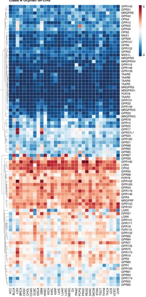
Specifically, as shown inFig. 5, an intriguing area of study is
the expression of orphan GPCRs in cancer. The endogenous ligands of more than 140 of these receptors remain unidentified and/or poorly understood, thus, their natural function is
cur-rently largely unknown (106). Nevertheless, according to our
pan-cancer analysis, orphan GPCRs are differentially expressed across cancer types, and they may exert multiple functions
dur-ing cancer progression (Fig. S1M). For example, since a
decrease in extracellular pH is a major tumor-promoting factor in the tumor microenvironment, an intriguing area of research is the group of proton-sensing GPCRs: GPR132, GPR65, GPR68, and GPR4, which are highly expressed in a large range of human cancers. Both GPR4 and TDAG8 (GPR65) have been shown to be overexpressed in many cancers and can cause
malignant transformation of cells in vitro (107). GPR132 (also
known as G2A) was previously shown to have tumor suppres-sor properties, as it prevents oncogenic transformations of pre-B cells by the BCR–ABL oncogene, similar to the role of GNA13in these cell types (108). However, GPR132 has been
shown to be highly transforming in fibroblasts (109). Thus,
pro-ton-sensing GPCRs may display tumor-promoting or -suppres-sive functions depending on the cancer cell of origin and may also display pro-tumorigenic activity when activated in the
tumor stroma (105). Interestingly, in our recent G protein–
coupling predictor trained by a large experimental dataset, orphan GPCRs tend to show a higher proportion of coupling
toward G␣12/13than other GPCR classes (79) further
suggest-ing potential importance of orphan GPCRs in cancers that
involve aberrant G␣12/13signaling.
The leucine-rich repeat-containing GPCRs (LGR) LGRs 4 – 8 are known for their role in development, bone formation, and remodeling, but LGR4 and LGR5 are also up-regulated in
sev-eral cancer types (110). These receptors are expressed in
mul-tiple tissue-resident stem cells, and their overexpression may reflect the expansion of this cellular compartment as well as the
establishment of cancer stem cell niches (110). Overexpression
at TOHOKU UNIVERSITY on May 19, 2020
http://www.jbc.org/
Figure 5. Expression of class A orphan receptors in cancer. Gene expression for class A orphan GPCRs from the UCSC TCGA PanCan Cohort RNA-seq dataset
is shown. Expression values are summarized by defining transcripts per million (TPM), which normalizes for both gene length and sequencing depth.
Expres-sion values are log2(TPM⫹ 0.001) averaged within the primary tumor samples of each cancer. GPCRs are clustered based on similarity across cancer types.
at TOHOKU UNIVERSITY on May 19, 2020
http://www.jbc.org/
of LGR4 and LGR5 in colon and ovarian tumors most likely
enhances cell proliferation and metastasis (111, 112).
Interest-ingly, many class A orphan GPCRs are rarely expressed across cancer types. These include the MAS oncogene, which can explain the limitations in analyzing its role in human cancer despite its initial identification during transfection experiments several decades ago. Others are expressed in a single cancer (e.g. GPR22 in pheochromocytoma and paraganglioma) or a few cancers (e.g. GPR17 and GPR37L1 that are expressed only in GBM and brain lower grade glioma), whereas others are expressed in most cancers, such as OPN3 and LGR4. These studies de-orphaning GPCRs and uncovering the function of additional overexpressed GPCRs may provide promising can-didates for therapeutic intervention in cancer.
The pan-cancer expression of each GPCR class is depicted in
Fig. S1, A–N. We hope that this information will be useful for hypothesis generation in our large community of scientists working in the field of GPCRs in academia and industry. Although this review will not provide a comprehensive analysis of each GPCR, a few concepts may be worth discussing. For
example, expression of the purinergic P2Y11and adenosine A2A
receptors is widespread in all cancers, whereas GBM tumors express high levels of ADORA1, ADORA2, and ADORA3, all of which can be activated by adenosine in the tumor
microenvi-ronment. Multiple lipid receptors for S1P (S1P1–3) and LPA
(LPA1, LPA2, and LPA6) are widely expressed as well. These
receptors are intriguing because ligands for these receptors have been shown to accumulate in the tumor
microenviron-ment (113, 114). Conserved residues in these receptors also
display a high mutational rate, which suggests that they may play vital roles in receptor signaling initiation, termination, and
coupling specificity (13).
This is also highly relevant for the 17 known GPCRs that specifically recognize intermediates or (by)products of cellular
metabolism, which are often involved in nutrient sensing (115).
These include receptors sensing amino acids and amino acid metabolites (GPR142, CasSR, GPR35, TAAR1, and FOPR1/2), bile acid (TGR5/GPBAR1), triglyceride metabolites (e.g. FFA1/ GPR40, FFA4/GPR120, and GPR119), products of the interme-diary metabolism and small carboxylic metabolites such as ace-tate and propionate (FFA2/GPR43 and FFA3/GPR41), butyrate
(FFA2/GPR43, FFA3/GPR41, and HCA2/GPR109A),
-hy-droxybutyrate (HCA2/GPR109A),-hydroxyoctanoate (HCA3/
GPR109B), lactate (HCA1/GPR81), succinate (GPR91), and capric acid (GPR84) receptors, as well as gut microbiota-derived prod-ucts (e.g. short-chain fatty acids, such as acetate, propionate,
and butyrate) (reviewed in Ref.115). These receptors are highly
expressed in multiple organs of the digestive tract and immune
cells (116), and they may be persistently activated in the tumor
microenvironment due to the high metabolic rate that charac-terizes most solid tumors.
The EP4(PTGER4) and EP2(PTGER2) receptors for the
typ-ical inflammatory mediator PGE2 (see below) are also widely
expressed, whereas EP3(PTGER3) is mainly expressed in
kid-ney cancer. PGE2 plays a critical role in epithelial regeneration following tissue injury and cancer growth, which occurs via
PI3K/Akt and-catenin pathways (63, 117). COX2
overexpres-sion and enhanced PGE2 production is most notable in
colo-rectal cancer, and COX2 blockade can help explain the cancer chemopreventive activity of aspirin and other nonsteroidal
anti-inflammatory drugs (NSAIDs) (118). However, direct roles
for PGE2 in tumorigenesis have been demonstrated for many other human malignancies, including breast, lung, liver, and gastric cancers, among others. For example, in laboratory mod-els of breast and gastric cancers, COX2 overexpression and alterations in Wnt signaling both led to increased
tumorigene-sis (119, 120). Moreover, EP3has been shown to be involved in
angiogenesis in lung cancer cell lines by increasing VEGF and
metalloproteinase-9 (MMP-9) expression (121).
Among the class of GPCRs for proteins (Fig. S1E), which
includes chemokine receptors, CXCR4 is the most widely expressed. This may include many cancers that express CXCR4 under hypoxic conditions, as well as in blood vessels and
immune cells (see below) (122–124). Other chemokine
recep-tors that are highly expressed in immune cells (see below) were less well-represented, suggesting a more limited impact of immune infiltrating cells to the overall mRNA expression pat-terns in our pan-cancer analysis. The analysis of GPCRs
acti-vated by peptides (Fig. S1F) show a clear widespread expression
in genes for thrombin PAR1 (F2R) and PAR2 (F2RL1) receptors and endothelin receptors (EDNRB), the latter with particularly higher expression in SKCM and uveal (UM) melanomas.
HRH1, encoding H1 histamine receptor, is the most widely
expressed aminergic GPCR (Fig. S1G), whereas M1 muscarinic
receptors (CHRM1) and1-adrenergic receptors (ADRB1) are
highly expressed in prostate cancer, the latter receptor being of unexpected importance for the most highly prevalent cancer among males (see below). Another interesting finding was the high level of expression of dopamine receptor 2 (DRD2) in a well-defined set of cancers, including GBM, considering that a new family of antagonists for this receptor has exhibited
encouraging anti-tumor activity in multiple cancer types (125,
126).
Interestingly, from our analysis of Frizzled GPCRs, SMO is widely expressed in most cancers, beyond its initial main role in BCC. This might be due to SMO being expressed in cancer
stromal cells that are present in most solid tumors (Fig. S1G)
(127, 128). There is also widespread expression of FZD6and a
more cancer-restricted expression of FZD1 and FZD4 (Fig.
S1H).
Intriguingly, analysis of the sensory GPCRs revealed a high level of expression of the taste receptor, TAS1R3, across most
cancer types, which has not been previously investigated (Fig.
S1J).
The adhesion GPCR family has mainly been studied in immunological and developmental functions, but they have
recently been linked to cancer (Fig. S1M). For example, EMR2
(ADGRE2) is overexpressed in human breast cancer, and increased nuclear expression of EMR2 is negatively correlated
with tumor grade (129). Additionally, CD97 (ADGRE5) and
GPR56 (ADGRG1) are the highest expressed adhesion GPCRs across all cancers, but they have only been studied in the con-text of melanoma, gastric, esophageal, and thyroid cancers (130 –132). Additionally, GPR65 (TDAG8) and GPR133 (ADGRD1) have also been associated with human cancers and
linked to tumor promotion (107, 133), but the role of this
at TOHOKU UNIVERSITY on May 19, 2020
http://www.jbc.org/
ly-expressed family of GPCRs in tumor initiation and metasta-sis is still not fully understood.
Overall, we expect that the emerging pan-cancer informa-tion on GPCR expression will ignite new interest on their study in human malignancies.
GPCRs in metastasis and angiogenesis
Metastasis is one of the cancer hallmarks, in which tumor cells can acquire the ability to migrate and disseminate from the tumor to distant tissues. Cancer cells spread from the primary organ to secondary sites through lymphatic vessels and blood and are the result of a sequential, highly-organized, and organ-selective process. The precise mechanisms determining the directional migration and invasion of tumor cells into specific organs remain to be fully established, but chemokine receptors, all of which are GPCRs, have been the most popular place to
look (134, 135). Chemokines are small, cytokine-like proteins
that induce directional migration for immune cells through interaction with GPCRs. Chemokines are secreted by multiple organs and act in a coordinated fashion with cell-surface pro-teins to direct homing of immune cells to specific anatomical
sites (136, 137). To serve a similar purpose, tumor cells can
hijack chemokine receptor networks and migrate toward spe-cific chemokines, facilitating metastasis to other organs, pri-marily the liver, lungs, brain, lymph nodes, and bone marrow
(134). In addition, the tumor microenvironment includes
chemokines that can enhance the motility and survival of can-cer cells in an autocrine and paracrine fashion, a process that we refer as oncocrine signaling.
There are 23 distinct chemokine receptors in humans, and they are divided into four classes according to the type of chemokine with which they interact (CC, CXC, CX3C, or XC) (135, 138, 139). CXCR4 and CCR7 represent the best-studied chemokine receptors driving cancer metastasis, as they play active roles in tumor growth, invasion, angiogenesis,
metasta-sis, and cancer relapse and therapeutic resistance (134, 140,
141). CXCL12, the chemokine for CXCR4, is highly expressed
in multiple tissues, mainly lungs, liver, and bone marrow, and it
is also secreted by tumor and stromal cells (134, 140). CXCL12
expression levels are highest in these common sites of metasta-sis, which could recruit cancer cells to these distant organs. CCR7 binds the ligands, CCL21 and CCL19, and guides the migration of lymphocytes and dendritic cells (DC) to lymph
nodes (142). Expression of CCR7 in tumor cells has emerged as
an important predictor of lymph node metastasis and poor prognosis in cervical cancer, gastric carcinoma, and breast
can-cer, among others (134).
CCR7 and CXCR4 are the main receptors typically present on metastasizing cells, but there are other chemokine receptors that may dictate a more organ-specific metastasis. For example, the small intestine is an organ that expresses high levels of
CCL25 physiologically to guide CCR9⫹ lymphocytes to this
tissue. Because melanoma, breast cancer, and ovarian cancer express high levels of CCR9, this receptor may play a pivotal role in the preferential metastasis of these tumors to the small
intestine (143–145). Additionally, malignant melanomas
express high levels of CCR10, a receptor that guides leukocytes to the skin, and consequently, CCR10 expression in melanoma
may drive metastasis to the skin (146, 147). CXCR3 and CXCR5
have both been shown to play a role in lymph node metastasis (148, 149).
With increasing nutrients and oxygen demands by the tumor cell, solid tumors produce angiogenic factors that promote the migration and proliferation of endothelial cells to form new vessels. Many of these factors exert their functions through GPCRs expressed on endothelial cells, including thrombin, prostaglandins, S1P, and many chemokines. In addition, chemokines, like CCL2, CCL5, and CXCL8/IL-8, can recruit leukocytes and macrophages to the tumor site, which leads to production of VEGF and other angiogenic factors that
contrib-ute to the growth of tumor-associated blood vessels (134, 135).
Production of inflammatory cytokines can also promote new vessel formation by elevating COX2 expression, and in turn prostaglandin E2 (PGE2) increases the expression of VEGF,
CXCL8, and CXCL5 by tumor cells (150, 151).
In the case of thrombin, this serine proteinase plays a vital role in regulating hemostasis by converting fibrinogen into
fibrin to stimulate platelet aggregation and coagulation (152).
Thrombin carries out its effects through the PAR family of receptors, which exhibit the unique property of harboring a tethered ligand within the receptor that becomes exposed upon cleavage of the N-terminal extracellular region by thrombin
(153). Thrombin promotes angiogenesis by increasing
metallo-proteinases and decreasing the ability of endothelial cells to
adhere to the extracellular matrix (154, 155), while promoting
the expression and activity of the VEGF receptor, VEGFR-2,
through G␣13and its target RhoA (156). However, S1P1
stimu-lates endothelial cell proliferation, survival, and migration and also regulates sprouting angiogenesis through cross-talk with VEGFR-2 and enhanced tissue hypoxia and VEGF production (157–159). The effects of S1P on angiogenesis largely depend
on the GPCR it binds (S1P1–5). S1P stimulates angiogenesis
mainly through S1P1and S1P3, and it mediates endothelial cell
migration and formation of capillary structures through G␣i(or
more likely its associated G␥ subunits) activation of the small
GTPase Rac1 (160). In contrast, S1P2has been shown to be
involved in the suppression of angiogenesis most likely through
the inhibition of Rac and cell migration (161). Altogether,
GPCRs participate in angiogenesis either by promoting the pro-liferation, migration, and sprouting of endothelial cells or by the release of pro-angiogenic factors for new blood vessel forma-tion, thereby increasing the blood supply to the growing tumors.
Key role for GPCRs in cancer immunology
In the last few years, cancer immunotherapy became one of the most exciting breakthroughs in cancer treatment. Recent revolutionary discoveries have highlighted the importance of the tumor microenvironment and its associated immune cells in cancer development and therapeutic resistance. Tumors can deploy multiple mechanisms to avoid immune recognition and an anti-tumor immune response, including the recruitment of myeloid-derived suppressor cells (MDSC) and conditioning of the surrounding microenvironment to become highly immune-suppressive by expressing cytokines, such as IL-6, IL-10, and
transforming growth factor (162). This can lead to the
at TOHOKU UNIVERSITY on May 19, 2020
http://www.jbc.org/