Doctoral Dissertation
Study on the Synthesis of Purification-free
67/68
Ga-Labeled Multivalent Probes for In Vivo
Imaging of Saturable Systems
標識反応後の精製なしに生体内微量分子の画像化を可能とする67/68Ga 標識多価プローブの作製に関する研究
Laboratory of Molecular Imaging and Radiotherapy
Graduate School of Pharmaceutical Sciences
Chiba University
August, 2018
[Abstract]
In the synthesis of radiometal-labeled probes, a large excess of ligands over radiometals are required to provide radiometal-labeled probes with high radiochemical yield in short reaction time. However, the unlabeled ligands hinder target accumulation of the radiometal-labeled probes by competing for target molecules. To circumvent the problem, our laboratory has developed a new design concept of “metal coordination-mediated synthesis” 99mTc-labeled bivalent and trivalent
probes. The 99mTc-labeled probes acquire higher binding affinities upon complexation with
monovalent ligand by multivalent effect, and the resulting probes visualized the target molecule by SPECT/CT without removing the unlabeled ligand. 68Ga is a positron emitter available from a
long-lived 68Ge/68Ga generator system, which allows for the cost-effective production of PET
probes without cyclotron facilities. The study was undertaken to develop the chemical strategy to apply the molecular design achieved with 99mTc-labeled probes to 68Ga-labeled ones. Since 67Ga-
and 68Ga-labeled probes were obtained under similar reaction conditions, 67Ga was used in this
study as a radionuclide of choice because of its longer half-life and availability.
Chapter 1 Chemical strategy to apply the chemical design developed with 99mTc-labeled
probes to 68Ga-labeled ones.
Based on the procedures of 99mTc-labeled bivalent and trivalent probes,
3-hydroxy-1,2-dimethyl-4-pyridone (Hdpp) and 2-acetylpyridine N,N-disubstituted thiosemicarbazone (PTSC) derivatives were selected as a bidentate and a tridentate chelator, due to a formation of thermodynamically stable complexes with the Ga3+ ion. Hdpp provided a single radioactive peak on TLC at an Rf
value identical to that of non-radioactive Ga-(Hdpp)3 complex verified by ESI-MS when the
developing solution contained 10 mM Hdpp. On RP-HPLC analysis of the reaction mixture, the majority of the radioactivity was adsorbed on the column. These results indicated that while Hdpp may provide [67Ga]Ga-(Hdpp)
3, the complex underwent rapid decomposition under low Hdpp
concentrations, which was not appropriate for in vivo applications. PTSC derivatives also provided the objective [67Ga]Ga-(PTSC)
2 complexes. The 67Ga-labeled complexes remained stable after
removing the unlabeled free ligand by RP-HPLC. However, the RP-HPLC-purified [67
Ga]Ga-(PTSC)2 derivatives were rapidly decomposed when incubated in 0.1 M bicarbonate buffer (pH
7.4) at 37 ºC. These results indicated that while the tridentate chelates increased the stability of the resulting 67Ga complexes, their stability was insufficient for in vivo applications. These results
called for an alternative approach to prepare multivalent 67Ga-labeled probes from monovalent
ligands. Free Schiff base chelators are present as equilibrium and are rapidly decomposed upon dilution. However, the Schiff base remains stable through metal coordination. From these considerations, a Schiff base chelator was designed and synthesized by reacting salicylaldehyde (Sal) with triamine compound TAMEol to prepare (Sal)3TAMEol. [67Ga]Ga-(Sal)3TAMEol was
prepared by adding [67Ga]Ga-citrate to a solution of (Sal)
3TAMEol at 80 ˚C for 15 min. [67
Ga]Ga-(Sal)3TAMEol remained stable after isolation from the excess of Sal and TAMEol. Furthermore,
[67Ga]Ga-(Sal)
3TAMEol was stable in the apo-transferrin challenge. As a result, [67
Ga]Ga-(Sal)3TAMEol provided 67Ga-complex possessing kinetic stability worth for further evaluation.
Chapter 2. Evaluation of the Schiff base ligand for in situ trivalent “1 to 3” design.
The reaction of [67Ga]Ga-citrate with a mixture of RGD-conjugated salicylaldehyde and triamine
provided a 67Ga-labeled trivalent probe [67Ga]Ga-(RGD-Sal)
3TAMEol with stability sufficient for
in vivo applications. The unlabeled ligands exhibited the much less inhibitory effect on the [67Ga]Ga-(RGD-Sal)
3TAMEol accumulation to the target molecules compared with a 67Ga-labeled
probe prepared under the conventional “3 to 3” approach, due to rapid decomposition of (RGD-Sal)3TAMEol to RGD-Sal and TAMEol after injection. As a result, [67Ga]Ga-(RGDsal)3TAMEol
visualized murine tumors without post-labeling purification, more clearly than that of 67Ga-labeled
trivalent probe from a trivalent ligand.
Conclusion.
Since the procedure achieved with 99mTc-labeled probes was not applicable to prepare 67Ga-labeled
probes, an alternative approach was developed utilizing Schiff base-stabilization upon metal coordination. This approach generated a 67Ga-labeled trivalent probe of high in vivo stability and
the resulting 67Ga-labeled probe provided clear tumor images without post-labeling purification.
These findings indicate the availability of Schiff base ligands to prepare 68Ga-labeled trivalent
probes by a simple radiolabeling procedure. The present findings would facilitate the applications of 68Ga-labeled probes in molecular imaging with PET.
Table of Contents
[Abstract] ... 1
[Background] ... 4
[Chapter 1]. Chemical strategy to apply the chemical design developed with 99mTc-labeled probes to 67/68Ga-labeled ones... 9
1-1. Bidentate or tridentate chelators based on the procedures applied to 99mTc ...9
[Introduction] ... 9
[Results] ... 10
[Discussion] ... 14
1-2. Schiff base chelator ... 16
[Introduction] ... 16
[Results] ... 17
[Discussion] ... 20
[Summary] ... 22
Chapter 2. To evaluate the applicability of the new design towards 67Ga after conjugation with a cyclic RGDfK (-Arg-Gly-Asp-D-Phe-Lys-: c(RGDfK)) peptide. ... 23
[Introduction] ... 23 [Results] ... 25 [Discussion] ... 33 [Summary] ... 36 [Conclusions] ... 37 [Experimental section] ... 38 [References] ... 50 [Associated article] ... 56 [Dissertation committee] ... 57 [Acknowledgment] ... 58
[Background]
Radiolabeled probes are radionuclide-containing pharmaceuticals and are used routinely in nuclear medicine for diagnosis or therapy of diseases. The radiolabeled diagnostic probe is a molecule labeled with a γ-emitting radionuclide for single photon emission computed tomography (SPECT) or with a positron-emitting radionuclide for positron emission tomography (PET).1–3
SPECT and PET are molecular imaging modalities and are powerful clinical and research tools for diagnosis, staging treatment planning, and therapeutic efficacy monitoring of patients. SPECT requires the use of an imaging probe labeled with a γ-emitting radionuclide. These γ rays are recorded by the detectors with a collimator to arrange the direction from multiple angles. SPECT instrument can be converted into the 3-D image identifying the localization of the radiotracer. The radionuclides used for SPECT is commercially available and used in SPECT imaging facility. Therefore, SPECT is more available and widely used and much cheaper than PET. On the other hand, PET is the molecular imaging instrument that requires a positron-emitting radionuclide labeled probe. The anti-parallel 511 keV γ rays emitted from the annihilation of positrons with surrounding electrons are recorded by detectors arranged in a ring around the subject. The data are then reconstructed using computer-based algorithms to yield the 3-D image of the radiotracer’s location. Compared with SPECT, PET has greater advantages regarding sensitivity and resolution since only two photons detected in coincidence are registered, eliminating unnecessary physical collimation to block scattered photons.4 However, due to the short half-lives of the PET
radioisotopes such as carbon-11 (20 min), nitrogen-13 (10 min), oxygen-15 (2 min) and fluorine-18 (110 min), the radiolabeled probes labeled by these radionuclides have traditionally been produced using an in-house cyclotron of the PET imaging facility.
Recently, Germanium-68 (68Ge)/Gallium-68 (68Ga) generators became commercially available
and are used in the clinical stages.5 The relation of 68Ge decay and 68Ga accumulation is described
by secular equilibrium since the half-life of the 68Ge (t
1/2 = 271 d) is over 100 times longer than
that of 68Ga (t
1/2 = 67.7 min). At the equilibrium, the daughter radionuclide (68Ga) reaches the
radioactivity identical to that of the parental radionuclide (68Ge). In addition, 4 h after the elution
of the daughter radionuclide, 91% of the maximum achievable radioactivity is generated, which allows the synthesis of 68Ga-labeled probes twice or three times within one working day. The
availability of 68Ga from a generator system also allows for the cost-effective production and the
advantage of radiometals such as 68Ga over non-metallic radionuclides (e.g., 11C and 18F) involves
the synthesis of the objective radiolabeled probes in radiochemical yields sufficient for administration to subjects without purification.3,7,8 These characteristics make 68Ga as an attractive
radionuclide for preparing PET imaging probes. Gallium possesses another radioisotope, 67Ga (t 1/2
= 78.3 h), that emits γ-rays available from a commercial source. Since 67Ga and 68Ga-labeled
probes are prepared under similar reaction conditions,9–11 the results obtained from 67Ga-labeled
probes can be applied to 68Ga-labeled probes and vice versa. Thus, the use of 67Ga compensates
the short half-life of 68Ga for developing 68Ga-labeled probes.
In the design of 68Ga-labeled probes, low molecular weight biomolecules such as peptides are
used as a targeting device to deliver 68Ga to the target molecules. Since most bioactive molecules
do not form stable complexes with metallic radionuclides such as 67/68Ga, technetium-99m (99mTc)
and indium-111, a chelating agent is conjugated to a biomolecule (referred to as “ligand”)4,12–14 to
provide a coordination site for the radiometals. The subsequent complexation reaction of the chelating agent-conjugated biomolecule with a metallic radionuclide provides a radiolabeled probe. A schematic illustration of a targeted radiometal-labeled probe is shown in Figure 1.
To ensure high radiochemical yields in short reaction time using extremely low concentration of radiometals (e.g. 68Ga: 37 MBq/mL corresponding to 4 × 10⁻¹⁰ M, 67Ga: 2.5 × 10⁻⁸ M)12 with the
ligand, the radiolabeled ligands are synthesized in the presence of a large excess of ligands over radiometals (Figure 2). The presence of a large excess of unlabeled ligands in the injectate impairs target uptake of the radiolabeled ligands by competing for target molecules, which results in poor images of the target molecules of low expression levels.7,15–1868Ga-labeled probes so far developed
need the removal of the non-chelated free ligand by HPLC or other purification methods. However, such manipulation significantly wastes time and radioactivity and impairs the practical utility and advantages of using radiometals, especially for 68Ga due to the short half-life. In other words, the
applications of 68Ga-labeled probes would increase significantly if 68Ga-labeled probes can be
obtained without post-labeling purification.
To ensure high radiochemical yields in short reaction time, due to the low concentration of radiometals (e.g. 68Ga: 37 MBq/mL corresponding to 4 × 10⁻¹⁰ M, 67Ga: 2.5 × 10⁻⁸ M),12 the
radiolabeled ligands are then synthesized in the presence of a large excess of ligands over radiometals (Figure 2). The presence of a large excess of unlabeled ligands in the injectate impairs target uptake of the radiolabeled ligands by competing for target molecules, which results in poor
Figure 1. A schematic illustration of radiometal-labeled probe. A biomolecule is conjugated
with a bifunctional chelator to produce a biomolecule chelator probe which called ligand. The complexation reaction of a radiometal and a ligand provides a radiolabeled probe with stability sufficient for in vivo studies.
images of the target molecules of low expression levels.7,15–18 Although the free ligands can be
removed from the radiolabeled-ligands by HPLC or other purification methods, such manipulation significantly wastes time and radioactivity and impairs the practical utility and advantages of the use of radiometals, especially for 68Ga due to the short half-life.
To circumvent the problem, our laboratory has recently developed a new design concept of “metal coordination-mediated synthesis” 99mTc-labeled trivalent probes using
[99mTc][Tc(CO)
3(OH2)3]+ and isonitrile-conjugated Arg-Gly-Asp (RGD) peptide (CN-RGD).17 In
this design, three water molecules in [99mTc][Tc(CO)
3(OH2)3]+ are displaced with the three
monovalent ligands (CN-RGD) to prepare a trivalent [99mTc][Tc(CO)
3(CN-RGD)3]+ probe. As a
result, [99mTc][Tc(CO)
3(CN-RGD)3]+ acquired higher integrin αvβ3 binding affinity than its
monovalent ligand (Figure 3A-1). When injected into mice, [99mTc][Tc(CO)
3(CN-RGD)3]+
showed higher tumor uptake and higher tumor to blood ratio. In SPECT imaging studies, [99mTc][Tc(CO)
3(CN-RGD)3]+ clearly visualized murine tumors without removing the unlabeled
ligands. In contrast, a 99mTc-labeled monovalent probe prepared from a monovalent ligand failed
to depict the tumors in the presence of the unlabeled ligands. Our laboratory has also reported the applicability of this design concept to 99mTc-labeled bivalent probes using D-penicillamine as a
Figure 2. A conceptual illustration of the conventional design synthesis of radiometal-labeled
probes. (A) Monovalent “1 to 1” design, (B) Trivalent “3 to 3” design. The presence of excess unlabeled ligands with similar affinity hinders target accumulation of the radiometal-labeled probes by competing for target molecules.
chelating molecule (Figure 3A-2).18 Based on the results of these studies, I investigated an
appropriate procedure to apply the chemical design developed with 99mTc-labeled probes to 68
Ga-labeled ones. I conducted the present studies with 67Ga because of its longer half-life and
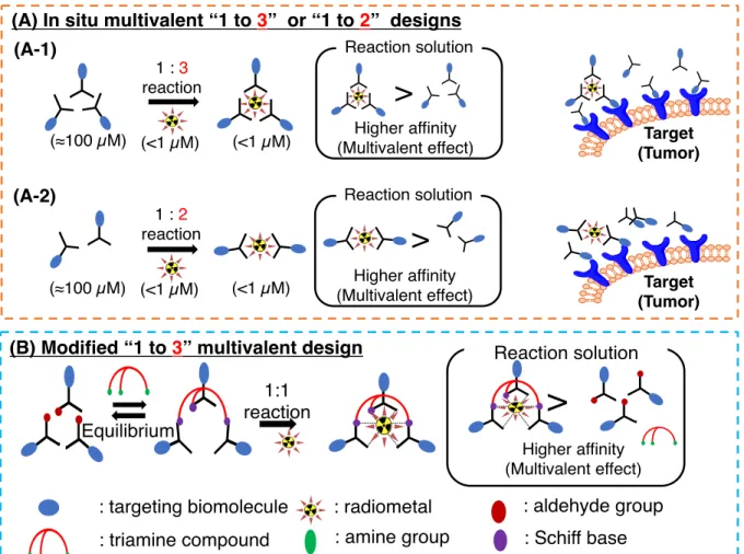
Figure 3. A conceptual illustration of the in situ molecular design of multivalent probes. (A) In
situ trivalent “1 to 3” design (A-1) and “1 to 2” divalent design (A-2). The radiolabeled probes prepared from the monovalent ligand acquired higher binding affinity than the monovalent ligand upon complexation by so-called multivalent effect. (B) Modified in situ “1 to 3” design. The radiolabeled probe was synthesized by reacting a radiometal ion with a trivalent Schiff base hexadentate ligand prepared from a monovalent ligand with an aldehyde group and a triamine molecule at the molar ratio of 3:1. While the Schiff base is stabilized upon metal coordination, the unlabeled Schiff base ligands present as equilibrium in the injectate are decomposed immediately after injection. As a result, the metal complex is an only trivalent compound and possess higher binding affinity than its unlabeled monovalent ligand.
[Chapter 1]. Chemical strategy to apply the chemical design developed with 99mTc-labeled
probes to 67/68Ga-labeled ones.
1-1. Bidentate or tridentate chelators based on the procedures applied to 99mTc
[Introduction]
Based on the procedures applied to 99mTc-labeled probes, bidentate or tridentate chelators were
used to investigate the applicability of the molecular design of the “metal coordination-mediated synthesis” of 67Ga-labeled multivalent probes. The hydroxypyridinone derivatives consist of a
six-membered aromatic N-heterocycle with a hydroxyl and a ketone functionality. At physiological conditions, they present the high affinity for hard trivalent metal ions such as Iron-(III) (Fe3+) and
the group 13 metal ions particularly for Ga,19,20 and provide a thermodynamically stable complex
with the Ga3+ ion.19,21,22 From these studies, I selected 3-hydroxy-1,2-dimethyl-4-pyridone (Hdpp)
as the bidentate chelator (Figure 4A).19,21
As for the tridentate chelators, 2-acetylpyridine N,N-disubstituted thiosemicarbazone (PTSC) derivatives were selected (Figure 4B).23,24 Thiosemicarbazones have been extensively studied as
toxic agents against bacterial and viral infections. Non-radioactive Ga complexes of thiosemicarbazone were also evaluated as ribonucleotide reductase inhibitors.25 The
4N-substituted α-N-heterocyclic thiosemicarbazone forms thermodynamically stable complex with Ga and the complex exhibits a potent antiproliferative action.25 Therefore, the applicability of these
[Results]
(1-1.1) Synthesis of non-radioactive Ga-(Hdpp)3.
To a suspension of 3-Hydroxy-1,2-dimethyl-4-pyridone (Hdpp) in water (5 mL) was added a solution of GaCl3 in water. After adjusting the pH of the solution to 9 with 2 N NaOH, the reaction
mixture was heated at 70 ˚C until the reaction volume was <2.5 mL. After cooling to room temperature, the precipitate was collected by filtration and Ga-(Hdpp)3 was obtained as an orange
solid (20.6 mg, 58%).
(1-1.2) Preparation of [67Ga]Ga-(Hdpp)
3.
A solution of Hdpp in 0.1 M acetate buffer was reacted with [67Ga]Ga-citrate at 80 ˚C for 30
min. The major radioactivity was observed at the origin in cellulose acetate electrophoresis (CAE) analysis (Figure 5B). Under these conditions, [67Ga]Ga-citrate migrated 4 cm towards the anode
(Figure 5A), suggesting a formation of a 67Ga-labeled Hdpp. A broad radioactivity peak from the
Rf values of 0 to 0.2 was observed in TLC analysis (Figure 5C). When Hdpp (10 mM) was added
Figure 4. In situ multivalent designs based on the design applied to 99mTc-labeled probes. (A)
In situ trivalent “1 to 3” design using bidentate Hdpp chelator. (B) In situ bivalent “1 to 2” design using tridentate PTSC derivatives chelators. Reactions with 67Ga provided its 67
to the developing solvent, a sharp radioactivity peak was observed at the Rf value of 0.3 (Figure 5D) similar to that of non-radioactive Ga-(Hdpp)3 characterized and verified by ESI-MS.
Figure 5. Analysis of [67Ga]Ga-(Hdpp)
3. CAE radioactivity traces of (A)[67Ga]Ga-(Hdpp)3 and
(B) [67Ga]Ga-citrate. TLC radioactivity traces of [67Ga]Ga-(Hdpp)
3 developed with
MeOH/water (C) without and (D) with 10 mM Hdpp.
0 50 100 150 mm TLC 0 50 100 150 mm 0 50 100 150 200 250 300 350 C/mm -5 0 5 10 ra d io a c ti v it y 0 50 100 150 mm TLC 0 50 100 150 mm 0 50 100 150 200 250 C/mm -5 0 5 10 ra d io a c ti v it y
Distance from origin (cm)
Distance from origin (cm) [67Ga]Ga-citrate [67Ga]Ga-(Hdpp) 3 (A) (B) 0 50 100 150 mm TLC 0 50 100 150 mm 0 1 2 3 4 5 6 7 8 9 10 11 12C/mm 0 50 100 150 mm TLC 0 50 100 150 mm 0 2 4 6 8 10 12 14 16 18 20 22 24 26C/mm 0 0.5 1 Rf value ra d io a c ti v it y 0 0.5 1 Rf value ra d io a c ti v it y [67Ga]Ga-(Hdpp) 3 [67Ga]Ga-(Hdpp) 3 (C) (D)
(1-1.3) Synthesis of tridentate PTSC derivatives.
The PTSC derivatives were synthesized by reacting 2-acetylpyridine with S-methyl dithiocarbazate26 in ethanol at 80 ºC, followed by the alkylation with secondary amines, as shown
in Scheme 1.
(1-1.4) Synthesis of non-radioactive Ga-(PTSC-1)2 and Ga-(PTSC-2)2.
A mixed solution of Ga(NO3)3/nH2O and PTSC-1 or PTSC-2 in EtOH was reacted at 40 ˚C for
1 h. A solution of ammonium hexafluorophosphate in EtOH was added to the solution. After mixing for 10 min, the precipitates were obtained by filtration to afford Ga-(PTSC-1)2 or
Ga-(PTSC-2)2.
(1-1.5) Preparation of [67Ga]Ga-(PTSC)2.
[67Ga]GaCl
3 was mixed with 0.1 M ammonium acetate, and the reaction mixture was left for 5
min. This solution was then mixed with PTSC derivatives in a mixture of EtOH and 0.1 M ammonium acetate pH 4.5 (1:8), and the reaction mixture was heated at 80 ˚C for 15 min. The RP-HPLC analyses of the reaction solution showed a single radioactivity peak at a retention time
Scheme 1. Synthesis of PTSC derivatives
Reagents : (a) bis(2-methoxyethyl)amine, (b) diisobutylamine.
N N N H S S N N N H S N O O N N N H S N (a) (b) 2, PTSC-1 3, PTSC-2
similar to those of the corresponding non-radioactive Ga-(PTSC)2 derivatives verified by ESI-MS
(Figure 6).
Figure 6. RP-HPLC UV (254 nm) traces of Ga-(PTSC-1)2 (A) and Ga-(PTSC-2)2 (B)
and radioactivity traces of [67Ga]Ga-(PTSC-1)
2 (C) and [67Ga]Ga-(PTSC-2)2 (D). Ga-(PTSC-1)2 Ga-(PTSC-2)2 [67Ga]Ga-(PTSC-1) 2 [67Ga]Ga-(PTSC-2)2 (A) (B) (D) (C) 0 10 20 30 U V ( 2 5 4 n m )
Retention time (min)
U V ( 2 5 4 n m )
Retention time (min)
0 10 20 30
Retention time (min)
0 10 20 30 40
Retention time (min)
0 10 20 30 40 ra d io a c ti v it y ra d io a c ti v it y
(1-1.6) Stability of [67Ga]Ga-(PTSC)2 derivatives.
67Ga-labeled complexes were purified by RP-HPLC to remove unlabeled ligands. The
radioactive peak was collected and the solvent was removed in vacuo. The residue was reconstituted in carbonate buffer (0.1 M, pH 7.4). Aliquots of the samples were collected after 1, 3 and 6 h incubation, and the radioactivity was analyzed by TLC (ammonium acetate : MeOH/1 : 1). [67Ga]Ga-(PTSC)
2 derivatives were rapidly decomposed after 1 h incubation at 37 ˚C (Table
1).
[Discussion]
When Hdpp was reacted with [67Ga]Ga-citrate at 80 ˚C for 30 min, a broad radioactivity peak
from the Rf values of 0 to 0.2 was observed in TLC analysis (Figure 5C). However, a sharp radioactivity peak was observed at an Rf value of 0.3 similar to that of non-radioactive Ga-(Hdpp)3
characterized and verified by ESI-MS when Hdpp (10 mM) was added to the developing solvent. On RP-HPLC analysis, the majority of the radioactivity was adsorbed on the column and was not eluted (data not shown). These results indicated that while Hdpp may provide an objective [67Ga]Ga-(Hdpp)
3 complex, the complex underwent rapid decomposition under low Hdpp
concentration, indicating that [67Ga]Ga-(Hdpp)
3 complex is not kinetically stable and is not
appropriate for in vivo applications. Contrary to [67Ga]Ga-(Hdpp)
3, both [67Ga]Ga-(PTSC-1)2 and [67Ga]Ga-(PTSC-2)2 remained
stable after isolation from the free ligands as observed in the RP-HPLC analyses. However, both [67Ga]Ga-(PTSC)2 were rapidly decomposed after incubation in 0.1 M bicarbonate buffer (pH 7.4)
Table 1. Stability of [67Ga]Ga-(PTSC-1)
2 and [67Ga]Ga-(PTSC-2)2 in 0.1 M bicarbonate
buffer (pH 7.4)a
percent of intact radiolabeled complex (%)
time (min) [67Ga]Ga-(PTSC-1)2 [67Ga]Ga-(PTSC-2)2
10 70.4 ± 9.67 31.0 ± 3.24
30 47.1 ± 4.83 5.60 ± 0.38
60 5.73 ± 1.55 0.71 ± 0.32
at 37 ºC (Table 1). These results indicated that while the tridentate ligands increased the stability of the resulting 67Ga complexes than that from the bidentate ligands, their stability was insufficient
for in vivo applications. These results called for an alternative approach to prepare multivalent
67Ga-labeled probes from monovalent ligands. These results also suggested that a hexadentate
1-2. Schiff base chelator [Introduction]
The results of a bidentate or a tridentate chelator suggested the use of hexadentate chelator to prepare 67/68Ga-labeled probes of high stability. To reduce the competitive inhibition of the free
ligand, the non-coordinated free chelator should be decomposed upon injection to subjects. To satisfy the criterion, a Schiff base chelator was selected as the next candidate.30 Schiff bases (also
known as an imine or azomethine) are condensation products of primary amines with carbonyl compounds such as aldehydes or a ketones. Free Schiff base chelators are present as equilibrium in the aqueous solution and are rapidly decomposed upon dilution while the Schiff base remains stable through metal coordination (Figure 3B)31 as well exemplified by 99mTc-labeled pyridoxal
isoleucine for hepatobiliary tract imaging.32,33 From these considerations, a Schiff base chelator
was designed and synthesized by reacting a salicylaldehyde (Sal) with a triamine compound, 2,2’,2”-tri(aminomethyl)ethanol (TAMEol), to prepare (Sal)3TAMEol (Figure 7). The
applicability of this approach was investigated.
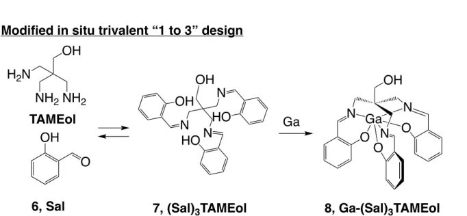
Figure 7. Modified in situ trivalent “1 to 3” design using hexadentate Schiff base
(Sal)3TAMEol with 67Ga. The Schiff base ligand (Sal)3TAMEol is stabilized upon 67Ga
coordination, while the free Schiff base ligand is decomposed to Sal and TAMEol after the change in the equilibrium state such as dilution.
[Results]
(1-2.1) Synthesis of Schiff base ligand (Sal)3TAMEol and the Ga-complex.
The Schiff base chelator was synthesized by reacting a Sal with a triamine compound TAMEol in MeOH (scheme 2) with 27% yield. The Ga-complex was prepared by refluxing the mixture of (Sal)3TAMEol and Ga-complex of acetylacetonate in ethanol solution with 54% yield.
Scheme 2. Synthesis of (Sal)3TAMEol
Reagents and conditions : (a) NaN3, DMF; (b) Pd/C, H2, MeOH; (c) Boc2O, NaHCO3,
MeCN; (d) HCl/ AcOEt; (e) Salycilaldehyde, MeOH.
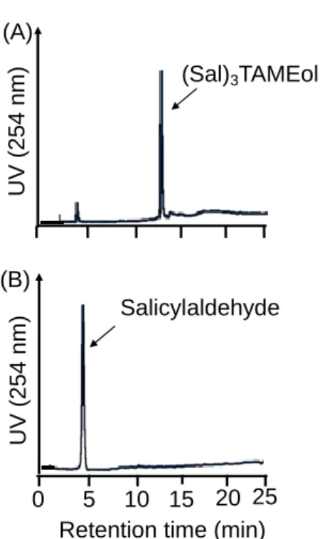
Figure 8.Analysis of (Sal)3TAMEol dissolved in ethanol solution (A) or phosphate buffer
solution (B) by RP-HPLC. 0 5 10 15 20 25 (A) (B) U V ( 2 5 4 n m ) U V ( 2 5 4 n m )
Retention time (min) Salicylaldehyde
(1-2.2) Evaluation of stability of (Sal)3TAMEol in aqueous solution.
(Sal)3TAMEol dissolved in a mixed solution of EtOH solution and phosphate buffer was
incubated at 37 ˚C for 1 h. RP-HPLC analyses revealed that while (Sal)3TAMEol was present in
the EtOH solution (Figure 8A), (Sal)3TAMEol was decomposed to Sal and TAMEol in the
phosphate buffer (Figure 8B).
(1-2.3) Preparation of [67Ga]Ga-(Sal)
3TAMEol.
[67Ga]Ga-(Sal)
3TAMEol was prepared in 95% radiochemical yield by reacting the EtOH
solution of (Sal)3TAMEol with [67Ga]Ga-citrate, followed by heating the reaction mixture at 80
˚C for 15 min (Figure 9A). The RP-HPLC retention time of the [67Ga]Ga-(Sal)3TAMEol was
similar to that of the corresponding non-radioactive Ga-complex characterized by ESI-MS (Figure 9B).
Figure 9. RP-HPLC radioactivity profile of [67Ga]Ga-(Sal)3TAMEol (A) and
69Ga-ion count profile of Ga-(Sal)
The RP-HPLC analysis of [67Ga]Ga-(Sal)3TAMEol by UV trace showed two peaks
corresponding to Sal and benzyl alcohol, a preservative in [67Ga]Ga-citrate, and the intact of
(Sal)3TAMEol was not observed (Figure 10).
Figure 10.RP-HPLC analyses of the labeling solution of [67Ga]Ga-(Sal)
3TAMEol
by UV (A) and radioactivity (B).
U V (2 5 4 n m ) R a d io a c ti v it y 0 5 10 15 0 5 10 15 [67Ga]Ga-(Sal) 3TAMEol Salicylaldehyde BnOH
(1-2.4) Stability study of [67Ga]Ga-(Sal)3TAMEol.
The stability of [67Ga]Ga-(Sal)
3TAMEol was evaluated in an apo-transferrin solution at 37 ˚C
for 6 h (Table 2). [67Ga]Ga-(Sal)
3TAMEol remained stable against the apo-transferrin challenge.
After 6 h incubation, over 95% of the radioactivity remained intact.
[Discussion]
Schiff base is present as an equilibrium of an amine and an aldehyde in aqueous solution. The hydrolysis of Schiff base begins with the binding of water molecules to carbon to form carbinolamine intermediate. If an unshared electron pair on the nitrogen atom of the Schiff base is used for coordination with a metal, the carbinolamine intermediate is not formed, and the Schiff base remains intact in an aqueous solution.34,35 Therefore, a hexadentate ligand, (Sal)
3TAMEol,
prepared from TAMEol and three molecules of a bidentate ligand Sal, was designed and synthesized.
The synthesis of [67Ga]Ga-(Sal)
3TAMEol was conducted by mixing an ethanolic solution of
(Sal)3TAMEol with [67Ga]Ga-citrate. To confirm the structure of the 67Ga-labeled compound,
non-radioactive Ga-(Sal)3TAMEol was synthesized and characterized by ESI-MS. Although I did not
analyze the compound by X-ray crystallography, the previous report indicated that the Ga-complex is formed by hexadentate N3O3 coordination.36 As mentioned above, Schiff base constitutes a
useful coordination molecule not only for Ga but for 99mTc as well.32,33 First, I evaluated the
retention time of non-radioactive Ga-(Sal)3TAMEol by RP-HPLC with UV. Since the solubility
of the non-radioactive Ga-(Sal)3TAMEol was too low to be detectable by the UV detector,
ICP-MS was used to determine the retention time of Ga-(Sal)3TAMEol (Figure 9B). The RP-HPLC
Table 2. Stability of [67Ga]Ga-(Sal)
3TAMEol in apo-transferrin solutiona
Time (h) Percent of intact radiolabeled complex (%)
1 97.0 ± 0.7
3 96.6 ± 0.6
6 95.3 ± 0. 7
analysis of [67Ga]Ga-(Sal)3TAMEol showed a retention time similar to that of the corresponding
non-radioactive Ga-complex analyzed by ICP-MS (Figure 9). These results indicated that [67Ga]Ga-(Sal)
3TAMEol was obtained with a radiochemical yield of over 95% at a ligand
concentration of 1 mM. When the labeling solution was analyzed by RP-HPLC with UV, Sal was mainly observed and the free (Sal)3TAMEol was not observed (Figure 10). These phenomena
indicated that the free Schiff base (Sal)3TAMEol was decomposed during the HPLC analysis and
[67Ga]Ga-(Sal)
3TAMEol was present as the only trivalent compound due to the stabilization of the
Schiff base by Ga-coordination. Since the administration of the 67Ga-labeled solution to subjects
forwards the equilibrium of the free Schiff base to dissociation, [67Ga]Ga-(Sal)
3TAMEol would
become the sole trivalent compound in the body. These results supported the hypothesis that the use of the metal-stabilized property of the Schiff base ligand would provide an alternative way to prepare 67/68Ga-labeled trivalent probes from monovalent ligands, as demonstrated with
[99mTc]Tc[(CO)3(CN-RGD)3]+ trivalent probe from the monovalent isonitrile-conjugated RGD.17
Another important issue to be evaluated is the plasma stability of the [67Ga]Ga-(Sal)
3TAMEol,
since an iron-binding protein, apo-transferrin, is a strong chelator for Ga present in the plasma. [67Ga]Ga-(Sal)3TAMEol remained stable during RP-HPLC analysis and after isolation from the
excess of Sal and TAMEol. Furthermore, [67Ga]Ga-(Sal)3TAMEol remained stable after
incubation with apo-transferrin (Table 2). These results confirmed that the 67Ga-complex has
[Summary]
[67Ga]Ga-(Hdpp)
3 complex synthesized from a bidentate ligand, Hdpp, could not be isolated
due to rapid decomposition during isolation process, while the [67Ga]Ga-(PTSC)
2 complexes from
tridentate ligands remained stable after isolation from the free ligands. However, the [67
Ga]Ga-(PTSC)2 complexes suffered from decomposition in bicarbonate buffer which called for further
modifications of PTSC structure to develop a chelator that forms a kinetically inert 67Ga-complex
for in vivo applications. Contrary to the bidentate and tridentate ligand, [67Ga]Ga-(Sal)
3TAMEol
remained stable after 6 h incubation in apo-transferrin solution while the free (Sal)3TAMEol
exhibited rapid decomposition during the HPLC analysis. These results indicated that [67
Ga]Ga-(Sal)3TAMEol synthesized from the Schiff base compound provided 67Ga-complex of high kinetic
Chapter 2. To evaluate the applicability of the new design towards 67Ga after conjugation with a cyclic RGDfK (-Arg-Gly-Asp-D-Phe-Lys-: c(RGDfK)) peptide.
[Introduction]
As mentioned in the Chapter 1, a Schiff base ligand, [67Ga]Ga-(Sal)
3TAMEol, provided a 67
Ga-labeled trivalent probe with stability sufficient for in vivo studies. The free Schiff base ligand present in the solution was decomposed rapidly into the monovalent Sal and TAMEol upon dilution. These results suggested that the present molecular design would constitute an alternative procedure to achieve the concept of the coordination-mediated multivalency. To further evaluate the molecular design, a cyclic RGDfK peptide (Arg-Gly-Asp-D-Phe-Lys) was selected as a targeting molecule, and a 67Ga-labeled trivalent c(RGDfK) probe was synthesized. The c(RGDfK)
peptide is a specific antagonist for the integrin αvβ3 that overexpressed on activated endothelial
cells during physiological and pathological angiogenesis and tumor cells.37 Moreover, this
targeting moiety was used in the previous studies to evaluate the coordination-mediated multivalent concept with 99mTc-labeled compounds.17,18 Thus, 67Ga-labeled Schiff base conjugated
with c(RGDfK)-peptides via aminohexanoic acid as a spacer was designed and synthesized ([67Ga]Ga-(RGD-Sal)3TAMEol) (Figure 11A). The stability, integrin αvβ3 binding affinity and
biodistribution studies in U87MG human glioma bearing nude mice was evaluated. A 67Ga-labeled
1,4,7-triazacyclononane-1,4,7-tris-(glutaric acid) (NOTGA) conjugated c(RGDfK) ([67
Ga]Ga-NOTGA(RGD)3) was synthesized and used as a reference of the conventional “3 to 3” approach
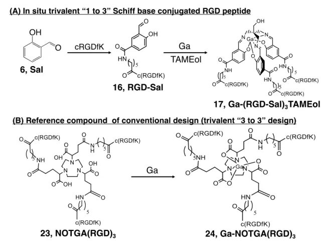
Figure 11. (A) In situ trivalent “1 to 3” Schiff base conjugated RGD peptide. Sal is
conjugated with cRGDfK to prepare monovalent RGD-Sal, subsequent reaction with TAMEol and Ga provides trivalent Ga-(RGD-Sal)3TAMEol (B) Reference trivalent
[Results]
(2.1) Synthesis of c[Arg(Pbf)-Gly-Asp(tBu)-D-Phe-Lys]
The synthesis of c[Arg(Pbf)-Gly-Asp(tBu)-D-Phe-Lys] was performed based on the previous
report.38 After Fmoc solid-phase peptide synthesis and cyclization in the liquid phase,
protected-c(RGDfK) peptide, c[Arg(Pbf)-Gly-Asp(tBu)-D-Phe-Lys], was obtained in 80% yields (Scheme
3).
Scheme 3. Synthesis of c[R(Pbf)GD(tBu)fK
Trt(2Cl)Resin-Gly-NH2
(a) Fmoc-Arg(Pbf)-OH, (i), (ii) ; (b) Fmoc-Lys(Z)-OH, (i), (ii) (c) Fmoc-D-Phe-OH, (i), (ii); (d) Fmoc-Asp(tBu)-OH, (i), (ii); (e) CH3COOH, TFE, CH2Cl2;
(f) DPPA, NaHCO3, DMF; (g) Pd/C, DMAC (i) DIC, HOBt, DMF
(ii) 20% piperidine/DMF Trt(2Cl)Resin-Gly-Arg(Pbf)-Lys(Z)-D-Phe-Asp(tBu)-NH2 (a) (b) (c) (d) (e) (f) (g)
cyclic -(Arg(Pbf)-Gly-Asp(tBu)-D-Phe-Lys) c(RGDfK)
Cleavage Cyclization
(2.1) Synthesis of RGD-conjugated Sal.
The synthetic procedure for RGD-Sal is illustrated in Scheme 4. Fmoc-protected aminohexanoic acid was activated to an active ester and then conjugated with the protected cRGDfK peptide. The resulting aminohexanoic acid-conjugated RGD was then conjugated with the aldehyde protected o-amino-phenol 10 to prepare compound 15. RGD-Sal (16) was obtained after removing the protecting groups and subsequent RP-HPLC purification (Figure 12).
Scheme 4. Synthesis of RGD-conjugated Sal.
Reagents : (a) Fmoc-OSu, (b) TFP, DCC, (c) c(R(Pbf)GD(tBu)fK) (d) 20% piperidine/DMF, (e)
TFA, hexamethylenetetramine, (f) o-aminophenol (g) DCC, HOAt, (h) TFA/water 9:1.
H2N OH O N H OH O Fmoc N H O O Fmoc F F F F 14 N H c[R(Pbf)GD(tBu)fK] O Fmoc H 2N c[R(Pbf)GD( tBu)fK] O (a) (b) (c) 11 12 13 OH OH O OH OH O O 9 OH OH O N OH 10 (d) (e) (f) N H c[R(Pbf)GD(tBu)fK] O OH O N OH (g) N H c(RGDfK) O OH O O (h) 15 16, RGD-Sal
Figure 12. RP-HPLC analysis of RGD-Sal (system 2). The purity of RGD-Sal was 98.3%.
U V(2 5 4 n m) 0 10 20 30
(2.2) Preparation of non-radioactive Ga-(RGD-Sal)3TAMEol.
A mixture of RGD-Sal, TAMEol in ammonium acetate was reacted with GaCl3 at pH 8 for 8 h
at 70 ˚C. The compound was purified by a preparative RP-HPLC to obtain the compound in 83% yields.
(2.3) Preparation of [67Ga]Ga-(RGD-Sal)
3TAMEol.
[67Ga]Ga-(RGD-Sal)
3TAMEol was prepared in 95% radiochemical yields by heating a mixed
solution of RGD-Sal, TAMEol and [67Ga]Ga-citrate at 70 ºC for 1 h. The final concentration of
RGD-Sal was 2.5 × 10⁻⁴ M.
The RP-HPLC analysis of [67Ga]Ga-(RGD-Sal)
3TAMEolshowed a single radioactive peak at
a retention time similar to that of the non-radioactive Ga-(RGD-Sal)3TAMEol characterized and
verified by ESI-MS (Figure 13).
Figure 13. RP-HPLC profiles of [67Ga]Ga-(RGD-Sal)
3TAMEol. (A) Non-radioactive
Ga-(RDG-Sal)3TAMEol verified by ESI-MS, (B) the radiotrace of [67Ga]Ga-(RGD-Sal)3TAMEol
and (C) the UV (254 nm) trace of the same sample. A single radioactivity peak was observed at a retention time similar to that of non-radioactive Ga-(RDG-Sal)3TAMEol. The UV trace
showed two peaks, corresponding to RGD-Sal and benzyl alcohol (BnOH).
RGD-sal
BnOH
0
U V (2 5 4 n m ) U V (2 5 4 n m ) R a d io a c ti v it y10
20
30
0
10
20
30
0
10
20
30
(B)
(C)
(A)
Retention time (min)
Retention time (min)
(2.4) Preparation of [67Ga]Ga-NOTGA(RGD)3. [67Ga]Ga-NOTGA(RGD)
3 was synthesized by the conventional method using
NOTGA(RGD)3 at pH 5, and the reaction proceeded in 20 min at room temperature. Under these
conditions, [67Ga]Ga-NOTGA(RGD)
3 was obtained over 95% radiochemical yields. The
RP-HPLC retention time of 67complex was identical to the corresponding non-radioactive
Ga-complex characterized and verified by ESI-MS separately (Figure 14).
Figure 14. RP-HPLC UV-chromatogram of Ga-NOTGA(RGD)3 (A) and radio-chromatograms
of [67Ga]Ga-NOTGA(RGD)
3 labeling solution (B). The retention time of radioactivity peak was
identical to that of Ga-NOTGA(RGD)3 verified by ESI-MS. The UV chromatograms of the
reaction mixture showed a peak corresponding to NOTGA(RGD)3 (C). Ga-NOTGA(RGD)3 U V (2 5 4 n m )
Retention time (min)
Reaction solution NOTGA(RGD)3 U V (2 5 4 n m ) R a d io a c ti v it y [67Ga]Ga-NOTGA(RGD) 3
Retention time (min)
(A)
(B) (C)
0
10
20
30
0
10
20
30
(2.5) Lipophilicity study.
The lipophilicity of [67Ga]Ga-(RGD-Sal)
3TAMEol was determined by measuring the Log D7.4
value using [67Ga]Ga-NOTGA(RGD)
3 as a control compound. The Log D7.4 values of [67
Ga]Ga-(RGD-Sal)3TAMEol and [67Ga]Ga-NOTGA(RGD)3 were -3.16 ± 0.03 and -4.10 ± 0.07,
respectively.
(2.6) Stability of [67Ga]Ga-(RGD-Sal)
3TAMEol.
The stability of [67Ga]Ga-(RGD-Sal)
3TAMEol was evaluated in the presence of
apo-transferrin at 37 ˚C. As shown in Table 3, over 95% of the radioactivity derived from [67
Ga]Ga-(RGD-Sal)3TAMEol remained intact after 6 h incubation, demonstrating that [67
Ga]Ga-(RGD-Sal)3TAMEol possessed stability sufficient for in vivo applications.
(2.7) Binding affinity to integrin αvβ3.
The binding affinity of Ga-(RGD-Sal)3TAMEol to integrin αvβ3 on human glioma U87MG
cells was evaluated with the competitive binding assay using [125I]I-c(RGDyV). The half-maximal
inhibitory concentration (IC50) values are summarized in Table 4, and the displacement curves of
[125I]I-c(RGDyV) are presented in Figure 15. The binding affinities of the trivalent probes
Ga-(RGD-Sal)3TAMEol, Ga-NOTGA(RGD)3, and NOTGA(RGD)3 were similar to each other (1.01,
2.31, and 2.26 nM, respectively) and significantly higher than those of the monovalent ligands. The IC50 of the trivalent probes also showed similar values to that of a 99mTc-labeled trivalent RGD
peptide reported previously.17 The binding affinity of the monovalent RGD-Sal (6.14 nM) was
higher than that of the monovalent c(RGDyV) (32.27 nM) (Table 4).
Table 3. Stability of [67Ga]Ga-(RGD-Sal)
3TAMEol in apo-transferrin solutiona
Time (h) Percent of intact radiolabeled complex (%)
1 98.0 ± 0.4
3 97.3 ± 0.6
6 95.7 ± 0. 3
Figure 15. In vitro inhibition curves of [125I]I-c(RGDyV) bound to U87MG glioma
cells by c(RGDyV), RGD-Sal, NOTGA(RGD)3, Ga-(RGD-Sal)3TAMEol, and
Ga-NOTGA(RGD)3.
Table 4. IC50 Values of each compound
design compound IC50, nM 95% C.I. a
“1 to 3” RGD-Sal 6.14 4.92-7.67
Ga-(RGD-Sal)3TAMEol 1.01 0.74-1.37
“3 to 3” NOTGA(RGD)3 2.26 1.81-2.82
Ga-NOTGA(RGD)3 2.31 1.80-2.97
reference c(RGDyV) 32.27 27.1-39.5
(2.8) Biodistribution studies.
The biodistribution of radioactivity after injection of [67Ga]Ga-(RGD-Sal)
3TAMEol in normal
mice is summarized in Table 5. [67Ga]Ga-(RGD-Sal)3TAMEol showed rapid blood clearance and
was excreted mainly in the urine through the kidney.
The biodistribution of radioactivity at 1 h postinjection in nude mice bearing U87MG xenografts was determined in the following 4 samples: (1) RP-HPLC-purified [67Ga]Ga-(RGD-Sal)
3TAMEol
(without RGD-Sal ligand), (2) unpurified [67Ga]Ga-(RGD-Sal)
3TAMEol (containing 10 nmol
ligand), (3) RP-HPLC-purified [67Ga]Ga-NOTGA(RGD)
3 (without NOTGA(RGD)3 ligand), (4)
unpurified [67Ga]Ga-NOTGA(RGD)3 (containing 10 nmol ligand). The results are summarized in
Table 6. The RP-HPLC-purified [67Ga]Ga-(RGD-Sal)
3TAMEol showed that the radioactivity
levels in the bone, a representative tissue of free gallium accumulation, was low. The tumor uptake of RP-HPLC-purified [67Ga]Ga-(RGD-Sal)3TAMEol (4.74 %ID/g) was similar to that of
RP-67 67
Table 5. Biodistribution of [67Ga]Ga-(RGD-sal)3TAMEol in ddY micea
Time (min) 10 30 60 180 360 blood 2.80 ± 0.14 1.21 ± 0.04 0.53 ± 0.06 0.07 ± 0.01 0.06 ± 0.01 liver 1.69 ± 0.07 1.64 ± 0.07 1.33 ± 0.13 1.40 ± 0.09 1.23 ± 0.10 spleen 2.54 ± 0.18 2.02 ± 0.06 1.63 ± 0.10 1.63 ± 0.17 1.67 ± 0.14 kidney 12.17 ± 0.64 9.48 ± 0.76 7.69 ± 0.39 5.48 ± 0.18 4.31 ± 0.16 pancreas 1.11 ± 0.02 0.79 ± 0.03 0.57 ± 0.04 0.45 ± 0.04 0.46 ± 0.02 heart 2.97 ± 0.22 1.18 ± 0.05 0.83 ± 0.04 0.59 ± 0.06 0.53 ± 0.02 lung 6.13 ± 0.26 3.34 ± 0.18 2.08 ± 0.10 1.24 ± 0.15 1.29 ± 0.07 stomachb 0.83 ± 0.05 0.69 ± 0.05 1.01 ± 0.08 0.54 ± 0.04 0.61 ± 0.08 intestineb 4.71 ± 0.17 3.71 ± 0.12 3.59 ± 0.18 4.07 ± 0.21 4.40 ± 0.41 muscle 1.12 ± 0.06 0.75 ± 0.12 0.51 ± 0.05 0.32 ± 0.02 0.36 ± 0.01 urine 48.08 ± 7.30 feces 0.90 ± 0.05 a
The results were expressed as percent injected dose per gram (%ID/g) ± SD (n = 5).
b
NOTGA(RGD)3 showed 60% reduction in the tumor accumulation when compared with the
RP-HPLC-purified [67Ga]Ga-NOTGA(RGD)
3 (4.19 %ID/g vs. 1.62 %ID/g). On the other hand, the
unpurified [67Ga]Ga-(RGD-Sal)
3TAMEol indicated only 25% reduction in the tumor
accumulation when compared with the RP-HPLC-purified [67Ga]Ga-(RGD-Sal)
3TAMEol (4.74
%ID/g vs. 3.59 %ID/g). As a result, the tumor uptake of the unpurified [67
Ga]Ga-(RGD-Sal)3TAMEol was two times higher than that of the unpurified [67Ga]Ga-NOTGA(RGD)3,
demonstrating the advantage of “1 to 3” design strategy over the conventional “3 to 3” design.
Table 6. Biodistribution at 1 h postinjection in tumor-bearing micea
[67Ga]Ga-(RGD-Sal) 3TAMEol (“1 to 3” design) [67Ga]Ga-NOTGA(RGD) 3 (“3 to 3” design)
purified unpurified purified unpurified
ligand (nmol) 0 10 0 10 blood 0.95 ± 0.01 1.07 ± 0.18 0.38 ± 0.14 0.43 ± 0.12 liver 4.17 ± 0.57c 2.46 ± 0.52 3.02 ± 0.52d 0.73 ± 0.13 kidney 11.85 ± 0.34 11.46 ± 0.99 7.90 ± 0.69 6.24 ± 1.16 spleen 5.01 ± 0.51c 2.19 ± 0.34 3.95 ± 0.44d 0.60 ± 0.26 intestine b 7.98 ± 1.02c 4.87 ± 0.71 9.91 ± 0.78d 7.95 ± 0.64 muscle 1.09 ± 0.27 0.72 ± 0.24 1.05 ± 0.40 0.62 ± 0.12 bone 2.48 ± 0.24c 1.26 ± 0.17 1.38 ± 0.23 n.d. tumor 4.74 ± 0.40c 3.59 ± 0.16 4.19 ± 0.24d 1.62 ± 0.35
aThe results were expressed as percent injected dose per gram (%ID/g) ± SD (n=4-5). bExpressed
as %ID. Statistical analysis was performed using one-way analysis of variance followed by Tukey’s multiple-comparison test; c(different from unpurified [67Ga]Ga-(RGD-Sal)
3TAMEol,
p<0.05), d(different from unpurified [67Ga]Ga-NOTGA(RGD)
(2.9) Small animal SPECT/CT imaging.
Figure 16 shows the SPECT/CT images of tumor-bearing mice of the unpurified [67
Ga]Ga-(RGD-Sal)3-TAMEol and the unpurified [67Ga]Ga-NOTGA(RGD)3 over 45-75 min postinjection.
The tumor was clearly visualized after injection of ([67Ga]Ga-(RGD-Sal)
3TAMEol) prepared from
the “1 to 3” design, while the tumor was barely visualized with [67Ga]Ga-NOTGA(RGD) 3 from
the “3 to 3” design. These images correlated well with the biodistribution results at the similar time point (Table 6).
[Discussion]
In the design of multivalent probes for targeting integrin αvβ3, the distance between each RGD
motif plays a crucial role in eliciting the multivalent effect. Previous studies indicated the optimum distances between each RGD moiety is 25-30 bonds.39–42 Therefore, I introduced a hexanoate
linker between RGD and Sal to adjust the recommended distance (28 bonds after complex formation).
Initially, I tried to isolate the trivalent Schiff base ligand, (RGD-Sal)3TAMEol. However, I could
not isolate (RGD-Sal)3TAMEol because of the instability of (RGD-Sal)3TAMEol in aqueous
solution. Therefore, [67Ga]Ga-(RGD-Sal)3TAMEol was prepared by mixing RGD-Sal, TAMEol,
and Ga, simultaneously. The RP-HPLC analysis of the reaction solution of [67
Ga]Ga-(RGD-Figure 16. SPECT/CT images of U87MG tumor-bearing nude mice over 45−75 min
post i.v. injection of (A) 1.8 MBq of the unpurified [67Ga]Ga-(RGD-Sal)
3TAMEol
(100 µL, [RGD-Sal] = 10 nmol) and (B) 2.2 MBq of the unpurified [67
Ga]Ga-NOTGA(RGD)3 (100 µL, [NOTGA(RGD)3] = 10 nmol). Images are shown at the
same signal intensity scale. Red arrow indicates the U87MG tumor.
High
Tumor
Tumor
Low
Sal)3TAMEol by UV trace showed two peaks corresponding to the monovalent RGD-Sal and
benzyl alcohol, a preservative in [67Ga]Ga-citrate, and the intact trivalent ligand,
(RGD-Sal)3TAMEol, was not observed (Figure 13C). As mentioned in Chapter 1, these results again
indicated that the Schiff base ligand (RGD-Sal)3TAMEol was rapidly decomposed to its
constituents by the changes in the equilibrium during RP-HPLC analysis, while the Schiff base stabilized by 67Ga coordination provided stable [67Ga]Ga-(RGD-Sal)
3TAMEol. These results
indicated that the metal coordination-mediated stabilization of Schiff base chelator constitutes a useful procedure to synthesize 67/68Ga-labeled trivalent probes. After removing the unlabeled
ligand by RP-HPLC, [67Ga]Ga-(RGD-Sal)
3TAMEol was obtained at the radiochemical purity over
99% and remained stable in apo-transferrin solution for 6 h (Table 3), confirming that [67
Ga]Ga-(RGD-Sal)3TAMEol provides a kinetically stable 67Ga-labeled complex.
The binding affinity of the monovalent RGD-Sal (6.14 nM) was higher than that of the monovalent c(RGDyV) (32.27 nM) (Table 4). After reducing the aldehyde of salicylaldehyde in RGD-Sal to alcohol, the binding affinity became similar to c(RGDyV) peptide (data not shown). Therefore, the high binding affinity of RGD-Sal would be attributable to the non-specific binding of the aldehyde group to amine groups of the protein. The trivalent Ga-(RGD-Sal)3TAMEol (1.01
nM) acquired higher integrin αvβ3 binding affinity than RGD-Sal (6.14 nM). Since the binding
affinity of Ga-NOTGA(RGD)3 (2.31 nM) was similar to that of NOTGA(RGD)3 (2.26 nM), the in
vivo competition of the monovalent RGD-Sal against the trivalent Ga-(RGD-Sal)3TAMEol would
be less than that of the trivalent NOTGA(RGD)3 against the trivalent Ga-NOTGA(RGD)3.
In the biodistribution study using normal mice, [67Ga]Ga-(RGD-Sal)
3TAMEol showed rapid
blood clearance and was excreted to urine via kidney (Table 5). This result was similar to other RGD compounds.43,44
When the 67Ga-labeled probes were administered into U87MG tumor-bearing nude mice, the
RP-HPLC-purified [67Ga]Ga-(RGD-Sal)
3TAMEol showed low radioactivity levels in bone (Table
6), a known tissue of free 67Ga accumulation.9,45 These results confirmed that [67
Ga]Ga-(RGD-Sal)3TAMEol possess stability sufficient for in vivo applications. The RP-HPLC-purified
[67Ga]Ga-(RGD-Sal)
3TAMEol and [67Ga]Ga-NOTGA(RGD)3 showed similar tumor uptakes
(4.74 ± 0.40 and 4.19 ± 0.24 %ID/g, respectively), reflecting the similar binding affinities to the U87MG cells (Table 4) and the high in vivo stability of the two 67Ga-labeled probes.
a scenario where a 67Ga-labeled probe is administered into subjects with excess of unlabeled
trivalent ligand was also evaluated. To allow direct comparison with [67
Ga]Ga-(RGD-Sal)3TAMEol, similar amount of the unlabeled ligand was co-injected into mice. According to the
previous reports, in many kind of chelators, 100 µM concentration of ligand is enough to obtain
67/68Ga-labeled compounds with over 95% radiochemical yield.22,46 Since 100 µL of radiolabeling
solution was injected into mouse, the amount of the unlabeled ligand was adjusted to 10 nmol/mouse. The tumor accumulation of [67Ga]Ga-NOTGA(RGD)
3 was inhibited by 60% in the
presence of 10 nmol of NOTGA(RGD)3. On the other hand, as low as 25% reduction of the tumor
accumulation was observed after injection of [67Ga]Ga-(RGD-Sal)
3TAMEol in the presence of 10
nmol of RGD-Sal and 2.7 nmol of TAMEol, which indicates that the free trivalent (RGD-Sal)3TAMEol present as an equilibrium in the injectate was rapidly decomposed to the monovalent
RGD-Sal and TAMEol upon injection, as also observed in the RP-HPLC analysis (Figure 13). Some RGD-Sal may have bound to plasma proteins through the aldehyde group, which may also reduce the inhibitory action of the monovalent RGD-Sal on tumor cells. Such characteristics of [67Ga]Ga-(RGD-Sal)
3TAMEol were well reflected in the SPECT/CT images where [67
Ga]Ga-(RGD-Sal)3TAMEol depicted clear tumor images in the presence of the unlabeled monovalent
ligand, RGD-Sal whereas [67Ga]Ga-NOTGA(RGD)3 showed poorly tumor images in the presence
of unlabeled trivalent ligand, NOTGA(RGD)3 (Figure 16). These results demonstrated that the
coordination-mediated stabilization of Schiff base chelator constitutes a useful procedure to apply the chemical design achieved with 99mTc-labeled trivalent probes to 68Ga-labeled ones.
[Summary]
The target binding affinity and the pharmacokinetics of the 67Ga-labeled Schiff base ligand
conjugated with RGD were conducted to evaluate the present molecular design of the coordination-mediated synthesis of multivalent 67/68Ga-labeled probes. [67
Ga]Ga-(RGD-Sal)3TAMEol possessed the stability sufficient for in vivo application and
Ga-(RGD-Sal)3TAMEol possessed the binding affinity to integrin αvβ3 similar to that of a trivalent probe,
Ga-NOTGA(RGD)3. Since the Schiff base ligand was rapidly decomposed upon injection, the
competitive inhibition of the unlabeled ligands against the trivalent 67Ga-labeled probes was low.
As a result, SPECT/CT imaging of [67Ga]Ga-(RGD-Sal)
3TAMEol visualized murine tumor
images more clearly compared to that of [67Ga]Ga-NOTGA(RGD)
3. The present study indicates
that the use of the Schiff base ligand would constitute a useful procedure to prepare 67Ga-labeled
[Conclusions]
I investigated the way to apply the coordination-mediated synthesis of multivalent probes developed with 99mTc-labeled probes to 67/68Ga-labeled ones. Hdpp, a bidentate chelator, failed to
prepare 67Ga-labeled probes since high Hdpp concentration was needed to maintain the intact
structure of the radiometal chelate. PTSC derivatives, tridentate chelators, provided 67Ga-labeled
probes of much higher stability than that from Hdpp. However, the radiolabeled probes still suffered from insufficient stability for in vivo applications, and further structural modification was required. Contrary to these approaches, the Schiff base chelator provided 67Ga-labeled trivalent
probes with stability sufficient for in vivo applications. Furthermore, (RGD-Sal)3TAMEol
exhibited the much less inhibitory effect on the [67Ga]Ga-(RGD-Sal)
3TAMEol accumulation to
the target molecules compared with [67Ga]Ga-NOTGA(RGD)
3 prepared under the conventional “3
to 3” approach, due to rapid decomposition of (RGD-Sal)3TAMEol to RGD-Sal and TAMEol after
injection. As a result, [67Ga]Ga-(RGD-Sal)3TAMEol visualized the murine tumor without
post-labeling purification. The present study indicates that the metal-coordination mediated synthesis of 67/68Ga-labeled probes was successfully achieved to a 67Ga-labeled probe through the use of a
Schiff base ligand. A new chemical procedure to prepare purification-free 67/68Ga-labeled probes
for imaging the saturable systems of the body was established. The present findings would facilitate the applications of 68Ga-labeled probes to molecular imaging with PET.
[Experimental section]
General. [67Ga]Ga-citrate and [67Ga]GaCl
3 was purchased from FUJIFILM RI Pharma Co., Ltd.
(Tokyo, Japan). Mass spectrometry was carried out using an Agilent 6130 Series Quadrupole LC/MS electrospray system (Agilent Technologies, Tokyo) or JMS-T100LP (JEOL Ltd., Tokyo).
1H-NMR spectra were recorded on a JEOL JNM-ECS-400 (400 MHz) spectrometer (JEOL Ltd.).
The melting points were measured using a MP-500D (Yanaco, Kyoto, Japan) and were reported uncorrected. Methyl 3-[1-(2-pyridyl)ethylidene]hydrazinecarbothioate were synthesized according to the procedures reported previously.26 All commercially available chemicals were of
analytical grade and used without further purification. All compounds purity used in affinity assays, biodistribution studies and SPECT studies was greater than 95%. Analytical RP-HPLC was performed with a Cosmosil 5C8 MS column (4.6 × 150 mm, Nacalai Tesque Inc., Kyoto, Japan) at a flow rate of 1 mL/min with a gradient mobile phase starting from 70% A (Milli-Q water containing 0.1% trifluoroacetic acid (TFA)) and 30% B (methanol containing 0.1% TFA) to 30% A and 70% B at 60 min (system 1), starting from 90% A and 10% B to 20% A and 80% B at 30 min (system 2), or starting from 100% C (10 mM acetate buffer pH 4.5) and 0% D (acetonitrile) to 95% C and 5% D at 10 min and to 90% C and 10% D at 20 min (system 3). Or analytical RP-HPLC was performed with a TSKgel TMS250 column (4.6 × 75 mm, TOSOH Inc, Tokyo) at a flow rate of 0.8 mL/min with a 0.1 M Tris-HNO3 buffer (pH 7.0)/acetonitrile (95/1) (system 4).
Or analytical RP-HPLC was performed with a Unison US-C18 (4.6 × 150 mm, Imtakt Inc., Kyoto, Japan) at a flow rate of 1 mL/min with a gradient mobile phase starting from 50% E (5 mM ammonium acetate) and 50% F (methanol) to 0% E and 100% F at 30 min (system 5) or starting from 30% E and 70% F to 0% E and 100% F at 30 min (system 6). Preparative RP-HPLC was performed with a Cadenza 5CD-C18 column (20 × 150 mm, Imtakt Inc.) with a guard column (Cadenza 5CD-C18 10 × 8 mm) at a flow rate of 5 mL/min with a gradient mobile phase starting form 60% A and 40% B to 20% A and 80% B in 30 min (system 7) or starting from 60% G (10 mM NH4HCO3/MilliQ water) and 40% F to 30% G and 70% F at 30 min (system 8). Or preparative
RP-HPLC was performed with a Mightysil RP-8 column (GP 20 × 250 mm, Kanto Chemical Inc., Tokyo, Japan) at a flow rate of 5 mL/min with a gradient mobile phase starting form 60% A and 40% B to 30% A and 70% B in 60 min (system 9). The eluent was monitored online with a UV/Visible single beam spectroscopy detector (L-7450, Hitachi Co. Ltd., Tokyo) coupled to a NaI(Tl) radioactivity detector (Gabi star, Raytest, Strubenhardt, Germany) or ICP-MS (Agilent
5400 ICP-MS, Agilent Technologies. TLC analyses were performed with silica plates (Silica gel 60F254, Merck Ltd., Tokyo). Cellulose acetate electrophoresis (CAE) strips were run in a veronal buffer (pH 8.5, I=0.06) at constant current of 1 mA/cm for 30 min. Radioactivity was measured using a MiniGita Star Gamma TLC Scanner (raytest) and an auto well gamma counter.
Synthesis of c[R(Pbf)GD(tBu)fK]
1. Solid phase peptide synthesis
Peptide-bound resin was prepared using H-Gly-Trt(2-Cl) Resin (376 mg, 0.3 mmol) as starting material for Fmoc coupling method, 2.5 equivalent of protected amino acid Fmoc-Arg(Pbf)-OH, 2.5 equivalent N,N’-diisopropylcarbodiimide (DIC) and 2.5 equivalent of 1-hydroxybenzotriazole (HOBt) was stirred in DMF (5 mL) for 3 hr. Completion of the coupling reaction was confirmed by Kaiser test. Deprotection of N-terminal of the Fmoc group was performed by 20% piperidine/DMF. Fmoc-Lys (Z)-OH, Fmoc-D-Phe-OH end by Fmoc-Asp(tBu)-OH sequentially
added by repeating the same operation. Resin bound linear peptide was obtained (A). 2. Cleavage of resin
Compound (A), a mixture of acetic acid, 2,2,2-trifluoroethanol and CH2Cl2 (3: 1: 6, v/v/v, in 5 ml)
was stirred at room temperature for 3 hours. After removal of the resin, vacuum evaporation of the filtrate, linear peptide (B) was obtained as a white solid (244.6 mg, 93%). ESI-MS, m/z: 1086.5 [(M + Na)+]; Found: 1086.45.
3. Cyclization of linear peptide
Compound (B) (311 mg, 0.292 mmol) was dissolved in DMF (58 mL). NaHCO3 (125 mg, 1.45
mmol) was added and stirred vigorously. Diphenylphosphoryl azide (DPPA, 188 μl, 0.87 mmol) was added slowly and stirred for 24 hours at room temperature. After removing the NaHCO3 by
filtration, the solvent was evaporated under reduced pressure. Residue afterwards precipitated by admixing of CH2Cl2 (5 mL) and sat. NaHCO3 (8 mL), and shaked vigorously. The resulting white
precipitate was collected by filtration, washed with water (3 x 10 mL) and Et2O (3 x 10 mL).
Cyclized peptides (C) as a white solid was obtained (252.4 mg, 82%) ESI-MS, m/z:1046.5 [(M + H)+], Found 1046.62.
4. Deprotection of Z group
Compound (C) (252 mg, 0.24 mmol) was dissolved in dimethylacetamide (DMAC, 5 mL), 10% palladium/carbon (Pd/C) (125 mg, 1.174 mmol) was added and the solution was stirred at room temperature for 4 h under H2 atmosphere. After reducing the solvent in vacuo, Pd/C was suspended
in MeOH and removed by filtration. The solvent was removed under reduced pressure, then admixing CH2Cl2 (5 mL) and hexane (5 mL). The mixture evaporated in vacuum to obtaine
c[R(Pbf)GD(tBu)fK] as white yellowish solid (201 mg, 92%). ESI-MS, m/z: 912.46 [(M + H)+] ;
Found 912.41.
Synthesis of 2,2’,2”-tri(aminomethyl)ethanol/3HCl (TAMEol). Pentaerythritol tribromide
(2.08 g, 6.40 mmol) and sodium azide (4.8 g, 73.8 mmol) were dissolved in dimethylformamide (DMF, 35 mL) and mixed at 80 ˚C for 24 h under N2 atmosphere. After the solvent was removed
in vacuo, the residue was resolved in H2O (20 mL) and then extracted with Et2O (30 mL). The
organic layer was dried over anhydrous MgSO4. After removing the solvent, pentaerithritol
triazide was obtained as a colorless oil. This product was used without further purification. Pentaerithritol triazide was dissolved in MeOH (10 mL), and then 10% Pd/C (1.03 g) was added portion-wise. After the mixture was stirred for 7 h, Pd/C was removed by filtration. After the solvent was evaporated in vacuo, a colorless transparent oil was obtained. The resulting oil was dissolved in the solution (20 mL, sat. NaHCO3 : acetonitrile/1 : 1) and then di-tert-butyl
dicarbonate (Boc2O, 5.79 g, 26.5 mmol) dissolved in acetonitrile (20 mL) was added dropwise on
ice. After mixing for 15 h, the solvent was evaporated in vacuo and then the residue was purified with open column chromatography using silica gel and subsequently eluted with CHCl3 : Et2O/20
: 1 to obtain N,N,N-(tert-butoxycarbonyl)-2,2’,2”-tri(aminomethyl)ethanol (TAMEol-Boc3).
TAMEol-Boc3 was dissolved in 4 M HCl/EtOAc (5 mL) and mixed for 150 min. After removing
the solvent in vacuo, HCl salt of compound TAMEol was obtained as a white solid (190 mg, 12%).
1H-NMR (D
2O), (ppm): 3.18 (t, 6H, CH2), 3.75 (s, 2H, CH2). ESI-MS, m/z: 134 [M+H]+, Found
134. M.p. 315-316 ˚C.
Synthesis of Ga-(3-hydroxy-1,2-dimethyl-4-pyridone)3 (Ga-(Hdpp)3) (1).
3-Hydroxy-1,2-dimethyl-4-pyridone (Hdpp, 30 mg, 0.22 mmol) was suspended in water (5 mL). GaCl3 (12 mg,
0.073 mmol) dissolved in water (50 µL) was added to the solution and then the solution became clear. After the pH of the solution was adjusted to 9 by 2 N NaOH, the reaction mixture was heated at 70˚C until the reaction volume was < 2.5 mL. After cooling to room temperature, the precipitate
was collected by filtration and then Ga-(Hdpp)3 was obtained as an orange solid (20.6 mg, 56%).
ESI-MS, m/z: 484 [M+H]+, Found 484.
Synthesis of 2-acetylpyridine thiosemicarbazone-1 (PTSC-1) (2). 0.89 mmol of
bis(2-methoxyethyl)amine was added to 200 mg (0.89 mmol) of methyl
3-[1-(2-pyridyl)ethylidene]hydrazinecarbothioate dissolved in 2.2 mL of EtOH. The solution was heated under reflux for 24 h. After cooling to room temperature, the resulting crystals were collected, washed with EtOH (30 mL) to afford PTSC-1 (2): (82.2 mg, 30.1%). 1H-NMR (CD3OD), 𝛿: 2.44
(m, 3H, CH3), 3.37 (m, 6H, OCH3), 3.75 (m, 4H, CH2O), 4.01 (s, 4H, NCH2), 7.47 (m, 1H, Pyr),
8.03 (m, 2H, Pyr), 8.62 (m, 1H, Pyr). ESI-MS, m/z: 310 [M+H]+, Found 310.
Synthesis of 2-acetylpyridine thiosemicarbazone-2. (PTSC-2) (3). 0.89 mmol of
diisobutylamine was added to 200 mg (0.89 mmol) of methyl
3-[1-(2-pyridyl)ethylidene]hydrazinecarbothioate dissolved in 2.2 mL of EtOH. The solution was heated under reflux for 24 h. After cooling to room temperature, the resulting crystals were collected, washed with EtOH (30 mL) to afford PTSC-2 (3): (101 mg, 37.5%) 1H-NMR (CD
3OD), 𝛿: 0.95
(m, 12H, CH3), 2.46 (s, 3H, CH3), 3.69 (m, 4H, CH2), 7.47 (m, 1H, Pyr), 7.94 (m, 2H, Pyr), 8.65
(m, 1H, Pyr). ESI-MS, m/z: 306 [M+Na]+, Found 307.
Synthesis of Ga-(PTSC-1)2 (4). Compound 2 (20 mg) and Ga(NO3)3/nH2O was dissolved in
EtOH (0.5 mL) and mixed at 40 ˚C for 1 h. A solution of ammonium hexafluorophosphate (13 mg, 0.08 mmol) in EtOH (200 µL) was added to the solution. After mixing for 10 min, the precipitates were obtained by filtration to afford compound 4 : 7.5 mg (17.1%). ESI-MS, m/z: 687 [M]+, Found
687.
Synthesis of Ga-(PTSC-2)2 (5). Compound 3 (20 mg) and Ga(NO3)3/nH2O was dissolved in
EtOH (0.5 mL) and mixed at 40 ˚C for 1 h. A solution of ammonium hexafluorophosphate (13 mg, 0.08 mmol) in EtOH (200 µL) was added to the solution. After mixing for 10 min, the precipitates were obtained by filtration to afford compound 5 : 4.3 mg (9.7%). ESI-MS, m/z: 679 [M]+, Found
679.
Synthesis of 2,2’,2”-tri(salicyliminoethyl)ethanol ((Sal)3TAMEol, 7). TAMEol/3HCl (170
mg, 0.70 mmol) and salicylaldehyde (6) (489 mg, 4.00 mmol) was dissolved in MeOH (10 mL). After standing overnight, the solvent was removed in vacuo. The residue was recrystallized from MeOH to afford (Sal) TAMEol (7) as a yellow solid (83 mg, 27%).1H-NMR (CD OD), (ppm):

![Figure 5. Analysis of [ 67 Ga]Ga-(Hdpp) 3 . CAE radioactivity traces of (A) [ 67 Ga]Ga-(Hdpp) 3 and (B) [ 67 Ga]Ga-citrate](https://thumb-ap.123doks.com/thumbv2/123deta/8490511.921599/12.918.128.556.177.633/figure-analysis-hdpp-cae-radioactivity-traces-hdpp-citrate.webp)
![Figure 6. RP-HPLC UV (254 nm) traces of Ga-(PTSC-1) 2 (A) and Ga-(PTSC-2) 2 (B) and radioactivity traces of [ 67 Ga]Ga-(PTSC-1) 2 (C) and [ 67 Ga]Ga-(PTSC-2) 2 (D)](https://thumb-ap.123doks.com/thumbv2/123deta/8490511.921599/14.918.202.688.202.667/figure-hplc-traces-ptsc-radioactivity-traces-ptsc-ptsc.webp)
![Table 1. Stability of [ 67 Ga]Ga-(PTSC-1) 2 and [ 67 Ga]Ga-(PTSC-2) 2 in 0.1 M bicarbonate buffer (pH 7.4) a](https://thumb-ap.123doks.com/thumbv2/123deta/8490511.921599/15.918.118.780.382.558/table-stability-ga-ga-ptsc-ptsc-bicarbonate-buffer.webp)
![Figure 9. RP-HPLC radioactivity profile of [ 67 Ga]Ga-(Sal) 3 TAMEol (A) and](https://thumb-ap.123doks.com/thumbv2/123deta/8490511.921599/19.918.136.703.536.796/figure-rp-hplc-radioactivity-profile-ga-sal-tameol.webp)
![Figure 10. RP-HPLC analyses of the labeling solution of [ 67 Ga]Ga-(Sal) 3 TAMEol by UV (A) and radioactivity (B)](https://thumb-ap.123doks.com/thumbv2/123deta/8490511.921599/20.918.132.446.257.556/figure-hplc-analyses-labeling-solution-sal-tameol-radioactivity.webp)