<原 著>
函館地区で発生した E 型急性肝炎に対する臨床的,
ウイルス学的,疫学的検討
―函館 4 病院における症例探索から―
梅村真知子
1)2)*渡邉
豊
2)小川 浩司
3)山本 義也
3)矢和田 敦
4)榮浪 克也
5)長佐古友和
6)川村 直之
6)工藤 峰生
6)松林 桂二
7)狩野 吉康
8)9)姜
貞憲
8)10)水尾 仁志
8)11)岡本 宏明
8)12)高橋 和明
8)13)安倍 夏生
8)13)新井 雅裕
8)13)三代 俊治
8)13) 要旨:2007∼10 年の 4 年間に,函館市内 4 病院で診療した 13 例,函館市内で感染し札幌で発症 した 1 例の計 14 例の E 型肝炎ウイルス(HEV)感染者を対象とし,E 型肝炎の臨床像,感染経 路の解析およびウイルス遺伝子の系統解析を行った.道南地区における有症状の非 A 非 B 非 C 型急性肝炎のうち E 型の頻度は 24.2% であった.E 型肝炎 14(男 8:女 6)例中,4(男 1:女 3)例が重症化しそのうち 3 例は劇症肝炎を呈し 2 例は死亡した.13 例から分離された HEV RNA ゲノムの ORF 1 326 塩基に対し遺伝子系統解析を行ったとこ ろ,3 株は genotype 3,10 株は genotype 4 に属した.genotype 4 の 7 株は既報の Kitami!Abashiri strain に,2 株は札幌圏小流行を起こした New Sapporo strain に属した.
患者の職業,食肉嗜好,居住地等は共通点に乏しかった.感染源として,従来から指摘され ている動物の内臓肉に加え,生の貝類が疑われた.
索引用語: E型肝炎ウイルス 劇症肝炎 遺伝子系統解析
New Sapporo strain Kitami!Abashiri strain はじめに E 型急性肝炎は,本邦では 2003 年以降 4 類感染症と され届け出が義務付けられているが,2011 年末に E 型肝炎ウイルス(HEV)感染検査が保険収載されるま では,感染実態の把握には困難を伴っていた.しかし, 国立感染症研究所感染症発生動向調査週報によると, 実臨床において HEV 感染診断が容易ではなかった 2011 年以前の期間も含む 1999 年 4 月から 2013 年 4 月まで に 613 例が全国から報告されている.2006 年から 2011 年度において北海道内での報告症例数は 11-39 例!年で あり,全国集計症例数に占めるその割合は,19-42% と, 群を抜いて高い1). 函館市内には比較的大規模な医療施設が集中し,渡 島地方の人口 43 万人をその医療圏としている2).しか し,本医療圏における E 型肝炎の発生実態は不明のま まであった. 北海道 E 型肝炎研究会(道 E 研)では 2007 年の発足 時より全道に協力施設を募り,全道的な HEV 感染実態 の把握を試みた.道南地域に位置する函館市内 4 病院 1)釧路労災病院消化器内科 2)函館中央病院内科 3)市立函館病院消化器病センター 4)函館五稜郭病院消化器内科 5)北海道社会事業協会函館病院内科消化器科 6)札幌北楡病院消化器内科 7)日本赤十字北海道ブロック血液センター 8)北海道 E 型肝炎研究会 9)札幌厚生病院第 3 消化器科 10)手稲渓仁会病院消化器病センター 11)勤医協中央病院内科 12)自治医科大学感染・免疫学講座ウイルス学部門 13)東芝病院研究部 * Corresponding author: [email protected] <受付日2014年2月18日><採択日2014年4月23日>
Fig. 1 Annual number of cases and frequencies of acute hepatitis E (AHE) among non A, non B, non C acute hepatitis (non ABC AH).
The numbers of patients with acute hepatitis E in four hospitals of Hako-date city were 0 to 6 per year. Dotted line indicates changes in the fre-quency of AHE in non ABC AH.
(計 2077 床)は,北海道 E 型肝炎研究会に発足当時よ り参加し,国内型 E 型肝炎症例の診療経験を蓄積して きた. 本論文では,2007∼2010 年の 4 年間に函館市を中心 とした医療圏で発生した国内型 E 型肝炎症例の臨床像, および原因ウイルスの遺伝子解析結果を検討し,道南 における HEV 感染の実態解明を目的とした. 対象と方法 2007∼2010 年の 4 年間に函館市内 4 病院で診療した 非 A,B,C 型急性肝障害のうち,道 E 研を介して診断 されたか,或は北海道ブロック赤十字血液センターに おける献血者 HEV スクリーニング陽性として紹介され た HEV 感染例を対象とした.これらの患者背景,臨床 像および HEV 感染リスク要因を検討した. HEV 感染診断は,肝炎初期に採取した患者血清中の HEV ORF1 と ORF2 領域における RT-PCR 法による HEV RNA 同定3)4),または enzyme linked immune
sorb-ent assay(ELISA)キット(特殊免疫研究所,東京)に よる抗 HEV IgM,IgA,の検出に拠った.さらに,HEV genome open reading frame(ORF)1 の metyl
trans-ferase 領域内 326 塩基に対する sequencing により遺伝 子型を特定し,neighbor-joining method(NJ 法)によ りウイルスの遺伝子系統解析を行った.HEV RNA 定量 を Real time PCR 法で行った5). 臨床病型では,PT 活性最低値が 40% 以下の急性肝 炎を重症型急性肝炎と定義し,劇症肝炎は肝性昏睡を 合併した重症型急性肝炎,通常型はそれ以外とした. 結 果 非 A,B,C 型急性肝障害における E 型肝炎の頻度 2007∼2010 年の 4 年間において函館市内 4 施設で診 療した非 A 非 B 非 C 型急性肝障害は 33 例であり,そ のうち 8 例(24.2%)が HEV 急性感染症と診断された. E 型肝炎の年次別症例数は,2007 年から順に 2,0,1, 5 例であり,非 A,B,C 型急性肝障害における E 型の 年次別頻度は 0-50% であった(Fig. 1).
HEV 感染診断根拠と HEV genotypes
上記 8 例に加え,そのうちの 1 例と函館市内で会食 後に発症した急性肝炎 1 症例,および献血者 HEV スク リーニング陽性を契機に紹介され受診した 5 例の合計 14 例が HEV 感染症と診断された(Table 1).
Table 1 Infection markers of hepatitis E virus in the cases of acute hepatitis E Case No. HEV Genotypes HEV RNA by Nested PCR isolated name of HEV Quantified level of HEV RNAa (Log copies/ml) Anti HEV
IgA IgM IgG
1 4 + JSK-Hak07 n.t.b + + 2 3 + HRC-HE103 (AB671039) 2.6 − − 3 4 + JMK-Hak07 4.8 − − 4 not determined − n.t + + + 5 3 + JSK-Hak08 3.2 − − 6 4 + JMS-Hak09R 4.6 − − 7 4 + JOM-Hak-FH09 n.t + + + 8 3 + HRC-HE185 2.5 − − 9 4 + JFS-Hak10 5.8 + + + 10 4 + JFI-Hak10 5.6 + + + 11 4 + JHK-Hak10 5.5 + + + 12 4 + JKO-HakSap10 3.3 + + + 13 4 + JSO-Hak10 5.9 + + − 14 4 + JKI-Hak10 n.t + + +
a: serum HEV RNA was quantified by TaqMan PCR; b: not tested.
このうち 13 例では初期血清に対する PCR で HEV RNA が同定され,genotype が決定された.その内訳は genotype 4 が 10 例,同 3 が 3 例 で あ り,genotype 3 HEV が検出された 3 症例は,いずれも献血症例であっ た.1 例(症例 4)は HEV RNA 陰性であったが,IgM, IgA クラス HEV 抗体陽性から診断された.
IgM,IgG クラス HEV 抗体両者が 14 症例全例で検討 され,それぞれ 9 例,8 例で陽性であった.IgA クラス HEV 抗体は 8 例で検討され,全例が陽性で,IgM クラ ス HEV 抗体との不一致例はなかった.献血者スクリー ニングで HEV RNA 陽性を指摘された 5 例(症例 2,3, 5,6,8)は,献血時 IgM,IgG クラス HEV 抗体陰性 であった. 遺伝子系統解析
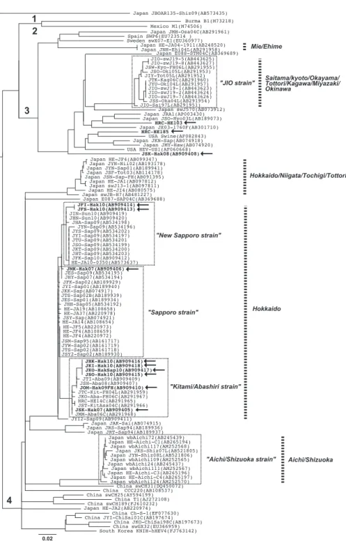
PCR で同定された HEV RNA 13 株の ORF1 326 塩基 における遺伝子系統解析結果を Fig. 2 に示す.13 株中 10 株が genotype 4 に属し,いずれも既報の Hokkaido strain に 含 ま れ た.な お,症 例 6 由 来 JMS-Hak09R では ORF1 326 塩基が増幅されなかったが,ORF1 821 nt にて増幅され,症例 7 から分離された JM-Hak-FH09 と ORF1 821 塩基において 100% 一致を認めた.Geno-type 4 の 10 株中 7 株は 2004 年北見,2006 年網走でそ れぞれ集団感染を起こした Kitami!Abashiri strain に属
しており6)∼8),残る 3 株のうち 2 株は 2009 年に札幌圏
で小流行を起こした New Sapporo strain に9)10),1 株は
1995-2009 年に札幌で多数同定された Sapporo strain に属した.症例 2,5,8 は genotype 3 に属したが,3 株間に高い相同性は認められなかった. 患者背景 E 型急性肝炎 14 症例のうち男性は 8 名,女性 6 名で あり,年齢中央値 52.7 歳であった(Table 2,3).11 名は函館市内に居住するも住所に一致を認めなかった. 14 例中,推定される HEV 感染期間に札幌ないし道東へ 旅行した患者は,後述する症例 12 以外いなかった.職 業は会社事務職 4 例,主婦 2 例で,畜産,食品加工, 調理等に従事する患者はいなかった.既往歴は糖尿病, 高血圧,関節リウマチ,脂肪肝各 1 例であった.4 例で 日常的な飲酒を認めたが,そのうち常習飲酒家は症例 3(純エタノール換算 72 g!day,5 年以上)のみであっ た.診断契機は,急性肝炎の症状発現による受診が 7 例で,献血時 HEV 陽性による紹介が 5 例であった.他, 職場(医療施設)での針刺し事故を契機に偶然肝機能 障害を指摘された 1 例(症例 4),HEV 感染症例と会食 歴を有したためスクリーニングが行われた 1 例(症例 14)であった. 検査成績,臨床経過と転帰 14 例の臨床経過と検査成績を Table 3,4 に示す.ALT 最高値は中央値 1777(47-6490)IU!l,T-bil 最高値は同
Fig. 2 Phylogenetic tree was constructed based on 326 nt sequence in open reading frame (ORF) 1 of HEV genotype 4 by neighbor-joining method. Two isolates, JFI-Hak10, JFS-Hak10, analyzed in the current study were segregated into New Sapporo strain, and 6 isolates, JSK-Hak07, JMK-Hak07, JHK-Hak10, JKI-Hak10, JKO-HakSap10, JSO-Hak10, were segregated into Kitami Abashiri/Hakodate strains. The strain isolated from case 6, JMS-Hak09R, was not amplified for 326 nt in ORF1, but amplified in 821 nt in ORF1, therefore, JMS-Hak09R was not described here.
Table 2 Back ground of the patients with acute infection of hepatitis E virus
Number of Patients, n 14 Number of male patients, n (%) 8 (57.1) Median age, years (range) 55.5 (28-73) Place of residence, n (%) Hakodate City 11 (78.6) N county 1 (7.1) Y county 1 (7.1) Sapporo City 1 (7.1) Occupation, n (%) Office worker 4 (28.6) House wife 2 (14.3)
Clerk of general store 1 (7.1)
Care worker 1 (7.1)
Sanitation worker in hospital 1 (7.1)
Taxi driver 1 (7.1)
Unknown 2 (14.3)
Habitual alcohol intakea, n (%) 1 (7.1)
Pre-Existing conditions, n (%)
Diabetes mellitus 1 (7.1)
Hypertension 1 (7.1)
Rheumatoid arthritis 1 (7.1)
Fatty liver 1 (7.1)
History of overseas travel, n none Medicine intake, n (%) 3 (21.4) Health foods intake (%) none
a: habitual intake of alcohol was defined as the con-sumption of 70 g/day or more ethanol over 5 years.
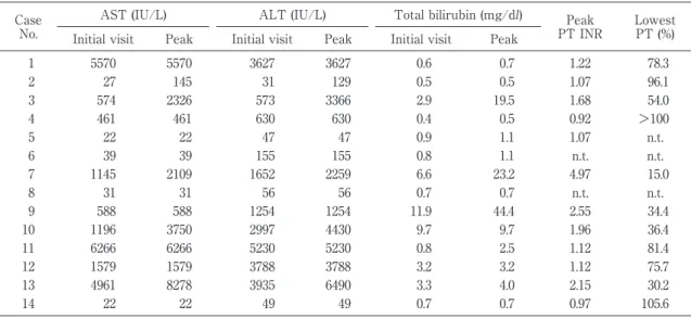
じく 1.8(0.5-44.4)mg!dl,プロトロンビン活性最低値 は 75.7(15-105)%,PT INR 最高値は 1.17(0.92-4.97) であった(Table 4a).臨床病型は,通常型が 10 例,重 症型 1 例,劇症型 3 例(急性型 1 例,亜急性型 2 例)で あった(Table 3).通常型のうち 4 例は経過観察のみで 自然軽快し,6 例は補液等の保存的加療で改善した.献 血時 HEV RNA 陽性,IgM,IgG クラス HEV 抗体陰性 であった 5 例(症例 2,3,5,6,8)は,経過観察中に 全例で通常型急性肝炎を発症した.経過中,IgM クラ ス HEV 抗体の陽性化を 3 例(症例 3,5,6)で,IgG クラス HEV 抗体の陽性化を全例で認めた.重症型およ び 劇 症 型 で は,methylprednisolone,cyclosporine A の投与による免疫抑制療法が行われ,肝性昏睡合併例 では高流量 CHF が施行された.劇症肝炎亜急性型 2 例(症例 7,9)は死亡した.14 例の入院期間は中央値 21.5 日(12-51 日)であった.
HEV genotype 別の生化学検査,HEV RNA 量の比較 病初期に採取した血清を用いた HEV RNA 定量は 10 株で可能であり,平均 3.9±1.6 Log copies!ml であった (Table 1).genotype 3 の 3 株では平均 2.5±0.2 Log cop-ies!ml,genotype 4 が同定された 7 株では平均 4.5±1.5 Log copies!ml で両者には有意差(p<0.05)を認めた (Table 4b).genotype 4 感染による 7 例では ALT,AST の最高値が genotype 3 感染例に比べ高いか或は高い傾 向を示した. HEV 感染リスクと感染経路の検討 会食歴調査により発端患者 2 例(症例 11,13)とそ の会食者 2 例(症例 12,14)で HEV 感染経路を検討し 得た(Table 5). 症例 11 は函館市在住の 44 歳営業職男性で,ブタ等 の内臓肉や生の魚介類の摂取頻度が高く,発症前 2-8 週間に会食歴のある人物の全数把握は困難であったが, 妻(会食者 1),元同僚である札幌市在住の男性(会食 者 2)の検討が可能であった.会食者 2 は症例 11 の発 症 25 日前(2010 年 5 月 19 日)に函館市内の焼鳥屋で 会食し,共に生貝類(ツブ貝,ホッキ貝等),動物種不 明レバー等を摂取した. 会食者 2 は症例 11 の入院日(6 月 14 日)と同日に 38 度台の発熱と黄疸を主訴に札幌市内の北楡病院を受 診し,急性肝炎の診断で入院した.当該病院では道 E 研を介して HEV 感染指標が検討され,HEV RNA 陽性, IgM,IgA クラス HEV 抗体陽性が判明した.会食者 2 (症例 12)は推定感染時期にこの会食以外には函館市内 で飲食をしておらず,また,同期間における札幌市内 でのレバー等内蔵肉の摂食はなかった.症例 11 と症例 12 から同定された HEV 株(JHK-Hak10,JKO-HakSap 10)は何れも genotype 4 に属し,ORF1 326 塩基にお ける配列は 100% 一致したため(Fig. 2),函館市内にお ける会食が感染契機と診断し,ツブ貝,ホッキ貝,動 物種不明レバーの何れかが感染源と考えられた(Table 5a). 症例 13 と会食歴のある人物を調査したところ,夫 (会食者 3),娘(会食者 4),同僚男性(会食者 5)が該 当した.この 3 名について各々症例 13 の入院当日,1 日後,9 日後に血液検査を施行したところ,会食者 5 (症例 14)のみ ALT 49 IU!L と軽度の肝機能異常を認 め,HEV RNA 陽 性,IgM,IgA,IgG ク ラ ス HEV 抗体陽性が判明した.症例 13 と 14 から同定された HEV 株(JSO-Hak10,JKI-Hak10)は共に genotype 4 に属し, ORF1 326 塩基が 100% 一致した(Table 5b,Fig. 2).
Table 3 Clinical features and outcomes of patients with acute hepatitis E Case No. Gender/ Age Opportunity of diagnosis Date of initial visiting Category of Disease Treatment Duration of Hospitalization (days) Out-come
1 F/47 Fatigue Aug-07 Self limited Conservative 12 Alive
2 M/57 Blood donationa Oct-07 Self limited Conservative 0 Alive
3 M/54 Blood donation Nov-07 Self limited Conservative 45 Alive 4 F/57 Needle stick accidentb Dec-07 Self limited Conservative 0 Alive
5 M/33 Blood donation May-08 Self limited Observation 0 Alive 6 F/59 Blood donation Mar-09 Self limited Observation 0 Alive
7 M/60 Fatigue Mar-09 FH, subacute High flow CHDF 23 Dead
8 M/63 Blood donation Dec-09 Self limited Observation 0 Alive
9 F/73 Jaundice Apr-10 FH, subacute mPSL, CsA 51 Dead
10 F/63 Jaundice, delirium Apr-10 FH, acute mPSL 20 Alive
11 M/54 Fever, fatigue Jun-10 Self limited Conservative 15 Alive
12 M/38 Fever, appetite loss Jun-10 Self limited Conservative 12 Alive
13 F/53 Fever, exanthema Jun-14 ASH mPSL, CsA 32 Alive
14 M/28 Dined with index patient Jun-21 Self limited Observation 0 Alive
a: Detection of HEV RNA in the screening for blood donation in Hokkaido Red Cross Blood Center. b: Abnormal liver function in the screening for a medical staff who had needle stick accident. FH; Fluminant hepatitis. ASH; Acute severe hepatitis, defined to show lowest prothrombin time index below 40%. CHDF; Continuous hemodiafiltration. mPSL; methylprednisolone pulsed therapy. CsA; Cyclosporine A. この 2 名は,症例 13 の発症 24 日前(2010 年 5 月 21 日)に函館市内の居酒屋で会食し,生の魚介類(マグ ロ,ツブ貝,ホヤ貝,ホッキ貝,ホタテ貝等),動物種 不明レバーの串焼きをほぼ同量ずつ摂取していた.症 例 14 はこの会食日以外に函館市内で飲食をしておらず, 豚内臓肉や魚介類も普段は全く摂取していなかった. 症例 13 は 6 月 14 日に発症しその後急性肝炎重症型を 呈したが,症例 14 は 6 月 13 日から 1 週間微熱,倦怠 感等を自覚したのみであった(Table 3). 共通する感染契機が特定された症例 11,12 及び症例 13,14 の 2 組より同定された 4 株(JHK-Hak10,JKO-HakSap10,JSO-Hak10,JKI-Hak10)は ORF1 326 塩基 及び 821 塩基の配列について完全に一致した(Fig. 2). 以上の会食者 2 組間の接触はなく,会食した飲食店 は函館市内の異なる地区にあった.これらの結果をも とに函館市保健所による飲食店への聞き取り調査が行 われたが,いずれの飲食店においても四足獣のレバー, レバー以外のブタ内臓肉ともに全く取り扱っていない と回答しており,汚染食品の特定には至らなかった. Table 6 に調査対象 14 例の潜伏期間(2-8 週)におけ る内臓肉及び魚介類摂取の有無を示す.全体の半数に あたる 7 例が動物の内臓肉を摂取していたが,そのう ち 6 例では生の貝の摂取歴も存在した.豚内蔵肉の摂 取歴がある 7 例中 3 例(21.4%)は豚ホルモン,豚レバー を加熱して摂取し, 残る 4 例(28.6%, 症例 11-14)は, 会食者間で内臓肉の動物種について記憶に相違を認め た.魚介類を摂取した 6 例では,全例が貝類を摂取し, うち 4 例はツブ貝とホッキ貝の両方を生食していた. 豚内臓肉・魚介類等のいずれも摂取する習慣がない のは 4 例(症例 1,9,10,14)であったが,症例 14 は同 13 との会食で喫食歴を確認したため潜伏期間中に これらの摂取歴の全くなかった症例は 3 例(21.4%)で あった. 検索対象 14 例のうち,聴取が可能であった 6 症例で は,食料品を購入する商店および普段外食をする飲食 店に重複はなかった. 考 案 函館では,2007 年以降の 4 年間に受診した非 A,B, C 型急性肝炎患者における E 型の頻度は 24.2%(8!33) であった.Yano らは 2002 年から 2008 年までに National Hospital Acute Hepatitis Study Group により集積され た非 A,B,C 型急性肝炎 228 例中 E 型急性肝炎は 25 例(11.0%)と報告しており11),函館におけるその頻度
は全国集計の倍に相当する.また,道南地区で感染し たと推測される E 型肝炎 14 例中女性は 6 例で,男女比
Table 4a Laboratory data in the patients with acute hepatitis E Case
No.
AST (IU/L) ALT (IU/L) Total bilirubin (mg/dl) Peak PT INR
Lowest PT (%) Initial visit Peak Initial visit Peak Initial visit Peak
1 5570 5570 3627 3627 0.6 0.7 1.22 78.3 2 27 145 31 129 0.5 0.5 1.07 96.1 3 574 2326 573 3366 2.9 19.5 1.68 54.0 4 461 461 630 630 0.4 0.5 0.92 >100 5 22 22 47 47 0.9 1.1 1.07 n.t. 6 39 39 155 155 0.8 1.1 n.t. n.t. 7 1145 2109 1652 2259 6.6 23.2 4.97 15.0 8 31 31 56 56 0.7 0.7 n.t. n.t. 9 588 588 1254 1254 11.9 44.4 2.55 34.4 10 1196 3750 2997 4430 9.7 9.7 1.96 36.4 11 6266 6266 5230 5230 0.8 2.5 1.12 81.4 12 1579 1579 3788 3788 3.2 3.2 1.12 75.7 13 4961 8278 3935 6490 3.3 4.0 2.15 30.2 14 22 22 49 49 0.7 0.7 0.97 105.6 a: not tested.
Table 4b Different clinical features in acute hepatitis E by genotype 3 or 4 of hepatitis E virus AST§, IU/L ALT§, IU/L T bil,§ mg/dl PT§, % PT
INR§ HEV RNA § Log copies/ml No. of ALF§§ HEV Gt 3 (n=3) 26.7±3.7 44.7±10.3 0.7±0.2 96.1±0 10.7±0 2.5±0.2 0 HEV Gt 4 (n=7) 2194±2295.1 2326±1728.5 4.05±3.8 70.3±18.6 1.3±0.3 4.5±1.5 3 P-value♭ 0.08 0.03 0.1 n.s. n.s. 0.04 n.s.
Abbreviations: Gt; genotype, PT; prothromnin time, INR: international ratio, ALF; acute liver failure.
§: mean±standard deviation were noted for their peak level in AST, ALT, total bilirubin, PT INR and HEV RNA. ¶: mean±standard deviation were noted for their nadir level in prothrombin time.
§§: acute liver failure was defined to be PT<_ 40% or PT INR>_ 1.5. ♭: by Student s U-test : not significant 3.4:1 とする既報12)と比較し性比は 1.3:1 と縮小してい た.極端な性比がみられず女性例が多い分,症例数が 増加し頻度も上昇したと思われるが,その背景には本 州とは異なり,日常的な食生活の中で女性にも HEV に感染する機会が存在することが推測され,後述する 感染経路の解明が重要と考える. 本研究では 14 例中 10 例で初診時血清中の HEV RNA を定量しえた.genoytpe 3 は 3 例中 2 例で献血時スク リーニングの際 ALT 最高値を示しその後無症状で経過 したが,genotype 4 では 10 例中 9 例が肝炎を発症し受 診していた.感染診断時の血清における比較では, geno-type 3 に対し genogeno-type 4 で ALT が有意に高く AST も高い傾向を認め,このことは阿部らの報告に合致し た12).一般的に genotype 3 感染では不顕性感染が多く, それに対して genotype 4 は急性肝炎発症例が多いと推 測されるが12),本研究では症例数は少ないものの同様の 傾向を示した.本研究では,HEV genoytpe 4 感染例で は,genotype 3 に比べ病初期の RNA 量が高いことが示 された.genotype 4 HEV は,潜伏期間中のウイルス増 幅が genotype 3 に比較し亢進し,宿主の免疫応答がよ り強く発現するため,より重い肝炎を発症する可能性 が示唆された.自験例では症例数が少なく,病初期血 液採取時期も一定ではなく,対象に献血時検査陽性例 も含む為,これらの背景が揃った血清を用いて genotype 別の HEV 増殖性と肝炎重症度の関係をさらに検討する 必要がある.
本研究では HEV RNA 陽性,IgM クラス HEV 抗体 陰性症例が 14 例中 5 例あり,HEV RNA 陽性患者での
Table 5 Retrospective search for HEV infection in the individuals who dined with the index patients with acute hepatitis E
a) Assessment of liver function and the serum markers of HEV infection in the individuals who dined with case 11 The individuals dined with case 11a Age of years Gender Association with case 11 Place of residence Duration from dining to onsetb (days) Duration from onset to blood samplingc (days) ALT (IU/L) Infection markers of HEV RNA IgM IgA IgG Attendant 1 37 F Wife Hakodate
city determinednot d 0 13 − − − −
Attendant 2, (case 12e) 38 M Former co-worker Sapporo city 28 0 3788 + + + +
a: The index patient, case 11, dueled in Hakodate city.
b: Duration from the dining with attendant 1 or 2 until the onset of hepatitis in the index patient, Case 11. c: Duration from onset of hepatitis in the index patient, Case 11, to the blood sampling in Attendant 1 or 2. d: Attendant 1 often had the opportunities of dinning together with the index patient, case 11.
e: Attendant 2, case 12, dined with Case 11 in Hakodate and was diagnosed as hepatitis E in Sapporo, on the same day when Case 11 was diagnosed in Hakodate.
b) Assessment of liver function and the serum markers of HEV infection in the individuals who dined with case 13 The individuals dined with case 13a Age of years Gender Association with case 13 Place of residence Duration from dining to onsetb (days) Duration from onset to blood samplingc (days) ALT (IU/L) Serum markers of HEV infection RNA IgM IgA IgG Attendant 3 55 M Husband N county not
determinedd 0 13 − − − −
Attendant 4 28 F Daughter Hakodate city
21 1 11 − − − −
Attendant 5,
(case 14e) 28 M Co-worker Y county 24 9 49 + + + +
a: The index patient, case 13, dueled in N county.
b: Duration from the dining with Attendant 3, 4 and 5, Case 14, until the onset of hepatitis in the index patient, Case 13. c: Duration from onset of hepatitis in the index patient, Case 13, to the blood sampling in Attendant 3, 4 and 5, Case 14. d: Attendant 3 often had the opportunities of dinning together with the index patient.
Table 6 Number of patients having the ingestion history of offalsa, raw fish or
shellfish at 2-8 weeks prior to the onset of hepatitis E.
Ingestion history of pork or unknown origin of offals, no. of patients (%)
Yesc No Unknowna
Ingestion history of raw fish or shellfish no. of patients (%)
Yes 6 (42.9)
No 4 (28.6)
Unknownb 1 (7.1) 3 (21.4)
a: offals were defined as large intestine, rectum and liver of pig or cow. b: the patient did not fulfilled the answer for the question of dietary history. c: out of 7 patients who ingested offals, 3 (21.4%) had intake pork offals.
IgM クラス HEV 抗体陽性率が 97.5% であったとする 報告と乖離を認めた13).IgM 抗体陰性はいずれも献血時
HEV RNA 陽性時の成績であり,その後の経過中に IgM ないし IgG クラス HEV 抗体が陽転しており,HEV
感染早期の IgM 抗体のウィンドウ期を捉えていたもの と考えられた.
北海道は日本国内において HEV 高浸淫地域とみなさ れているが,豚内臓肉喫食で伝搬した genotype 4 株に
よる集団感染事例が北見(2004 年),網走(2006 年)か ら報告されている7)8).地理的に隣接する北見,網走市
で2年を経て集団発生を起こしたKitami!Abashiri strain は,地域内に循環し HEV 感染を発生させたと推測され る7).2009 年秋には genotype 4 の同一系統株による札
幌圏の E 型肝炎小流行が発生したが,遺伝子系統解析 では既報の HEV 株とは異なる New Sapporo strain の 感染によると報告された9)10).これらの分子疫学的事実 から,ある地域に同一株の E 型肝炎が一定数発生する 場合,この地域内で循環する同一系統 HEV 株が関与す る可能性が示唆される. 4 年間に道南地区で診断した HEV genotype 4 感染 10 症例からは,系統解析で異なった cluster を形成する Ki-tami!Abashiri strain 7 株,New Sapporo strain 2 株が同 定された.Kitami!Abashiri strain により,2007 年に症 例 1(JSK-Hak07),2009 年には症 例 6,7(JMS-Hak 09R,JOM-Hak-FH0914)),3 年後の 2010 年には症例
11- 14(JHK-Hak10,JKI-Hak10,JKO-HakSap10,JSO-Hak10)がそれぞれ発症した.また,New Sapporo strain (JFS-Hak10,JFI-Hak1015)16))により症例 9,10 の 2 症例 が発生している(Table 1,3,Fig. 2). 函館地区から札幌,北見はそれぞれおよそ 300 km, 600 km 遠隔であるが,北海道から本州に至るより広域 でのウイルスの拡散を報告した既報に留意すると,HEV に汚染された生鮮食品が全道規模で流通した結果,函 館地区でも複数の感染者を発生させたと推測すること は可能である17).他方,これまで北見・網走,札幌で観 察された地域限定的な HEV 感染の知見からは,函館近 隣地域に Kitami!Abashiri strain や New Sapporo strain で汚染された感染源が恒常的に存在し,汚染食材が道 南で流通している可能性も排除できない.函館医療圏 で複数系統の HEV genotype 4 株が存在し,一系統株の 感染が反復した事実は,日本国内型 HEV 感染の実態を 把握する上で重要な示唆を含むと考えられる. 阿部らの全国集計に拠ると HEV 感染が豚などの食肉 に関連すると推測される症例は全体の半数以下であり 感染経路には不明な点が多い12).道南地区の E 型肝炎で は全例で輸血歴あるいは畜産,食肉販売,調理などの 職業歴はなく,感染源は摂取された食物にほぼ限定で きた.潜伏期間中に豚もしくは動物種不明内臓肉の摂 取歴があると回答したのは 7 例と半数を占めたが,6 例が同時期に生の魚介類摂取歴も有しており,そのう ち 5 例では生の貝類の摂取を認めた点は注目に値する (Table 6). 石田らは,全道で豚・エゾジカの肝臓計 468 検体, 処理前下水・処理後下水・海水・河川水・牡蠣等を含 む 954 検体を収集し, HEV 株の分離同定を行った18). その結果,豚肝臓 390 検体中 4 検体,海水 37 検体中 1 検体,処理前下水 1 検体より HEV 株を分離した.海水, 処理前下水より分離された genotype 3 HEV 株は,同時 期にヒト血清から分離された 3 株と部分塩基配列で 99.7-100%,1 株とは 93-94% の相同性を示した.HEV が分 離された環境水検体からは同時にノロウイルスも検出 されており,HEV 感染状態にあるヒトもしくは動物に より水系環境の HEV 汚染が引き起こされている可能性 が示唆された18).国産ヤマトシジミから HEV が分離さ れた事実を考え合わせると19),今後道内沿岸部で水揚げ された貝類における HEV 汚染の実態が確認される可能 性は低くないと考えられる. 生の貝類は道内では比較的安価で,男女を問わず広 く生食が好まれるため生産地に近い地域で消費される 傾向がある.本研究の事例では貝類の生食と HEV 感染 との関連は不明であったが,貝類が有する HEV 感染源 としての可能性について今後検討する必要があると考 えられる. 食物自給率 191%(カロリーベース,平成 23 年度概 算値)を誇り20),多くの食肉・水産加工品を道外に供給 している北海道で HEV 浸淫度が高いことは看過できな い事実である.しかし風評被害を恐れるあまり精査を 疎かにするきらいがないとは言えず,また 2011 年後半 まで E 型肝炎の診断法が保険収載されていなかったこ とが障壁となり,未だ HEV 感染経路を解明するには至っ ていない.迅速な診断は勿論のこと,詳細な疫学研究 を行うための各医療機関,研究施設と行政機関の連携 が感染予防対策を考える上で重要である. 結 語 道南地域を医療圏とする函館市内 4 病院では 4 年間 に 3 例の劇症例を含む 14 例の E 型急性肝炎を経験した. 既報に比して症例の男女比は小さく,感染 HEV の geno-type は 3,4 であったが,4 では複数系統の HEV 株が 発症に関与し,そのうち Kitami!Abashiri strain は 3 年間にわたり断続的に繰り返し 7 症例を発生させてい た.HEV 感染経路は不明であるが豚など既報の食肉経 路に加え生の貝類摂取に対し注目する必要があると考 えられた.
HEV 株のうち,JIN-Sun10(AB909419),JHN-Sun10(AB 909420)は,砂川市立病院内科吉田行範先生が北海道 E 型肝炎研究会に登録された E 型肝炎症例から分離同定され た事実をここに記し,本論文に対するご協力に対し,この場 を借りて深く御礼申し上げます. 文 献 1)国立感染症研究所感染症疫学センター;感染症発生 動向調査 週 報 http:!!www.nih.go.jp!niid!ja!fro m-idsc.html 2)平成 22 年度国勢調査
3)Takahashi K, Kang JH, Ohnishi S, et al. Genetic het-erogeneity of hepatitis E virus recovered from Japanese patients with acute sporadic hepatitis. J Infect Dis 2002; 185: 1342―1345
4)Mizuo H, Suzuki K, Takikawa Y, et al. Polyphyletic strains of hepatitis E virus are responsible for spo-radic cases of acute hepatitis in Japan. J Clin Micro-biol 2002; 40: 3209―3218
5)Jothikumar N, Cromeans TL, Robertson BH, et al. A broadly reactive one-step real-time RT-PCR as-say for rapid and sensitive detection of hepatitis E virus. J Virol Methods 2006; 131: 65―71
6)加藤 将,種市幸二,松林圭二.焼き肉店での会食 後に発生した E 型肝炎ウイルス集団感染:うち 1 例は劇症肝炎で死亡.肝臓 2004;45:688 7)Matsubayashi K, Kang JH, Sakata H, et al. A case
of transfusion-transmitted hepatitis E caused by blood from a donor infected with hepatitis E virus via zoonotic food-borne route. Transfusion 2008; 48: 1368―1375
8)Miyashita K, Kang JH, Saga A, et al. Three cases of acute or fulminant hepatitis E caused by inges-tion of pork meat and entrails in Hokkaido, Japan: Zoonotic food-borne transmission of hepatitis E vi-rus and public health concerns. Hepatol Res 2012; 42: 870―878 9)姜 貞憲,松居剛志,佐賀啓良,他.Genotype 4 単一系統株による札幌圏内 E 型肝炎小流行.肝臓 2010;51:51―53 10)小関 至,姜 貞憲,水尾仁志,他.2009 年秋に 札幌圏で発生した E 型肝炎小流行の臨床的・ウイ ルス学的・分枝疫学的解析.肝臓 2012;53:78― 89
11)Yano K, Tamada Y, Yatsuhashi H, et al. Dynamic epidemiology of acute viral hepatitis in Japan. In-tervirol 2010; 53: 70―75 12)阿部敏紀,相川達也,赤羽賢浩,他.本邦に於ける E 型肝炎ウイルス感染の統計学的・疫学的・ウイル ス学的特徴:全国集計 254 例に基づく解析.肝臓 2006;47:384―391 13)岡本宏明.新規に保険収載された検査法「IgA-HE 抗体価(定性)」.モダンメディア 2012;58:12― 17 14)菅原伸明,矢和田敦,高橋和明,他.劇症 E 型肝 炎:“Kitami!Abashiri strain”が検出された第 3 例目.肝臓 2009;50:473―474 15)小川浩司,梅村真知子.北海道 E 型肝炎研究会症 例報告:2010 年春に函館地区で発症し,“09 年秋札 幌小流行”起因 new Sapporo strain が分離された E 型肝炎 2 重症例について.「厚生労働科学研究費 補助金(肝炎等克服緊急対策研究事業)経口感染す る肝炎ウイルス(A 型,E 型)の感染防止,遺伝多 様性,および治療に関する研究平成 22 年度総括・ 分担研究報告書」2011,p26―27 16)小川浩司,山本義也,梅村真知子.2010 年函館地 区で発症し,09 年秋札幌小流行起因“new Sapporo strain”が分離された E 型劇症肝炎の 2 例.肝臓 2012;53:206―215
17)Takahashi K, Okamoto H, Abe N, et al. Virulent strain of hepatitis E virus genotype 3, Japan. Emerg Infect Dis 2009; 15: 704―709
18)Ishida S, Yoshizaki S, Ikeda T, et al. Detection and molecular characterization of hepatitis E virus in clinical, environmental and putative animal sources. Arch Virol 2012; 158: 2363―2368 19)Li TC, Miyamura T, Takeda N. Detection of
Hepati-tis E virus RNA from the bivalve Yamato-Shijimi (Corbicula japonica) in Japan. Am J Trop Med Hyg 76: 170―172
20)農林水産省;平成 23 年度都道府県別食物自給率 http:!!www.maff.go.jp!index.html
Occurrence of acute hepatitis E virus infection in the Hakodate district:
A prospective study of four hospitals in Hakodate City
Machiko Umemura1)2)*, Yutaka Watanabe2), Koji Ogawa3), Yoshiya Yamamoto3), Atsushi Yawata4),
Katsuya Einami5), Tomokazu Nagasako6), Naoyuki Kawamura6), Mineo Kudo6), Keiji Matsubayashi7),
Yoshiyasu Karino8)9), Jong-Hon Kan8)10), Hitoshi Mizuo8)11), Hiroaki Okamoto8)12), Kazuaki Takahashi8)13),
Natsumi Abe8)13), Masahiro Arai8)13), Shunji Mishiro8)13)
Clinical presentations, infection routes, and viral genomes were analysed in 14 cases with hepatitis E virus (HEV) infection that occurred in the Hakodate district between 2007 and 2010. Among these, eight cases were diagnosed with HEV infection by the Hokkaido Hepatitis E Research Group and were treated at one of four hos-pitals in Hakodate City, one case developed an acute HEV infection after dining in Hakodate City, and five cases were diagnosed as HEV-positive during routine screening of blood donors by the Hokkaido Red Cross Blood Center and were referred to hospitals in the Hakodate district. The patients differed in terms of their occupa-tions, meat consumption, and the neighbourhoods in which they lived. Raw shellfish and offal meat were sus-pected to be the sources of the infections. Viral RNAs were detected in 13 of the cases, and phylogenetic analy-sis of 326 base pairs of HEV open reading frame 1 (ORF1) revealed that three patients had HEV genotype 3 and the other ten patients had HEV genotype 4. Further phylogenetic analysis of 821 base pairs of ORF1 of HEV genotype 4 revealed that six of nine sequences matched the Kitami Abashiri strain. This strain was identified in a group of people who developed HEV infections between 2004 and 2006 in the Kitami and Abashiri districts. The remaining two sequences matched the New Sapporo strain, which infected a small number of people in Sapporo City in 2009.
Key words: hepatitis E virus fulminant hepatitis phylogenetic analysis New Sapporo strain Kitami!Abashiri strain
Kanzo2014; 55: 349―359 1)Department of Gastroenterology, Kushiro Rosai Hospital
2)Department of Internal Medicine, Hakodate Central General Hospital 3)Department of Gastroenterology, Hakodate Municipal Hospital 4)Department of Gastroenterology, Hakodate Goryoukaku Hospital 5)Department of Gastroenterology, Hakodate Kyokai Hospital 6)Department of Gastroenterology, Sapporo Hokuyu Hospital 7)Hokkaido Red Cross Blood Center
8)Hokkaido Study Group of Hepatitis E
9)Department of Hepatology, Sapporo Kosei General Hospital 10)Center of Gastroenterology, Teine Keijinkai Hospital 11)Department of Internal Medicine, Kin-ikyo Chuo Hospital
12)Division of Virology, Department of Infection and Immunity, Jichi Medical University School of Medicine 13)Department of Medical Sciences, Toshiba General Hospital
*Corresponding author:[email protected]