Fukushima Medical University
福島県立医科大学 学術機関リポジトリ
This document is downloaded at: 2021-11-08T00:09:08Z
Title Neutralizing epitopes of RSV and palivizumab resistance in Japan
Author(s) Hashimoto, Koichi; Hosoya, Mitsuaki
Citation Fukushima Journal of Medical Science. 63(3): 127-134
Issue Date 2017
URL http://ir.fmu.ac.jp/dspace/handle/123456789/665
Rights © 2017 The Fukushima Society of Medical Science
DOI 10.5387/fms.2017-09
Text Version publisher
Vol. 63, No. 3, 2017
[Review]
Neutralizing epitopes of RSV and palivizumab resistance in Japan
Koichi Hashimoto and Mitsuaki Hosoya
Department of Pediatrics, School of Medicine, Fukushima Medical University (Received May 2, 2017, accepted July 20, 2017)
Abstract
Respiratory Syncytial Virus (RSV) is one of the most important viral pathogen related to acute lower respiratory infection in young children. The virus surface envelope contains the G, F, and SH pro- teins as spike proteins. The F protein is considered to be a major antigenic target for the neutraliz- ing (NT) epitope as only the F protein is essential for cell infection among the three viral envelope proteins, and it is more highly conserved than the G protein. Recently, four antigenic targets relat- ed to NT activity have been reported ; site I, site II, site IV, and site zero (0). Site II is the target for palivizumab used throughout the world to suppress severe RSV infection as passive immunity in high-risk children since 1998. Under the recent conditions in which indications for palivizumab ad- ministered subjects are being expanded, palivizumab-resistant mutations have been confirmed over- seas in children with RSV infection, although they remain infrequent. Therefore, continuous ge- netic analysis of the palivizumab-binding region of the F protein is necessary. In addition, as vaccine development progresses, RSV infection control is expected to improve greatly over the next decade.
Key words: RSV, neutralizing epitope, palivizumab, resistance, Japan
Introduction
Respiratory Syncytial Virus (RSV) is one of the most important viral pathogen related to acute lower respiratory infection (ALRI) in young children
1). RSV infection accounts for 1/3 of deaths from ALRI in infants aged under 1 year and is the second larg- est cause of death from infectious diseases world- wide after malaria, with up to 200 thousand children, mainly in developing countries, losing their lives due to RSV infection each year
2). In addition, the sever- ity of infection in the high
-risk group
3,4)as well as the onset of airway hyperresponsiveness/asthma af- ter RSV infection are problems
5,6). Patients with RSV infection usually receive symptomatic treat- ment, including oxygen administration, fluid replace- ment, and respiratory management
7). RSV mono- clonal antibodies have been administered to high
-risk children as a prophylactic measure
8). RSV monoclonal antibodies have been administered to high
-risk children as a prophylactic measure, on the
other hand, there are several ongoing trials in pre- clinical, Phase
-I, Phase
-II, or Phase
-III clinical stag- es for RSV vaccine development
9). Analysis of the RSV epitope is important for the development of more effective RSV monoclonal antibodies and vac- cines.
Virological structure
RSV is a single
-stranded (-) RNA virus classi- fied as a member of the genus Orthopneumovirus in the family Pneumoviridae, and is composed of 10 genes including NS1, NS2 encoding nonstructural protein, N encoding nucleoprotein, P encoding phos- phoprotein, M/M2 encoding matrix protein, SH en- coding small hydrophobic protein (SH protein), G encoding large glycoprotein (G protein), F encoding fusion protein (F protein), and L encoding large poly- merase protein. The virus surface envelope con- tains the G, F, and SH proteins as spike proteins.
RSV is classified into two subgroups, A and B, due
Corresponding author : Koichi Hashimoto, MD. PhD E-mail : [email protected]https://www.jstage.jst.go.jp/browse/fms http://www.fmu.ac.jp/home/lib/F-igaku/
127
128 K. Hashimoto et al.
to differences in reactivity with monoclonal antibod- ies to the G protein
10).
F protein
The F protein of RSV is a 574 amino acids class I fusion glycoprotein similar to influenza virus hem- agglutinin and HIV
-1 envelope glycoprotein
11,12). It is synthesized as a precursor F
0and cleaved by the furin
-like host protease to produce F
1and F
2sub- units
13,14). The mature protein contains three cop- ies of two polypeptides (F
1and F
2). After initially folding into a metastable prefusion conformation (pre
-F), RSV F undergoes a structural change natu- rally or in the process of infection, and eventually acquires a stable postfusion conformation (post
-F)
12). Recently, cryoelectron tomography of cell culture
-grown RSV has revealed that pre
-F and post
-F are present on the virion surface. In addition, The pre
-F and post
-F are about 11 nm and 16 nm high, with the distribution dependent on the age and state of the virus
15,16).
Neutralizing epitope
Of the 11 proteins that comprise RSV, only the F and G proteins are targeted by neutralizing anti-
bodies
17). The F protein is considered to be a major antigenic target for the neutralizing (NT) epitope as only the F protein among the three viral envelope proteins is essential for cell infection, and F protein is more highly conserved than G protein
18). Four antigenic targets related to NT activity have been reported. Site I is an antigenic target for monoclo- nal antibodies (mAbs), such as 2F, 44F, or 45F
19), with weak or negligible NT activity. Site II is rec- ognized by murine 47F and 1129
20,21), with human- ization of the latter resulting in palivizumab
22)and its more potent derivative motavizumab
23). Palivizum- ab is licensed and widely used throughout the world to suppress severe RSV infection in high
-risk chil- dren
24). Site IV is recognized by mAb 19
25)or 101 F
26)with moderate NT activity. A recently isolated human mAb, 54G10, shows NT activity for human metapneumovirus (hMPV), which belongs to genus paramyxovirus as RSV
27). The 54G10 mAb is pre- dicted to recognize antigenic site IV. Of these three epitopes, site I exists only in post
-F, and site II and site IV exist in both pre
-F and post
-F. All the mAbs that recognize these three sites can bind to the stable post
-F conformation
28). Studies using RSVIG, an immunoglobulin product enriched for high RSV NT activity, showed that adsorbing the product with post
-F did not remove the NT activity
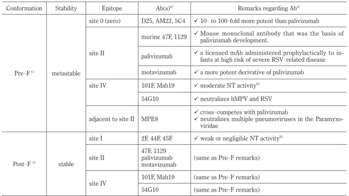
Table 1. Conformation of the F protein and epitopes
Conformation Stability Epitope Ab(s)a) Remarks regarding Aba)
Pre-F c) metastable
site 0 (zero) D25, AM22, 5C4 10- to 100-fold more potent than palivizumab
site II
murine 47F, 1129 Mouse monoclonal antibody that was the basis of palivizumab development.
palivizumab a licensed mAb administered prophylactically to in- fants at high risk of severe RSV-related disease motavizumab a more potent derivative of palivizumab site IV 101F, Mab19 moderate NT activityb)
54G10 neutralizes hMPV and RSV
adjacent to site II MPE8 cross-competes with palivizumab
neutralizes multiple pneumoviruses in the Paramyxo- viridae
Post-F c) stable
site I 2F, 44F, 45F weak or negligible NT activityb)
site II 47F, 1129
palivizumab
motavizumab (same as Pre-F remarks) site IV 101F, Mab19 (same as Pre-F remarks) 54G10 (same as Pre-F remarks) a) Ab(s) shows antibody or antibodies.
b) NT activity means neutralizing activity.
c) Pre-F, and Post-F indicate prefusion conformation F, and post-fusion conformation F, respectively.
from the immunoglobulins elicited by natural infec- tion
17), suggesting that there are other NT
-sensitive targets on the pre
-F. Site zero (0) was recently re- vealed to exist in the pre
-F as the major fourth anti- genic target
28). Monoclonal antibodies against site 0 include D25, AM22, and 5C4, all of which are pre- fusion
-specific and have NT activity 10
-100 fold greater than that of palivizumab
28,29). MPE8 is an additional prefusion
-specific antibody that which cross
-competes with palivizumab for binding to pre
-F
30). MPE8 can neutralize not only human and bo- vine RSV, but also hMPV and pneumonia virus in mice, suggesting that the MPE8 epitope is particu- larly well
-conserved among pneumoviruses
30)(Table 1). Based on the recently revealed characteristics of neutralizing epitope against RSV, not only new monoclonal Abs but also vaccines as the final goal of overcoming infectious diseases are expected in the future.
Palivizumab
For use in providing passive immunity to RSV infection, a humanized monoclonal antibody palivi- zumab (Synagis
®) targeting the antigenic site II (also called site A) region of the RSV F protein has been developed by genetic recombination technology
22,31)and is now used throughout the world. There are many antigenic variations in the G protein among subtypes, whereas there are few for the F protein.
Therefore, an antibody targeting the F protein was created. On the other hand, the G protein is the most variable RSV protein, making it useful for RSV evolution studies
12). Palivizumab has been ap- proved and used in the United States since 1998 with the aim of suppressing the severity of RSV in- fection in high
-risk children, such as preterm in- fants, and those with congenital heart disease or im- munodeficiency
32). It is currently used in more
than 60 countries and has been shown to be effective in preventing severe infection in the high
-risk group and subsequently alleviating airway hyperrespon- siveness and asthma episodes
33-35).
The indications for palivizumab administration of USA is different from that of Japan. The Ameri- can Academy of Pediatrics guidance for palivizumab use has become more restrictive against otherwise healthy preterm infants born at or after 29 weeks of gestation
36). In Japan, palivizumab was listed as a prescribed drug from 2002, with the subjects for ad- ministration being preterm infants (newborns and infants under 12 months of age with preterm birth from 29 weeks to 35 weeks or newborns and infants under 6 months of age with premature birth of 28 weeks or less of gestation), neonates, infants and young children under 24 months of age who have re- ceived treatment for bronchopulmonary dysplasia within the past 6 months. In 2005, palivizumab dosing indications were added for newborns, infants and young children with congenital heart disease with abnormal hemodynamics below 24 months of age. Furthermore, indications for neonates, infants and young children with immunodeficiency or Down’s syndrome less than 24 months of age were added in 2013
37-39).
Palivizumab resistance
Recently, there have been reports on the detec- tion of viruses showing palivizumab neutralizing
-re- sistant amino acid mutations. Clinical isolates N262D
40), K272E/M/Q
41,42), and S275F/L
40,42,43)were reported as possessing mutations in the F protein region palivizumab
-binding site (amino acids 258
-275) and exhibiting resistance to palivizumab neu- tralization, while laboratory induced isolates N268I, and K272N/T/M/Q have also been reported
43-46). The N276S mutation adjacent to the palivizumab
-Fig. 1. RSV genome and palivizumab-binding site
Amino acids (aa) and base sequences are shown based on the Long strain, which is a standard RSV-A strain.
Bold and italic characters indicate palivizumab-binding site, aa 258-275. Mutations at aa 276 (underlined) have been increasing in recent years.
130 K. Hashimoto et al.
binding site in RSV
-A was confirmed in Israel and Turkey in the 2007
-2008 season
40)and was dominant in Canada between 2008 and 2010
41). Adams
et al.showed that the N276S mutation in RSV
-A leads to a decrease in NT activity against palivizumab
47). This mutant virus was isolated from preterm infants 4 months old who were administered palivizumab but were treated in an intensive care unit for RSV infection. Furthermore, when the isolate virus was cultured in the presence of palivizumab, a second amino acid mutation, K272E, occurred in the palivi- zumab
-binding site abolishing its ability to bind to palivizumab. On the other hand, Papenburg et al.
conducted F gene analysis of 23 RSV strains isolated from patients with palivizumab and RSV 100 strains isolated from children without palivizumab during the 4 seasons from 2006 to 2010
41). The N276S mutation was detected in RSV
-A at a rate of 44.4%
in 2008
-2009 and 100% in 2009
-2010. Further- more, they reported that the N276S mutation was observed in children both with and without a history of palivizumab treatment
41). In addition, Zhu et al.
demonstrated that the N276S mutation in RSV
-A and the S276N mutation in RSV
-B did not result in palivizumab neutralization resistance based on neu- tralization tests using clinical isolates and recombi- nant virus
40). However, in a mouse experiment, mutation of the 276th amino acid in the F protein was shown to induce MARMs (Monoclonal Anti- body
-Resistant Mutants) showing resistance to the mouse monoclonal antibody against the antigenic site II region
20,44), indicating that the 276th amino acid is susceptible to selection pressure by the anti- body.
Zhu
et al. investigated natural gene polymor-phisms involving the F protein in RSV isolates from patients with respiratory disease without palivizum- ab treatment
40,43). The detection frequency of palivizumab neutralization resistant mutations was 0.7%, and these mutations were reported to be ran- dom mutations that occurred due to selective pres- sure by maternal or the patient’s own NT antibodies against site II of the RSV F protein. On the other hand, they also reported that the detection frequen- cy of palivizumab
-resistant mutations in RSV isolat- ed from patients receiving palivizumab treatment was 5.4%
43). The amino acid at position 276 of the F protein is generally asparagine (N) for RSV
-A and serine (S) for RSV
-B
40,47). It is possible that the N276S mutation in RSV
-A and S276N mutation in RSV
-B appeared due to hybridization between the subgroups, natural mutation and so on. However, since the N276S mutation has not been reported in
RSV isolated before the use of palivizumab, the pre- dominance of the N276S mutation may be related to some selective pressure attributable to palivizum- ab
20,44). Since the palivizumab binding ability is completely lost when a second amino acid mutation is introduced following the N276S mutation, the N276S mutation is considered to be the first amino acid mutation inducing palivizumab neutralization resistance.
Recently, the ON1 strain of RSV
-A
48,49)and BA strain of RSV
-B
50)have spread to various parts of the world over several seasons. These strains pos- sess mutations in the G protein associated with ad- sorption to host cells, indicating that the immunoge- nicity of the G protein is involved in the epidemic expansion. On the other hand, the N276S mutation is a mutation in the F protein. The global domi- nance of the N276S mutation in the short term is thought to be due to some factors positively affect- ing virus growth. Viruses having palivizumab neu- tralization resistance mutations (K272 E/Q and S275 F/L) have a lower proliferative capacity than do wild type strains
40). It is necessary to investigate the relation of virus proliferation ability and infectivity to amino acid mutations in segments other than anti- body recognition segments, such as the N276S mu- tation.
The situation in Japan
Apart from our previous report
51), there have been no reports on palivizumab resistance in Japan.
We analyzed palivizumab resistance using clinical
isolates or PCR products derived from 116 airway
secretion specimens from patients diagnosed with
RSV infection in Miyagi prefecture between 2004
and 2008, and in Fukushima prefecture between
2008 and 2013. Only one RSV patient in 2013 re-
ceived prophylaxis with palivizumab. The nucleo-
tide sequence and amino acid sequence of amino ac-
ids 215
-313 including the F protein region
palivizumab
-binding site were analyzed for all 116
strain. A comparison of the RSV
-A and RSV
-A
Long strain revealed 23 silent mutations and 9 mu-
tations (N228I/T, E236A, V239I, L258I, N276S,
V281L, R282S, S290P, and D310H). A comparison
of the RSV
-B and RSV
-B 9320 strain revealed 35 si-
lent mutations and 4 mutations (S228N/T, L247V,
S276N, and I305L). Among the detected amino
acid mutations, the only mutation in the palivizum-
ab
-binding site was the L258I mutation in RSV
-A,
which was detected from a specimen from a patient
not receiving palivizumab. The frequency of muta-
tions other than N276S in RSV
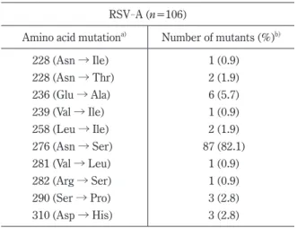
-A was 6% or less (0.9 to 5.7%). The frequency of N276S was 82.1%
(82 out of 106 strains) in RSV
-A (Table 2
-1), and the frequency of the S276N mutation in RSV
-B was 50%
(5 out of 10 strains) (Table 2
-2). The frequency of theN276S mutation in RSV
-A is 0% (0/2) in 2004, 0% (0/3) in 2005, 0% (0/3) in 2006, 60% (3/5) in 2007, 22.2% (2/9) in 2009, 95.6% (22/23) in 2009, 96.8% (31/32) in 2010, 100% (4/4) in 2011, 100%
(7/7) in 2012, and 100% (18/18) in 2013. In other words, the N276S mutation began to appear around 2007
-2008 and was found in over 90% of the ana- lyzed strains after 2009 (Table 3). Thus, the re- sults in Japan are similar to those reported over- seas
40). Furthermore, the K272E mutation as second mutation was not reported in Japan and was not observed in our analysis. Analysis in children with RSV infection administered with palivizumab is necessary in the future.
Conclusion
Under the recent conditions in which indica- tions for palivizumab administration are being ex- panded, palivizumab
-resistant mutations have been confirmed overseas in children with RSV infection, although they remain infrequent at present. In Ja- pan, the N276S mutation thought likely to lead to palivizumab neutralization resistance is becoming dominant. Therefore, continuous genetic analysis of the F protein palivizumab
-binding region is neces- sary. On the other hand, site 0, which leads to strong NT activity was also identified, and the devel- opment of monoclonal antibodies with site 0 as an epitope is certain to advance in the future. Vaccine development is also progressing, so that the control of RSV infection is expected to change markedly over the next decade.
Table 2-1. Mutations and detection rates in the F pro- tein of RSV-A
RSV-A (n=106)
Amino acid mutationa) Number of mutants (%)b)
228 (Asn → Ile) 1 (0.9)
228 (Asn → Thr) 2 (1.9)
236 (Glu → Ala) 6 (5.7)
239 (Val → Ile) 1 (0.9)
258 (Leu → Ile) 2 (1.9)
276 (Asn → Ser) 87 (82.1)
281 (Val → Leu) 1 (0.9)
282 (Arg → Ser) 1 (0.9)
290 (Ser → Pro) 3 (2.8)
310 (Asp → His) 3 (2.8)
a) Amino acid mutations were identified in comparison with the Long strain (Accession No. AY911262) of RSV-A.
b) The detection frequency of mutants was obtained by dividing the number of mutant strains by the total number of strains analyzed.
c) The palivizumab-binding site is in region of amino acid 258-275.
d) The table was cited from the reference 51.
Table 2-2. Mutations and detection rates in the F pro- tein of RSV-B
RSV-B (n=10)
Amino acid mutationa) Number of mutants (%)b)
228 (Ser → Asn) 9 (90)
228 (Ser → Thr) 1 (10)
236 (Glu → Ala) 2 (20)
247 (Leu → Val) 10 (100)
276 (Ser → Asn) 5 (50)
305 (Ile → Leu) 10 (100)
a) Amino acid mutations were identified in comparison with the 9320 strain (Accession No. AY353550) of RSV-B.
b) The detection frequency of mutants was obtained by dividing the number of mutant strains by the total number of strains analyzed.
c) The palivizumab-binding site is in region of amino acid 258-275.
d) The table was cited from the reference 51.
Table 3. Annual change in N276S mutations in RSV-A Year Number of N276S mutations (%)a)
2004 0/2 (0)
2005 0/3 (0)
2006 0/3 (0)
2007 3/5 (60.0)
2008 2/9 (22.2)
2009 22/23 (95.6)
2010 31/32 (96.8)
2011 4/4 (100)
2012 7/7 (100)
2013 18/18 (100)
a) One-hundred and sixteen RSV clinical strains or PCR products of RSV-A were analyzed to detect N276S. The detection frequency for N276S was obtained by dividing the number of strains with N276S by the total number of strains analyzed in each year.
b) The table was cited from the reference 51 with slight modifications.
132 K. Hashimoto et al.
Acknowledgements
We are grateful for the cooperation of the pedi- atric department at the Shirakawa Kosei Hospital and pediatrics department of the Soma Public Hospi- tal in providing the airway secretions from children with RSV. We also thank the Sendai City Hospital Virus Center for providing the RSV clinical isolates.
Finally, we thank Ms. Toshiko Minegishi and Mr.
Ken Honzumi for their technical support in virus isolation. This work was supported by JSPS Grant
-in
-Aid for Science Research (C), Grant Number JP15K09658.
Conflicts of interest
The authors have no conflict of interest to dis- close with respect to this manuscript.
References
1. Nair H, Nokes DJ, Gessner BD, et al. Global bur- den of acute lower respiratory infections due to re- spiratory syncytial virus in young children : a sys- tematic review and meta-analysis. Lancet, 375: 1545-1555, 2010.
2. Lozano R, Naghavi M, Foreman K, et al. Global and regional mortality from 235 causes of death for 20 age groups in 1990 and 2010 : a systematic analysis for the Global Burden of Disease Study 2010. Lancet, 380: 2095-2128, 2012.
3. Hall CB, Simoes EA, Anderson LJ. Clinical and epidemiologic features of respiratory syncytial vi- rus. Curr Top Microbiol Immunol, 372: 39-57, 2013.
4. Sommer C, Resch B, Simoes EA. Risk factors for severe respiratory syncytial virus lower respirato- ry tract infection. Open Microbiol J, 5: 144-154, 2011.
5. Kneyber MCJ, Steyerberg EW, de Groot R, Moll HA. Long-term effects of respiratory syncytial virus (RSV) bronchiolitis in infants and young children : a quantitative review. Acta Paediatr, 89: 654-660, 2000.
6. Mohapatra SS, Boyapalle S. Epidemiologic, ex- perimental, and clinical links between respiratory syncytial virus infection and asthma. Clin Micro- biol Rev, 21: 495-504, 2008.
7. Ralston SL, Lieberthal AS, Meissner HC, et al.
Clinical practice guideline : the diagnosis, manage- ment, and prevention of bronchiolitis. Pediatrics, 134: e1474-1502, 2014.
8. Subramanian KN, Weisman LE, Rhodes T, et al.
Safety, tolerance and pharmacokinetics of a human-
ized monoclonal antibody to respiratory syncytial virus in premature infants and infants with bron- chopulmonary dysplasia. MEDI-493 Study Group. Pediatr Infect Dis J, 17: 110-115, 1998.
9. Rezaee F, Linfield DT, Harford TJ, Piedimonte G.
Ongoing developments in RSV prophylaxis : a cli- nician’s analysis. Curr Opin Virol, 24: 70-78, 2017.
10. Collins PL, Fearns R, Graham BS. Respiratory syncytial virus : virology, reverse genetics, and pathogenesis of disease. Curr Top Microbiol Im- munol, 372: 3-38, 2013.
11. Collins PL, Huang YT, Wertz GW. Nucleotide se- quence of the gene encoding the fusion (F) glyco- protein of human respiratory syncytial virus. Proc Natl Acad Sci U S A, 81: 7683-7687, 1984.
12. McLellan JS, Ray WC, Peeples ME. Structure and function of respiratory syncytial virus surface glycoproteins. Curr Top Microbiol Immunol, 372: 83-104, 2013.
13. Gruber C, Levine S. Respiratory syncytial virus polypeptides. III. The envelope-associated pro- teins. J Gen Virol, 64(Pt 4): 825-832, 1983.
14. Day ND, Branigan PJ, Liu C, et al. Contribution of cysteine residues in the extracellular domain of the F protein of human respiratory syncytial virus to its function. Virol J, 3: 34, 2006.
15. Liljeroos L, Krzyzaniak MA, Helenius A, Butcher SJ. Architecture of respiratory syncytial virus re- vealed by electron cryotomography. Proc Natl Acad Sci U S A, 110: 11133-11138, 2013.
16. Graham BS, Modjarrad K, McLellan JS. Novel antigens for RSV vaccines. Curr Opin Immunol, 35: 30-38, 2015.
17. Magro M, Mas V, Chappell K, et al. Neutralizing antibodies against the preactive form of respiratory syncytial virus fusion protein offer unique possibili- ties for clinical intervention. Proc Natl Acad Sci U S A, 109: 3089-3094, 2012.
18. Melero JA, Moore ML. Influence of respiratory syncytial virus strain differences on pathogenesis and immunity. Curr Top Microbiol Immunol, 372: 59-82, 2013.
19. Calder LJ, Gonzalez-Reyes L, Garcia-Barreno B, et al. Electron microscopy of the human respiratory syncytial virus fusion protein and complexes that it forms with monoclonal antibodies. Virology, 271: 122-131, 2000.
20. Beeler JA, van Wyke Coelingh K. Neutralization epitopes of the F glycoprotein of respiratory syncy- tial virus : effect of mutation upon fusion function.
J Virol, 63: 2941-2950, 1989.
21. Arbiza J, Taylor G, Lopez JA, et al. Characteriza- tion of two antigenic sites recognized by neutraliz- ing monoclonal antibodies directed against the fu- sion glycoprotein of human respiratory syncytial
virus. J Gen Virol, 73(Pt 9): 2225-2234, 1992.
22. Johnson S, Oliver C, Prince GA, et al. Develop- ment of a humanized monoclonal antibody (MEDI- 493) with potent in vitro and in vivo activity against respiratory syncytial virus. J Infect Dis, 176: 1215-1224, 1997.
23. Wu H, Pfarr DS, Johnson S, et al. Development of motavizumab, an ultra-potent antibody for the pre- vention of respiratory syncytial virus infection in the upper and lower respiratory tract. J Mol Biol, 368: 652-665, 2007.
24. Rogovik AL, Carleton B, Solimano A, Goldman RD.
Palivizumab for the prevention of respiratory syn- cytial virus infection. Can Fam Physician, 56: 769-772, 2010.
25. Taylor G, Stott EJ, Furze J, Ford J, Sopp P. Pro- tective epitopes on the fusion protein of respiratory syncytial virus recognized by murine and bovine monoclonal antibodies. J Gen Virol, 73(Pt 9): 2217-2223, 1992.
26. Magro M, Andreu D, Gomez-Puertas P, Melero JA, Palomo C. Neutralization of human respiratory syncytial virus infectivity by antibodies and low- molecular-weight compounds targeted against the fusion glycoprotein. J Virol, 84: 7970-7982, 2010.
27. Schuster JE, Cox RG, Hastings AK, et al. A broadly neutralizing human monoclonal antibody exhibits in vivo efficacy against both human meta- pneumovirus and respiratory syncytial virus. J Infect Dis, 211: 216-225, 2015.
28. McLellan JS, Chen M, Leung S, et al. Structure of RSV fusion glycoprotein trimer bound to a prefu- sion-specific neutralizing antibody. Science, 340: 1113-1117, 2013.
29. Kwakkenbos MJ, Diehl SA, Yasuda E, et al. Gen- eration of stable monoclonal antibody-producing B cell receptor-positive human memory B cells by genetic programming. Nat Med, 16: 123-128, 2010.
30. Corti D, Bianchi S, Vanzetta F, et al. Cross-neu- tralization of four paramyxoviruses by a human monoclonal antibody. Nature, 501: 439-443, 2013.
31. Wu H, Pfarr DS, Losonsky GA, Kiener PA. Im- munoprophylaxis of RSV infection : advancing from RSV-IGIV to palivizumab and motavizumab.
Curr Top Microbiol Immunol, 317: 103-123, 2008.
32. Palivizumab, a humanized respiratory syncytial vi- rus monoclonal antibody, reduces hospitalization from respiratory syncytial virus infection in high- risk infants. The IMpact-RSV Study Group. Pe- diatrics, 102: 531-537, 1998.
33. Simoes EA, Groothuis JR, Carbonell-Estrany X, et al. Palivizumab prophylaxis, respiratory syncytial
virus, and subsequent recurrent wheezing. J Pe- diatr, 151: 34-42, 42 e31, 2007.
34. Yoshihara S, Kusuda S, Mochizuki H, et al. Effect of palivizumab prophylaxis on subsequent recur- rent wheezing in preterm infants. Pediatrics, 132: 811-818, 2013.
35. Wegzyn C, Toh LK, Notario G, et al. Safety and Effectiveness of Palivizumab in Children at High Risk of Serious Disease Due to Respiratory Syncy- tial Virus Infection : A Systematic Review. Infect Dis Ther, 3: 133-158, 2014.
36. American Academy of Pediatrics Committee on In- fectious D, American Academy of Pediatrics Bron- chiolitis Guidelines C. Updated guidance for palivizumab prophylaxis among infants and young children at increased risk of hospitalization for re- spiratory syncytial virus infection. Pediatrics, 134: 415-420, 2014.
37. Kusuda S, Takahashi N, Saitoh T, et al. Survey of pediatric ward hospitalization due to respiratory syncytial virus infection after the introduction of palivizumab to high-risk infants in Japan. Pediatr Int, 53: 368-373, 2011.
38. Mori M, Kawashima H, Nakamura H, et al. Na- tionwide survey of severe respiratory syncytial vi- rus infection in children who do not meet indica- tions for palivizumab in Japan. J Infect Chemother, 17: 254-263, 2011.
39. Mori M, Morio T, Ito S, et al. Risks and preven- tion of severe RS virus infection among children with immunodeficiency and Down’s syndrome. J Infect Chemother, 20: 455-459, 2014.
40. Zhu Q, Patel NK, McAuliffe JM, et al. Natural polymorphisms and resistance-associated muta- tions in the fusion protein of respiratory syncytial virus (RSV) : effects on RSV susceptibility to palivizumab. J Infect Dis, 205: 635-638, 2012.
41. Papenburg J, Carbonneau J, Hamelin ME, et al.
Molecular evolution of respiratory syncytial virus fusion gene, Canada, 2006-2010. Emerg Infect Dis, 18: 120-124, 2012.
42. Boivin G, Caouette G, Frenette L, Carbonneau J, Ouakki M, De Serres G. Human respiratory syn- cytial virus and other viral infections in infants re- ceiving palivizumab. J Clin Virol, 42: 52-57, 2008.
43. Zhu Q, McAuliffe JM, Patel NK, et al. Analysis of respiratory syncytial virus preclinical and clinical variants resistant to neutralization by monoclonal antibodies palivizumab and/or motavizumab. J In- fect Dis, 203: 674-682, 2011.
44. Crowe JE, Firestone CY, Crim R, et al. Monoclo- nal antibody-resistant mutants selected with a re- spiratory syncytial virus-neutralizing human anti- body fab fragment (Fab 19) define a unique epitope on the fusion (F) glycoprotein. Virology, 252:
134 K. Hashimoto et al.
373-375, 1998.
45. Zhao X, Chen FP, Sullender WM. Respiratory syncytial virus escape mutant derived in vitro re- sists palivizumab prophylaxis in cotton rats. Vi- rology, 318: 608-612, 2004.
46. Zhao X, Liu E, Chen FP, Sullender WM. In vitro and in vivo fitness of respiratory syncytial virus monoclonal antibody escape mutants. J Virol, 80: 11651-11657, 2006.
47. Adams O, Bonzel L, Kovacevic A, Mayatepek E, Hoehn T, Vogel M. Palivizumab-resistant human respiratory syncytial virus infection in infancy.
Clin Infect Dis, 51: 185-188, 2010.
48. Agoti CN, Otieno JR, Gitahi CW, Cane PA, Nokes DJ. Rapid spread and diversification of respirato- ry syncytial virus genotype ON1, Kenya. Emerg
Infect Dis, 20: 950-959, 2014.
49. Tsukagoshi H, Yokoi H, Kobayashi M, et al. Ge- netic analysis of attachment glycoprotein (G) gene in new genotype ON1 of human respiratory syncy- tial virus detected in Japan. Microbiol Immunol, 57: 655-659, 2013.
50. Trento A, Viegas M, Galiano M, et al. Natural his- tory of human respiratory syncytial virus inferred from phylogenetic analysis of the attachment (G) glycoprotein with a 60-nucleotide duplication. J Virol, 80: 975-984, 2006.
51. Sato T, Hashimoto K, Sato M, Miyazaki K, Hosoya M. Amino Acid Substitutions-Associated Palivi- zumab-Resistant RSV in Japan, 2004-2013. Clini- cal Virology (Japanese), 43: 296-303, 2015.