Fukushima Medical University
福島県立医科大学 学術機関リポジトリ
This document is downloaded at: 2021-11-07T23:38:17Z
Title
Ectopic expression of a tight-junction molecule in podocytes is associated with childhood onset nephrotic syndrome( 本文 )
Author(s)
菅野, 修人
Citation
Issue Date
2017-03-24
URL
http://ir.fmu.ac.jp/dspace/handle/123456789/956
Rights
This is the pre-peer reviewed version. Published version
"Pediatr Res. 2019 Oct;86(4):485-491. doi: 10.1038/s41390- 019-0423-7. © 2019, Springer Nature"
DOI
Text Version
ETD
1 1
学 位 論 文
2
3
Ectopic expression of a tight-junction molecule in podocytes is
4
associated with childhood onset nephrotic syndrome
5
6
(
ポドサイトにおけるタイト結合分子の異所性発現は7
小児ネフローゼ症候群に関連している
)
8 9 10 11 12 13
14
医学研究科
(
平成25
年度入学)
小児科学分野15
学籍番号
135010
16
菅野 修人
17
福島県立医科大学小児科学講座
18
2
【略語】
19
AR: after remission (
寛解後) 20
BR: before remission (
寛解前) 21
BSA: bovine serum albumin (
ウシ血清アルブミン) 22
CLDN: Claudin (
クローディン) 23
FSGS: focal segmental glomerulosclerosis (
巣状分節性糸球体硬化症) 24
HSPG: Heparan Sulfate Proteoglycan (
へパラン硫酸プロテオグリカン) 25
IgA-N: IgA nephritis (IgA
腎症) 26
mAb: monoclonal antibody (
モノクローナル抗体) 27
MCD: minimal change disease (
微小変化群) 28
NS: Nephrotic syndrome (
ネフローゼ症候群) 29
pAb: polyclonal antibody (
ポリクローナル抗体) 30
PBS: phosphate-buffered saline (
リン酸緩衝生理食塩水) 31
PECs: parietal epithelial cells (
壁側上皮細胞) 32
SDs: slit diaphragms (
スリット膜) 33
TJs: tight junctions (
タイト結合) 34
35
36
37
38
39
40
41
42
43
44
45
46
47
48
49
50
51
52
53
54
3
【
Introduction
】55
Nephrotic syndrome (NS) is a complex disorder characterized by severe proteinuria 56
along with hypoalbuminemia, edema and hyperlipidemia. The primary NS in children is 57
most frequently caused by minimal change disease (MCD) and focal segmental 58
glomerulosclerosis (FSGS). In both diseases, podocyte injury initiates foot process 59
effacement, whereas the change in podocyte morphology and the resulting proteinuria 60
are usually reversible and irreversible in MCD and FSGS, respectively. However, the 61
pathogenesis of these diseases remains obscure, and the majority of cases cannot be 62
explained by mutations in various podocyte genes (1, 2). In addition, it is unresolved 63
whether MCD and FSGS are distinct types of one disease or two different diseases (3).
64
During early stage of glomerulogenesis, immature podocytes represent columnar 65
epithelia with tight junctions (TJs) (4, 5, 6). On the other hand, mature podocytes lack 66
TJs and form slit diaphragms (SDs) between opposing foot processes, establishing the 67
final barrier to urinary protein loss. Interestingly, in several animal models for NS, 68
TJ-like structures are generated in instead of decreased or disappeared SDs (7, 8, 9, 10).
69
The SD-TJ transition is also observed in human MCD cases (11). Nevertheless, it is 70
indefinite by which mechanism the SD-TJ transition occurs in both MCD and FSGS.
71
Claudins (CLDNs) are capable of forming TJ strands (12) and thereby the backbone 72
of TJs. The CLDN family consists of 27 members in mammals, and shows distinct 73
expression patterns in tissue- and cell-type specific manners (13, 14, 15, 16). Among 74
CLDNs expressed in normal renal corpuscle, CLDN1 and CLDN2 are known to be 75
observed in parietal epithelial cells (PECs), which cover the inner surface of Bowman’s 76
capsule, but not in podocytes (17, 18, 19). During the SD-TJ transition in MCD and 77
FSGS, however, it is unknown which CLDN subtype is responsible for newly formed 78
TJs in injured podocytes.
79
On the other hand, CLDN2 is also detected in epithelial cells of the proximal tubule 80
and the thin descending limb of Henle along the normal renal tubule (17). CLDN2 is 81
one of the pore-forming CLDNs, and in the proximal tubule, it has a role in the bulk 82
reabsorption of salt and water (20). Therefore, I focused on CLDN2 and found that 83
ectopic expression of CLDN2 existed in glomeruli of primary NS. In the present study, 84
I show ectopic expression of CLDN2 in podocytes of pediatric MCD and FSGS. I also 85
demonstrate that CLDN2 is associated with their pathogenesis, suggesting that both 86
diseases are “the CLDN2-related podocytopathies”. Moreover, I discuss the possible 87
mechanism by which CLDN2 expression in podocytes lead to their dysfunction.
88
89
90
4
【
Methods
】91
Patients
92
Renal frozen specimens were obtained by needle biopsy from 49 pediatric patients:
93
21 subjects (8 subjects before remission [BR] and 13 after remission [AR]) with MCD, 94
18 (8 BR and 10 AR) with FSGS, and 10 with IgA nephritis (IgA-N) as disease controls.
95
This study was approved by the Ethical Committee of Fukushima Medical University 96
(approval number: 1809).
97
Clinical data for the subjects were documented at the time of biopsy, and were 98
summarized in Tables 1 and 2. Proteinuria and urinary occult blood were 99
semi-quantitatively scored as follows: (−)=0, (±)=0.5, (1+)=1, (2+)=2, (3+)=3, and 100
(4+)=4.
101 102
Antibodies
103
Rabbit polyclonal antibody (pAb) against CLDN2 was the gift from Dr. Furuse 104
(National Institute for Physiological Sciences, National Institutes of Natural Sciences) 105
(21). Rabbit pAbs against CLDN1 and podocin were purchased from IBL (Gunma, 106
Japan) and SIGMA (St. Louis, MO, USA), respectively. Mouse monoclonal antibodies 107
(mAbs) against CD34 (clone NU-4A1), podocalyxin (clone #222328) and synaptopodin 108
(clone G1D4) were obtained from Nichirei Bioscience (Tokyo, Japan), R&D Systems 109
(Minneapolis, MN, USA) and Progen Biotechnik (Heidelberg Germany), respectively.
110
A rat anti-Heparan Sulfate Proteoglycan (HSPG) (Perlecan) mAb (clone A7L6) was 111
purchased from Merck Millipore (Temecula, CA, USA). The secondary antibodies used 112
were as follows: AlexaFluor488-labeled donkey anti-rabbit IgG (H+L) (Invitrogen, 113
Waltham, MA, USA), Cy3-conjugated AffiniPure donkey anti-mouse IgG (H+L) 114
(Jackson ImmunoResearch, West Grove, PA, USA), AlexaFluor647-labeled AffiniPure 115
donkey anti-rat IgG (H+L) (Jackson ImmunoResearch) and immunogold conjugate EM 116
goat anti-rabbit IgG (BBI Solutions, Cardiff, UK).
117 118
Immunohistochemistry
119
Renal biopsy specimens were frozen on dry ice and kept at −80°C until use. They 120
were sectioned at a thickness of 5 µm and fixed in ice-cold methanol for 15 min at 121
−20°C. After washing with phosphate-buffered saline (PBS), sections were blocked in
122
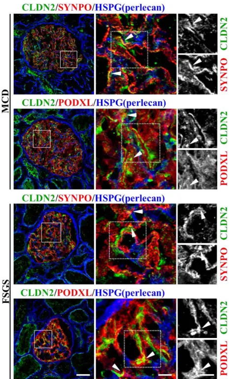
2% bovine serum albumin (BSA) for 1 h at room temperature. After washing, they were 123
subsequently incubated with primary antibodies overnight at 4°C and rinsed again with 124
PBS followed by a reaction for 1 h at room temperature with appropriate secondary 125
antibodies. They were then mounted after washing with PBS. All samples were
126
5
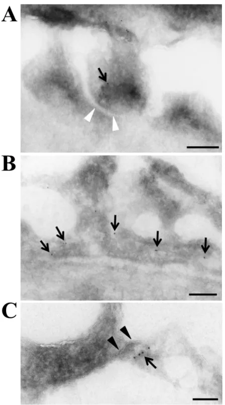
examined using a laser-scanning confocal microscopy (FV1000, OLYMPUS, Tokyo, 127
Japan).
128 129
Calculations
130
The CLDN2-positive area was calculated using image processing software (ImageJ, 131
Java). The images stained with CLDN2 and HSPG (Perlecan) were set the threshold 132
from 100 to 255, in order to exclude background signals. A circle was drawn by free 133
hand along the inside of PECs, and the total area in the circle (A) and the 134
CLDN2-expression area in the circle (B) were determined. The CLDN2-positive area 135
was defined as B/A × 100 (%), and represented using box-and-whisker plots.
136 137
Immunoelectron microscopy
138
Renal biopsy tissues were fixed with periodate-lysine-paraformaldehyde for 2 h at 139
4°C, and, after washing with PBS, they were incubated with 140
polyvinylpyrrolidone-sucrose overnight at 4°C. They were then frozen by liquid 141
nitrogen and ultrathin cryosections were prepared using a Leica Ultracut UCT 142
microtome equipped with the FCS cryoattachment (Wien, Austria) at −20°C. They were 143
transferred to nickel grids (150 mesh) with coating in formvar and carbon, and 144
subsequent incubation steps were carried out by floating grids on droplets of the filtered 145
soluction. After quenching free aldehyde groups with PBS/0.01 M glycine, sections 146
were incubated with rabbit anti-CLDN2 pAb overnight at 4°C, and reacted for 1 h at 147
room temperature with 10 nm gold-labeled goat anti-rabbit IgG followed by a fixation 148
with 2.5% glutaraldehyde buffered with 0.1 M PBS (pH 7.4). They were subsequently 149
contrasted with 3% uranyl acetate solution for 40 min, and absorption-stained with 3%
150
polyvinyl alcohol containing 0.2% acidic uranyl acetate for 40 min. Micrographs were 151
captured using an electron microscope (JEM1230, JOEL).
152 153
Statistical analysis
154
All values are shown as the mean ± standard deviation (SD) except for those of the 155
CLDN2-positive area. Statistical analysis was performed by IBM SPSS statistics 23 156
software (Chicago, IL, USA). Results were analyzed using two-sample t-test and one 157
way analysis of variance (ANOVA).
158
159
160
161
162
6
【
Results
】163
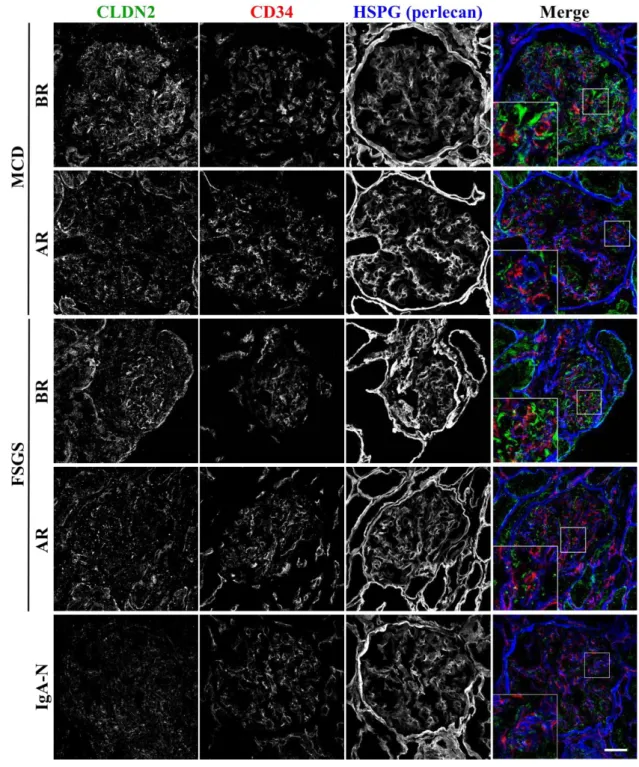
CLDN2 is ectopically detected in MCD and FSGS glomeruli 164
I first examined, by immunohistochemistry, the expression of CLDN2 in glomeruli 165
obtained from pediatric MCD and FSGS patients, as well as in those from IgA-N 166
subjects as disease controls. To distinguish the overall structure of glomeruli, the 167
endothelial marker CD34 and the basement membrane marker HSPG (Perlecan) were 168
co-immunostained with CLDN2. As shown in Figure 1, strong filamentous signals for 169
CLDN2 appeared to be detected in the before remission cases with MCD and FSGS, but 170
not in subjects with IgA-N. CLDN2 was also occasionally observed within whole cell 171
bodies. These CLDN2 signals were generally distributed close to the basement 172
membrane and separated from endothelial cells, implying that CLDN2-expressing cells 173
correspond to podocytes. By contrast, in the after remission cases with the MCD and 174
FSGS, the CLDN2 expression was strikingly decreased, and the filamentous and 175
cytoplasmic staining disappeared.
176
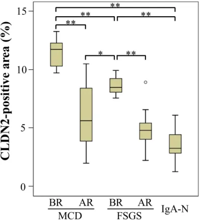
I also quantitatively evaluated the CLDN2 expression by calculating the positive area 177
in glomeruli (Figure 2). The CLDN2-stained region in MCD and FSGS glomeruli 178
before remission was significantly greater than that after remission and in IgA-N 179
patients. In addition, the abundance of CLDN2 expression was well correlated with the 180
amounts of proteinuria of each group at the time of biopsy (Table 1 and 2). Interestingly, 181
among before remission subjects, CLDN2 was expressed in MCD glomeruli at high 182
levels compared with that in FSGS.
183 184
CLDN2 is expressed in MCD and FSGS podocytes 185
To determine whether CLDN2-expressing cells represent podocytes, I next performed 186
multiple immunostaining using the podocyte markers synaptopodin (SYNPO) and 187
podocalyxin (PODXL) (22, 23) (Figure 3). In both MCD and FSGS glomeruli before 188
remission, CLDN2 was at least in part colocalized with SYNPO and PODXL, 189
suggesting that CLDN2 expression is observed in podocytes.
190
I subsequently verified, by immunogold immunoelectron microscopy, the nature of 191
CLDN2-positve cells in MCD glomeruli before remission, as well as the detailed 192
subcellular localization of CLDN2 (Figure 4). The CLDN2 labeling was detected not 193
only in residual foot processes of podocytes (Figure 4A) but also in fused foot processes 194
(Figure 4B). Importantly, CLDN2 was also concentrated along newly formed TJs in 195
podocytes (Figure 4C).
196 197
CLDN1 is not expressed in MCD glomeruli but segmentally observed in FSGS
198
7
I also evaluated the CLDN1 expression in renal corpuscle of MCD and FSGS cases 199
before remission (Figure 5). As expected, PECs were positive for CLDN1 in both 200
diseases. By contrast, in glomeruli, the CLDN1 signals were not apparently detected for 201
MCD subjects. In addition, CLDN1 was only focally and segmentally expressed in 202
FSGS glomeruli with a trabecular pattern.
203 204
Newly formed TJs constructed by CLDN2 are generated together with decrease of 205
SDs in MCD and FSGS glomeruli before remission 206
To confirm the SD-TJ transition, I performed multiple immunostaining using the SDs 207
marker podocin (24), and compared CLDN2 and podocin expression in MCD and FSGS 208
glomeruli (Figure 6). In the before remission cases with MCD and FSGS, the 209
filamentous signals for podocin were decreased and changed to the granulated signals 210
together with the expression of strong filamentous signals for CLDN2. By contrast, in 211
the after remission cases with MCD and FSGS, the filamentous signals for podocin 212
were recovered together with decreased expression for CLDN2.
213
214
8
【
Discussion
】215
In the present study, I found that CLDN2, which is detected in epithelial cells of 216
Bowman’s capsule, the proximal tubule and the thin descending limb of Henle along the 217
normal nephron (17), was ectopically expressed on injured podocytes in pediatric MCD 218
and FSGS. In both diseases, CLDN2 was distributed along the glomerular basement 219
membrane maker HSPG, and apart from the vascular endothelial marker CD34. In 220
addition, CLDN2 was at least in part colocalized with the podocyte markers SYNPO 221
and PODXL in MCD and FSGS. Moreover, CLDN2-immunogold signals was observed 222
in podocytes, especially in residual and fused foot processes as well as at TJs, 223
definitively indicating that CLDN2-expressing cells represent podocytes. Hence, 224
immunofluorescence and immunoelectron studies for CLDN2 appear to be a powerful 225
tool for diagnosis of these primary NS. Since no substantial abnormality in glomerular 226
structure is detected in MCD by light microcopy, CLDN2 should be a novel diagnostic 227
marker especially for these patients who are resistant to steroid therapy and underwent 228
renal biopsy (25).
229
Although both MCD and FSGS are typical podocyte diseases (26, 27), information 230
on the pathophysiological basis for these diseases is still fragmentary. In this regard, it 231
is noteworthy that ectopic expression of CLDN2 on podocytes was observed not only in 232
MCD but also in FSGS as far as we determined. Thus, both MCD and FSGS could be 233
regarded as “the CLDN2-related podocytopathies”. Lower expression levels of CLDN2 234
in FSGS before remission compared with that in MCD most probably reflect podocyte 235
loss in FSGS. The CLDN2-immunoreactive area in podocytes of both diseases after 236
remission was significantly decreased to levels similar to that of the disease control 237
group, further supporting that the CLDN2 expression is involved in their pathogenesis.
238
Since circulating glomerular permeability factors, including angiopoietin-like-4 and 239
urokinase plasminogen activator receptor in MCD and FSGS, respectively, are expected 240
to result in the onset of these diseases (3, 28, 29, 30), it is intriguing to elucidate 241
whether and how these factors are associated with the CLDN2 expression in damaged 242
podocytes.
243
The CLDN1 expression, which is restricted in PECs along the healthy nephron (17, 244
18), is induced in glomerulus from humans and animals with diabetic nephropathy (31).
245
On the other hand, we demonstrated that CLDN1 was not principally observed in 246
glomerular tuft of pediatric MCD. In FSGS glomeruli before remission, the CLDN1 247
signals displayed a cord-like array in focal and segmental patterns, which are totally 248
different from those of CLDN2. In FSGS, PECs are activated on Bowman’s capsule and 249
migrate onto the glomerular capillary to substitute or dislocate podocytes (32).
250
9
Activated PECs in glomerulosclerotic lesions are also known to positive for CLDN1 251
(33). Taken collectively, CLDN1-expressing cells in glomeruli of our FSCG cases may 252
correspond to activated PECs.
253
Podocin is one of the proteins forming SDs, and its mutations (NPHS2 gene) are 254
responsible for the autosomal recessive form of steroid-resistant NS (24). The strong 255
filamentous signals for CLDN2 were appeared together with decrease of the 256
filamentous signals and change to the granulated pattern for podocin in the before 257
remission cases with MCD and FSGS. These changes are suggested that SDs are 258
displaced to TJs constructed by CLDN2 in the before remission cases of MCD and 259
FSGS. In the after remission cases with MCD and FSGS, the filamentous signals for 260
podocin were recovered. It is seemed that the SDs related molecules containing podocin 261
accumulate and form SDs again.
262
Several TJ proteins such as junctional adhesion molecule-A, coxsackie and 263
adenovirus receptor, ZO-1 and cingulin, are concentrated at the SD in mature podocytes 264
(34, 35, 36). Among them, ZO-1 is indispensable for the interdigitation of foot 265
processes and the formation of SDs (37), even though the precise role of other TJ 266
components in glomerular filtration barrier remains elusive. Therefore, I speculate that 267
the ectopically expressed CLDN2 could recruit these TJ constituents from the SD pool 268
and disrupt the architecture of foot processes and SDs, resulting in being 269
dedifferentiated into immature podocytes with glomerular dysfunction. To prove this 270
idea, CLDN2-knockin mice, in which CLDN2 is podocyte-specifically expressed under 271
human podocin promoter, were generated, and their characterization is under analysis 272
(Ichikawa-Tomikawa et al., unpublished data).
273
In conclusion, I showed that both MCD and FSGS in children possessed the same 274
pathological findings in terms of ectopic CLDN2 expression on podocytes. I also 275
demonstrated that the abundance of CLDN2 was diminished after remission, indicating 276
that the levels of CLDN2 expression are related to the disease state. Further studies are 277
required to clarify the functional relevance of CLDN2 expression in the pathogenesis of 278
these diseases.
279 280
【
Acknowledgments
】281
I thank Prof. M Hosoya, assistant Prof. Y Kawasaki, and Prof. H Chiba (Fukushima 282
Medical University) for their advice. I am also grateful to Drs. N Ichikawa-Tomikawa 283
and M Mizuko (Fukushima Medical University) for immunohistochemical study; Dr. K 284
Sugimoto (Fukushima Medical University) for preparing figures; and Dr. H Kurihara 285
(Juntendo University) for immunoelectron microscopy analysis.
286
287
10
【
References
】288
1. Allison A Eddy, Jordan M Symons: Nephrotic syndrome in childhood. Lancet 362:
289
629-639, 2003 290
2. D'Agati VD, Kaskel FJ, Falk RJ: Focal segmental glomerulosclerosis. N Engl J Med.
291
365: 2398-2411, 2011 292
3. Floege J, Amann K: Primary glomerulonephritides. Lancet 387: 2036-2048, 2016 293
4. Reeves W, Caulfield JP, Farquhar MG: Differentiation of epithelial foot processes 294
and filtration slits: sequential appearance of occluding junctions, epithelial 295
polyanion, and slit membranes in developing glomeruli. Lab Invest. 39: 90-100, 296
1978 297
5. Grahammer F, Schell C, Huber TB: The podocyte slit diaphragm⎯from a thin grey 298
line to a complex signalling hub. Nat Rev Nephrol. 9: 587-598, 2013 299
6. Schell C, Wanner N, Huber TB: Glomerular development⎯shaping the 300
multi-cellular filtration unit. Semin Cell Dev Biol. 36: 39-49, 2014 301
7. Pricam C, Humbert F, Perrelet A, Amherdt M, Orci L: Intercellular junctions in 302
podocytes of the nephrotic glomerulus as seen with freeze-fracture. Lab Invest. 33:
303
209-218, 1975 304
8. Ryan GB, Leventhal M, Karnovsky MJ: A freeze-fracture study of the junctions 305
between glomerular epithelial cells in aminonucleoside nephrosis. Lab Invest. 32:
306
397-403, 1975 307
9. Caulfield JP, Reid JJ, Farquhar MG: Alterations of the glomerular epithelium in 308
acute aminonucleoside nephrosis. Evidence for formation of occluding junctions 309
and epithelial cell detachment. Lab Invest. 34: 43-59, 1976 310
10. Kurihara H, Anderson JM, Kerjaschki D, Farquhar MG: The altered glomerular 311
filtration slits seen in puromycin aminonucleoside nephrosis and protamine 312
sulfate-treated rats contain the tight junction protein ZO-1. Am J Pathol. 141:
313
805-816, 1992
314
11
11. Lahdenkari AT, Lounatmaa K, Patrakka J, Holmberg C, Wartiovaara J, Kestilä M, 315
Koskimies O, Jalanko H: Podocytes are firmly attached to glomerular basement 316
membrane in kidneys with heavy proteinuria. J Am Soc Nephrol. 15: 2611-2618, 317
2004 318
12. Furuse M, Sasaki H, Fujimoto K, Tsukita S. A single gene product, claudin-1 or -2, 319
reconstitutes tight junction strands and recruits occludin in fibroblasts. J Cell 320
Biol.143: 391–401, 1998 321
13. Tsukita S, Furuse M, Itoh M. Multifunctional strands in tight junctions. Nat Rev 322
Mol Cell Biol. 2: 285–293, 2001 323
14. Van Itallie CM, Anderson JM: Claudins and epithelial paracellular transport. Annu 324
Rev Physiol. 68: 403-429, 2006 325
15. Chiba H, Osanai M, Murata M, Kojima T, Sawada N: Transmembrane proteins of 326
tight junctions. Biochim Biophys Acta. 1778: 588-600, 2008 327
16. Günzel D, Yu AS: Claudins and the modulation of tight junction permeability.
328
Physiol Rev. 93: 525-569, 2013 329
17. Kiuchi-Saishin Y, Gotoh S, Furuse M, Takasuga A, Tano Y, Tsukita S: Differential 330
expression patterns of claudins, tight junction membrane proteins, in mouse nephron 331
segments. J Am Soc Nephrol. 13: 875-886, 2002 332
18. Ohse T, Pippin JW, Vaughan MR, Brinkkoetter PT, Krofft RD, Shankland SJ:
333
Establishment of conditionally immortalized mouse glomerular parietal epithelial 334
cells in culture. J Am Soc Nephrol. 19: 1879-1890, 2008 335
19. Ohse T, Pippin JW, Chang AM, Krofft RD, Miner JH, Vaughan MR, Shankland SJ:
336
The enigmatic parietal epithelial cell is finally getting noticed: a review. Kidney Int.
337
76: 1225-1238, 2009 338
20. Yu AS: Claudins and the kidney. J Am Soc Nephrol. 26: 11-19, 2015 339
340
12
21. Kubota K, Furuse M, Sasaki H, Sonoda N, Fujita K, Nagafuchi A, Tsukita S:
341
Ca
2+-independent cell-adhesion activity of claudins, a family of integral membrane 342
proteins localized at tight junctions. Curr Biol. 9: 1035-1038, 1999 343
22. Kerjaschki D, Sharkey DJ, Farquhar MG: Identification and characterization of 344
podocalyxin⎯the major sialoprotein of the renal glomerular epithelial cell. J Cell 345
Biol. 98: 1591-1596, 1984 346
23. Mundel P, Heid HW, Mundel TM, Krüger M, Reiser J, Kriz W: Synaptopodin: an 347
actin-associated protein in telencephalic dendrites and renal podocytes. J Cell Biol.
348
139: 193-204, 1997 349
24. Caridi G, Bertelli R, Carrea A, Di Duca M, Catarsi P, Artero M, Carraro M, 350
Zennaro C, Candiano G, Musante L, Seri M, Ginevri F, Perfumo F, Ghiggeri GM:
351
Prevalence, genetics, and clinical features of patients carrying podocin mutations in 352
steroid-resistant nonfamilial focal segmental glomerulosclerosis. J Am Soc Nephrol.
353
12: 2742-2746, 2001 354
25. Gulati S, Sharma AP, Sharma RK, Gupta A, Gupta RK: Do current 355
recommendations for kidney biopsy in nephrotic syndrome need modifications?
356
Pediatr Nephrol. 17: 404-408, 2002 357
26. Wiggins RC: The spectrum of podocytopathies: a unifying view of glomerular 358
diseases. Kidney Int. 71: 1205-1214, 2007 359
27. D'Agati VD: The spectrum of focal segmental glomerulosclerosis: new insights.
360
Curr Opin Nephrol Hypertens. 17: 271-281, 2008 361
28. Clement LC, Avila-Casado C, Macé C, Soria E, Bakker WW, Kersten S, Chugh SS:
362
Podocyte-secreted angiopoietin-like-4 mediates proteinuria in 363
glucocorticoid-sensitive nephrotic syndrome. Nat Med. 17: 117-122, 2011 364
29. Wei C, El Hindi S, Li J, Fornoni A, Goes N, Sageshima J, Maiguel D, Karumanchi 365
SA, Yap HK, Saleem M, Zhang Q, Nikolic B, Chaudhuri A, Daftarian P, Salido E, 366
Torres A, Salifu M, Sarwal MM, Schaefer F, Morath C, Schwenger V, Zeier M, 367
Gupta V, Roth D, Rastaldi MP, Burke G, Ruiz P, Reiser J: Circulating urokinase
368
13
receptor as a cause of focal segmental glomerulosclerosis. Nat Med. 17: 952-960, 369
2011 370
30. Chugh SS, Clement LC, Macé C: New insights into human minimal change disease:
371
lessons from animal models. Am J Kidney Dis. 59: 284-292, 2012 372
31. Hasegawa K, Wakino S, Simic P, Sakamaki Y, Minakuchi H, Fujimura K, Hosoya 373
K, Komatsu M, Kaneko Y, Kanda T, Kubota E, Tokuyama H, Hayashi K, Guarente 374
L, Itoh H: Renal tubular Sirt1 attenuates diabetic albuminuria by epigenetically 375
suppressing Claudin-1 overexpression in podocytes. Nat Med. 19: 1496-1504, 2013 376
32. Shankland SJ, Smeets B, Pippin JW, Moeller MJ: The emergence of the glomerular 377
parietal epithelial cell. Nat Rev Nephrol. 10: 158-173, 2014 378
33. Smeets B, Kuppe C, Sicking EM, Fuss A, Jirak P, van Kuppevelt TH, Endlich K, 379
Wetzels JF, Gröne HJ, Floege J, Moeller MJ: Parietal epithelial cells participate in 380
the formation of sclerotic lesions in focal segmental glomerulosclerosis. J Am Soc 381
Nephrol. 22: 1262-1274, 2011 382
34. Schnabel E, Anderson JM, Farquhar MG: The tight junction protein ZO-1 is 383
concentrated along slit diaphragms of the glomerular epithelium. J Cell Biol. 111:
384
1255-1263, 1990 385
35. Nagai M, Yaoita E, Yoshida Y, Kuwano R, Nameta M, Ohshiro K, Isome M, 386
Fujinaka H, Suzuki S, Suzuki J, Suzuki H, Yamamoto T: Coxsackievirus and 387
adenovirus receptor, a tight junction membrane protein, is expressed in glomerular 388
podocytes in the kidney. Lab Invest. 83: 901-911, 2003 389
36. Fukasawa H, Bornheimer S, Kudlicka K, Farquhar MG: Slit diaphragms contain 390
tight junction proteins. J Am Soc Nephrol. 20: 1491-1503, 2009 391
37. Itoh M, Nakadate K, Horibata Y, Matsusaka T, Xu J, Hunziker W, Sugimoto H: The 392
structural and functional organization of the podocyte filtration slits is regulated by 393
Tjp1/ZO-1. PLoS One 9: e106621, 2014 394
395
14 396
397 398 399 400 401 402 403
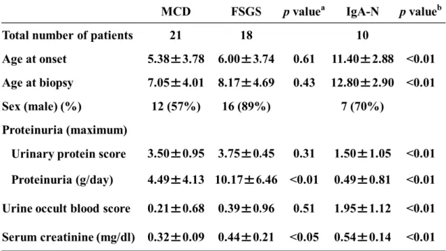
Table 1: Clinical characteristics of patients in this study.
404
405
The values represent the mean ±SD.
406
a, b
: Two-sample t-test.
407
a
: p value compared the values in the MCD group with those in the FSGS group.
408
b
: p value compared the values in the MCD group with those in the IgA-N group.
409
410
411
412
413
414
415
416
417
418
15 419
420 421 422 423 424 425
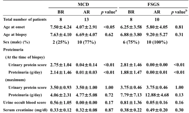
Table 2: Comparison of clinical characteristics between the before and after remission 426
cases in patients with MCD and FSGS.
427
428
BR: before remission, AR: after remission. The values represent the mean ±SD.
429
a, b
: Two-sample t-test.
430
a
: p value compared the values in the BR group with those in the AR group of MCD.
431
b