Fukushima Medical University
福島県立医科大学 学術機関リポジトリ
This document is downloaded at: 2021-11-07T23:37:09Z
Title DPP4 inhibition ameliorates cardiac function by blocking the cleavage of HMGB1 in diabetic mice after myocardial infarction( 本文 )
Author(s) 佐藤, 彰彦
Citation
Issue Date 2017-03-24
URL http://ir.fmu.ac.jp/dspace/handle/123456789/950
Rights © 2017 by the International Heart Journal Association. Int Heart J. 2017 Oct 21;58(5):778-786. doi: 10.1536/ihj.16-547
DOI
Text Version ETD
DPP4 Inhibition Ameliorates Cardiac Function by Blocking the Cleavage of HMGB1 in Diabetic Mice After Myocardial Infarction
Akihiko Sato,1 MD, Satoshi Suzuki,1MD, Shunsuke Watanabe,1 MD, Takeshi Shimizu,1 MD, Yuichi Nakamura,1MD, Tomofumi Misaka,1 MD, Tetsuro Yokokawa,1 MD, Tetsuro Shishido,2MD,
Shu-ichi Saitoh,1MD, Takafumi Ishida,1 MD, Isao Kubota,2MD and Yasuchika Takeishi,1MD
Summary
High mobility group box 1 (HMGB1), a ubiquitous DNA-binding protein, promotes angiogenesis and tis- sue repair, resulting in restored cardiac function after myocardial infarction (MI). Although dipeptidyl peptidase 4 (DPP4) degrades certain peptides, it remains unclear as to whether HMGB1 is a substrate of DPP4 and whether DPP4 inhibition prevents the cleavage of HMGB1.
In transgenic mice with cardiac-specific overexpression of HMGB1 (TG) and wild-type mice (WT), a dia- betic state was induced by streptozotocin, and MI was created by ligation of the left anterior descending coro- nary artery. To inhibit DPP4 activity, a DPP4 inhibitor anagliptin was used. The plasma levels of HMGB1, in- farct size, echocardiographic data, angiogenesis, and vascular endothelial growth factor (VEGF) expression in the peri-infarct area were compared among non-diabetic MI WT/TG, diabetic MI WT/TG, and anagliptin-treated diabetic MI WT/TG mice.
DPP4 activity was increased in the diabetic state and blocked by anagliptin administration. The HMGB1 plasma levels were reduced in the diabetic TG compared with the non-diabetic TG mice, but DPP4 inhibition with anagliptin increased HMGB1 plasma levels in the diabetic TG mice. The infarct area was significantly larger in the diabetic TG than in the non-diabetic TG mice, and it was reduced by DPP4 inhibition. Cardiac function, angiogenesis, and VEGF expression were impaired in the diabetic TG mice, but they were ameliorated by the DPP4 inhibition to levels similar to those found in the non-diabetic TG mice.
The DPP4 inhibitor ameliorated cardiac function by inhibiting the inactivation of HMGB1 in diabetic mice after MI.
(Int Heart J 2017; 58: 778-786) Key words:Angiogenesis, Ischemic heart disease, Diabetes mellitus
A
cute myocardial infarction (MI) is an emergent disease caused by occlusion of the coronary ar- tery. The morbidity and mortality of MI remain high despite the advancement of emergency medical serv- ices and the development of optimal therapies, including pharmacological treatment and percutaneous coronary in- tervention, and surgical technique.1) Infarct size is known as an important prognostic predictor; thus, many studies of ischemic heart disease have been performed to deter- mine how to limit the extent of infarction, focusing on the mechanisms of collateral development, ischemic reperfu- sion injury and ischemic preconditioning, among others.2-4) High mobility group box 1 (HMGB1) is a ubiquitous non-histone DNA-binding protein that mediates gene tran- scription.5-7) HMGB1 secreted from necrotic cells into the extracellular space triggers tissue repair as a cytokine.8-11) Moreover, several studies have proven that HMGB1 is in- volved in limiting the size of the infarct area and preserv-ing cardiac function by promoting angiogenesis.12,13)In ad- dition, we have previously demonstrated that HMGB1 promotes angiogenesis and tissue repair by enhancing mo- bilization and differentiation of endothelial progenitor cells from bone marrow, resulting in preserving cardiac function after MI.14)These data suggest that HMGB1 is a possible cardio-protective protein.
Diabetes mellitus (DM) is a major coronary risk fac- tor, and MI patients with DM are likely to have a poor prognosis compared with those without DM.15) Over the past few years, many researchers have shown an interest in the pleiotropic effect of the inhibition of dipeptidyl peptidase 4 (DPP4), whose activity is upregulated in a diabetic state.16,17) A DPP4 inhibitor has an anti-diabetic effect by suppressing the degradation of incretin hormones such as glucagon-like peptide-1 (GLP-1) and gastrointesti- nal peptide (GIP).18) It has recently been reported that DPP4 degrades not only incretin hormones but also some
From the 1Department of Cardiovascular Medicine, Fukushima Medical University, Fukushima and 2First Department of Internal Medicine, Yamagata University School of Medicine, Yamagata, Japan.
Address for correspondence: Satoshi Suzuki, MD, Department of Cardiovascular Medicine, Fukushima Medical University, 1 Hikarigaoka, Fukushima 960- 1295, Japan. E-mail: [email protected]
Received for publication October 30, 2016. Revised and accepted December 2, 2016.
Released in advance online on J-STAGE September 30, 2017.
doi: 10.1536/ihj.16-547
All rights reserved by the International Heart Journal Association.
778
Int Heart J
September 2017 CARDIOPROTECTION OF DPP4 INHIBITOR VIA HMGB1 779
Figure 1. The schemas of the experimental protocols for each of the six groups used in this study. The mice were divided into six groups (WT, WT-DM, WT-DM-DPP4I, TG, TG-DM, and TG-DM-DPP4I) and experimental myo- cardial infarction (MI) was induced in all groups. Anagliptin administration was started at five days before induction of experimental MI and then continued through the experimental periods in the WT-DM-DPP4I and TG-DM-DPP4I groups.
WT TG
WT-DM TG-DM
WT-DM-DPP4I TG-DM-DPP4I
Sodium citrate buffer
STZ (50 mg/kg/day)
(experimental days) Anagliptin (300 mg/kg/day)
1 5 19 24
Induction of MI
28 52
Echocardiography
1 5 19 24 28 52
1 5 19 24 28 52
STZ (50 mg/kg/day) Control MI
Diabetic MI
Diabetic MI with DPP4I
sacrifice sacrifice
peptides with a role in the regulation of the cardiovascular system, such as stromal-cell-derived factor-1 (SDF-1), B- type natriuretic peptide (BNP) and substance P, as well as other peptides.19)Although some reports have claimed that HMGB1 can also be degraded by DPP4,20) it still remains unclear as to whether DPP4 cleaves HMGB1in vivo.
In the present study, we tested our hypothesis that the inhibition of DPP4 restores HMGB-1-induced angio- genesis and tissue repair in diabetic mice after MI.
Methods
Animals and ethical statements: Transgenic male mice with cardiac-specific HMGB1 overexpression (TG mice) on a BDF-1 background12) and wild-type littermate male mice (WT mice) (10-12 weeks of age) were used for the experiments. All mice genotypes were confirmed by PCR analyses of tail DNA before the experiments. The mice were housed under pathogen-free conditions in isolated cages on a 12-hour light/dark cycle. They could freely ac- cess standard rodent food and water.
The investigations conformed to the Guide for the Care and Use of Laboratory Animals, published by the US National Institutes of Health (NIH publication, 8th Edition, 2011). The Fukushima Medical Research Com- mittee approved our research protocol. All animal experi- ments were performed in accordance with the guidelines of the Fukushima Medical University Animal Research Committee, with efforts made to minimize the suffering of the animals.
Experimental protocol: The experimental protocol is shown in Figure 1. We divided the mice into six groups;
control MI groups (WT and TG), diabetic MI groups
(WT-DM and TG-DM), and diabetic MI treated with DPP 4 inhibitor groups (WT-DM-DPP4I and TG-DM-DPP4I).
To induce a diabetic state, streptozotocin (STZ, 50 mg/kg/
day, Sigma, St. Louis, MO, USA), which was dissolved in 0.1 M sodium citrate buffer (pH 4.5) as described in pre- vious reports,21) was administered for five consecutive days by intraperitoneal injection. Sodium citrate buffer without STZ was administered to control MI group for five consecutive days by intraperitoneal injection. Two weeks after injection of STZ, whole blood glucose levels were measured by a glucometer (GLUCOCARD , ARKRAY Inc., Kyoto, Japan). A diabetic state was de- fined as when blood glucose levels were elevated to more than 16 mmol/L (288 mg/dL).21) Experimental MI was in- duced as previously described12,14)in all groups at day 24.
Briefly, the mice under general anesthesia by intraperito- neal injection of tribromoethanol (0.25 mg/g of body weight) were intubated with a 20-gauge polyethylene catheter and ventilated using a rodent ventilator (Shinano Manufacturing, Tokyo, Japan). A chest incision was per- formed at the level of the fourth rib and along the left sternal border, and the left anterior descending coronary artery (LAD) was ligated with 8-0 prolene sutures. Induc- tion of MI was considered successful when the color of the anterior wall, including the apical portion, turned pale.
After LAD ligation, the chest wall was closed using 6-0 nylon sutures. Western blotting was performed on the six groups and two sham groups (WT-sham and TG-sham).
The sham operation procedure was the same as induction of MI except for LAD ligation. In the WT-DM-DPP4I and TG-DM-DPP4I groups, administration of a DPP4 inhibi- tor, anagliptin (supplied by SANWA KAGAKU KENKY- USHO CO. LTD, Aichi, Japan), via the oral route was
started at five days before MI induction, and was then continued throughout the experimental periods. Anagliptin (300 mg/kg/day) was dissolved in drinking water. The dose of anagliptin was determined from previous re- ports.22-24) In order to determine whether the dose of ana- gliptin (300 mg/kg/day) was adequate to inhibit DPP4 ac- tivity, DPP4 activity was measured as described in the next section, and we found DPP4 activity in this study was inhibited in nearly equal level compared with those of previous experiments (80-90% inhibition of DPP4 activ- ity).22,25,26) Echocardiography was performed in order to evaluate the cardiac function of the mice at day 52 (four weeks after MI induction). To obtain mice heart tissue samples, sacrifice was performed at one week and four weeks after induction of MI in each group.
Fluorometric assay for measurement of DPP4 activity:
Plasma DPP4 activities at baseline, two weeks after STZ injection, five days after starting administration of ana- gliptin, and four weeks after MI operation, were measured in whole blood using a Fluorometric assay kit based on fluorescent substrates (BioVision Inc, Milpitas, CA, USA) according to the lab manual.
Enzyme-linked immunosorbent assay (ELISA): Plasma levels of HMGB1 at four days after induction of MI in each group were measured using an ELISA kit (Shino Test Corporation, Tokyo, Japan), according to the lab manual.12,14)
Echocardiographic measurements: Transthoracic echo- cardiography was performed under light inhaled anesthe- sia with isoflurane in each group at four weeks after in- duction of MI using a Vevo 2100 High-Resolution In Vivo Imaging System (Visual Sonics Inc., Toronto, Canada) with a high-resolution 40-MHz imaging transducer.27)With the use of the M-mode images, interventricular septal thickness, posterior wall thickness, left ventricular end- diastolic dimension (LVDd), and left ventricular end- systolic dimension (LVDs) were measured. The percentage of left ventricular fractional shortening (FS) was calcu- lated as 100×((LVDd-LVDs)/LVDd).
Histological examinations: To observe morphological changes of heart tissue, mice from all groups were sacri- ficed by cervical dislocation at four weeks after induction of MI. The heart was excised and weighed after flushing with 1 × PBS buffer. The paraffin-embedded heart tissues of each group were prepared and sliced serially at the papillary muscle level. The obtained slices were then stained with hematoxylin-eosin and Masson’s trichrome stains. The infarct size was expressed as the ratio of the infarct length to the total left ventricular length calculated by averaging the endo- and epi-cardial perimeters.12,14)The above analyses of the infarct size were performed using Image J software (NIH, Bethesda, MD, USA).
On immunohistochemical analysis, the paraffin sec- tions of the heart tissue at four weeks after MI were stained by anti-platelet endothelial cell adhesion molecule (PECAM-1) antibodies (Santa Cruz, CA, USA) to identify endothelial cells and by anti-α smooth muscle actin (αSMA) antibodies (Santa Cruz, CA, USA) to identify αSMA positive cells. The paraffin sections of the heart tissue were stained with horseradish peroxidase- conjugated secondary antibodies (Histofine Simple Stain
Mouse MAX PO, Nichirei Bioscience Inc., Tokyo, Japan) and diaminobenzidine tetrahydrochloride, then counter- stained with hematoxylin. To assess the number of PECAM-1- and αSMA-positive cells as indicators of an- giogenesis, digital photomicrographs of the border zone (1-2 mm from the edge of the infarct area) were obtained, and the numbers of PECAM-1- andαSMA-positive cells in the border zone were counted in a randomly selected high-power field (HPF, × 400) by two independent re- searchers. The counts were repeated ten times, and the number of PECAM-1- andαSMA-positive cells were ob- tained by calculating the average of the ten measure- ments.12,14)
Western blotting:The heart tissue samples were obtained from each group at four days after induction of MI. The total protein was extracted from the snap-frozen left ven- tricle using Cell Lysis Buffer (Cell Signaling Technology, Beverly, MA) with Protease Inhibitor Cocktail (BD Bi- osciences, San Jose, CA, USA).27) The protein concentra- tion of the myocardial sample was determined by protein assay (DC protein assay kit, Bio-Rad Laboratories, Hercu- les, CA). Equal amounts (20 μg) of the protein samples were subjected to electrophoresis onto sodium dodecyl sulfate polyacrylamide gel electrophoresis (SDS-PAGE, 10%) and transferred onto polyvinylidene difluoride mem- branes (ATTO Co., Tokyo, Japan). The signals from the immunoreactive bands were visualized by an Amersham ECL system (Amersham Pharmacia Biotech UK Ltd., Buckinghamshire, UK) and quantified using Image J Soft- ware (NIH). The antibodies used in the present study were rabbit polyclonal anti-vascular endothelial growth factor (VEGF) antibody (used at dilution of 1:1000, Santa Cruz) and mouse polyclonal anti-βactin antibody (1:5000, Santa Cruz). VEGF expression was normalized byβactin.
Statistical analysis: All data were presented as mean±
standard error. Statistical significance was evaluated using one-way analysis of variance for comparisons among the six groups, followed by multiple comparisons with the Bonferroni post hoc test. AP < 0.05 was considered sta- tistically significant. All analyses were performed using a statistical software package (SPSS ver. 22.0, IBM, Ar- monk, NY, USA).
Results
Measurements of DPP4 enzyme activity: We first con- firmed whether the DPP4 activity was suppressed by oral administration of anagliptin (300 mg/kg/day). Whole blood samples were obtained from the WT and TG mice at baseline, two weeks after induction of STZ-induced DM, five days after administration of anagliptin (before induction of MI), and four weeks after induction of MI (n
= 5-8). As shown in Figure 2, DPP4 enzyme activity was increased in the diabetic state and was decreased by ad- ministration of anagliptin in both the WT and TG mice, in a similar pattern. These results showed that a dose of 300 mg/kg/day of anagliptin adequately inhibited DPP4 activ- ity in both the WT and TG mice.
Measurements of plasma HMGB1 level:We next exam- ined the plasma levels of HMGB1 using the ELISA kit in order to verify our hypothesis that HMGB1 is one of the
Int Heart J
September 2017 CARDIOPROTECTION OF DPP4 INHIBITOR VIA HMGB1 781
Figure 2. Plasma DPP4 enzyme activity of WT and HMGB-1-TG mice. Representative evalua- tions of DPP4 enzyme activity at baseline, two weeks after induction of STZ-induced DM, five days after administration of anagliptin, and four weeks after induction of MI in WT and TG mice. The number of mice in each group was 5-8. *P < 0.05 versus baseline, #P < 0.05 versus two weeks after STZ injection in the same group.
2 6
4 12
10
8
0
DPP4 enzyme activity (ȝM/min)
Baseline (day 1)
2 weeks after STZ injection (day 19)
5 days after
administration of anagliptin (day 24)
4 weeks after induction of MI (day 52)
*
* *
TG WT
# #
Figure 3. Plasma levels of HMGB1 at four days after induction of MI. The number of mice in each group was 8-10. *P < 0.05 versus WT group, #P < 0.05 versus TG group, †P < 0.05 versus TG-DM group.
5 15
10 25
20
0
Plasma levels of HMGB1 (ng/ml)
WT-DM TG-DM
WT WT-DM-DPP4I TG TG-DM-DPP4I
*
#
†
potential substrates degraded by DPP4 enzyme, and that inhibition of DPP4 activity increases the plasma levels of HMGB1, resulting in cardio-protective effects in the dia- betic state (Figure 3). At four days after induction of MI, HMGB1 plasma levels were significantly higher in the TG group than in the WT group (15.7 ± 3.8 ng/mL versus 8.4
± 1.6 ng/mL,P < 0.05). The TG-DM group showed lower plasma HMGB1 levels (5.5 ± 1.4 ng/mL) than the TG group (P < 0.05), and this decline was restored by inhibi- tion of DPP4 activity (10.1 ± 2.2 ng/mL) (P < 0.05). No significant differences were observed in the plasma levels of HMGB1 among the WT, WT-DM, and WT-DM-DPP4I groups.
Gravimetric and echocardiographic data at four weeks after MI induction: The gravimetric and echocar-
diographic data at four weeks after induction of MI among the six groups are shown in the Table. Blood sugar (BS) was significantly higher in the DM and DM-DPP4I groups than in the non-DM groups (P< 0.01) in both the WT and TG mice. We used a type 1 DM model induced by STZ in the present study, so BS was not significantly decreased by administration of the DPP4 inhibitor. Body weight tended to be decreased by the induction of diabe- tes, and did not significantly change with administration of the DPP4 inhibitor. The ratio of heart weight to body weight did not show any significant differences among the six groups. The echocardiographic data showed that FS was significantly higher in the TG group than in the WT group (15.5 ± 0.84% versus 11.3 ± 0.91%) and deterio- rated with the diabetic state (11.9 ± 0.64%, P < 0.05);
Figure 4. The size of the infarct area at four weeks after induction of MI. Representative photomicrographs show myocardial cross sections stained with Masson’s trichrome. The number of mice in each group was 8-10. The scale bar shows 2 mm. *P < 0.05 versus WT group, #P < 0.05 versus TG group, †P < 0.05 versus TG-DM group.
10 60
30 50
20 40 70
0
Infarct size (%)
WT-DM TG-DM
WT WT-DM-DPP4I TG TG-DM-DPP4I
*
#
†
WT WT-DM WT-DM-DPP4I TG TG-DM TG-DM-DPP4I
Table. Comparison of Baseline Characteristics and Echocardiographic Data in Each Group at Four Weeks After MI
Parameter WT WT-DM WT-DM-DPP4I TG TG-DM TG-DM-DPP4I
Baseline characteristics
BW (g) 27.3 ± 0.71 25.2 ± 1.59 27.1 ± 0.85 27.1 ± 0.76 25.3 ± 1.46 26.7 ± 0.81 BS (mg/dL) 150.2 ± 8.6 420 ± 17.7** 402.6 ± 24.9** 143.9 ± 9.53 418.6 ± 27.8## 389.7 ± 24.3##
HW/BW (mg/g) 8.08 ± 0.24 7.99 ± 0.23 7.94 ± 0.34 7.20 ± 0.18 8.29 ± 0.36 7.35 ± 0.33 Echocardiography
IVST (mm) 0.30 ± 0.04 0.32 ± 0.02 0.33 ± 0.02 0.34 ± 0.04 0.33 ± 0.02 0.33 ± 0.03 PWT (mm) 0.82 ± 0.04 0.76 ± 0.03 0.78 ± 0.05 0.78 ± 0.05 0.85 ± 0.05 0.83 ± 0.05 LVDd (mm) 5.87 ± 0.02 5.88 ± 0.07 5.85 ± 0.08 5.66 ± 0.15 5.90 ± 0.09 5.66 ± 0.04 LVDs (mm) 5.2 ± 0.17 5.1 ± 0.23 5.1 ± 0.15 4.90 ± 0.18 5.00 ± 0.19 4.78 ± 0.07 FS (%) 11.3 ± 0.91 10.5 ± 0.5 11.1 ± 1.24 15.5 ± 0.84* 11.9 ± 0.64# 14.2 ± 0.44† BW indicates body weight; BS, blood sugar; HW, heart weight; IVST, interventricular septal thickness; PWT, posterior wall thick- ness; LVDd, left ventricular end-diastolic dimension; LVDs, left ventricular end-systolic dimension; and FS, fractional shortening.
Data are presented as mean ± SE. The number of mice in each group was 8-10. *P < 0.05 versus WT group, **P < 0.01 versus WT group, #P < 0.05 versus TG group, ##P < 0.01 versus TG group, †P < 0.05 versus TG-DM group.
however, it was ameliorated by the inhibition of DPP4 ac- tivity (14.2 ± 0.44%,P< 0.05).
Assessment of infarct size: We then investigated the im- pact of DPP4 inhibition on the infarct area at four weeks after induction of MI (Figure 4). The size of the infarct area was significantly smaller in the TG mice than in the WT mice (44.8 ± 2.4% versus 55.2 ± 2.6%,P < 0.05) as previously reported.12) The TG-DM group had a signifi- cantly broader infarct area compared with the TG group (56.3 ± 2.4%) (P < 0.05). The size of the infarct area was reduced by the inhibition of DPP4 enzyme activity (41.8
± 3.4%) (P < 0.05). No significant differences were ob- served among the WT, WT-DM, and WT-DM-DPP4I groups.
Evaluation of capillary and arteriole density in the MI border zone: We evaluated the number of PECAM-1-
andαSMA-positive cells in the MI border zone as indica- tors of angiogenesis (Figures 5, 6). As shown in Figure 5, capillary density, defined by the number of PECAM-1- positive cells in the MI border zone, was higher in the TG group than in the WT group (54.4 ± 2.3 versus 38.2 ± 2.1/HPF,P < 0.05). In the TG mice, the capillary density in the MI border zone was significantly reduced by induc- tion of DM (40.9 ± 3.2/HPF,P < 0.05); however, this re- duction was restored to levels nearly equal to those of the TG group by administration of the DPP4 inhibitor (53.1 ± 3.8/HPF). There were no significant differences in capil- lary density among the WT, WT-DM, and WT-DM-DPP4I groups (Figure 5).
Arteriole density was assessed by the number of αSMA-positive cells, which were smooth muscle cells of the arterioles in the MI border zone. Arteriole density had
Int Heart J
September 2017 CARDIOPROTECTION OF DPP4 INHIBITOR VIA HMGB1 783
Figure 5. Capillary density in the border zone at four weeks after induction of MI. Representative photo- micrographs show myocardial tissue with immunohistochemical staining with anti-PECAM-1 antibody.
PECAM-1-positive cells indicate microvascular endothelium. The number of mice in each group was 7-9.
The scale bar shows 50 μm. *P < 0.05 versus WT group, #P < 0.05 versus TG group, †P < 0.05 versus TG- DM group.
10 30
20 60
50
40
0
PECAM-1 positive cells / HPF
WT-DM TG-DM
WT WT-DM-DPP4I TG TG-DM-DPP4I
*
#
†
WT WT-DM WT-DM-DPP4I TG TG-DM TG-DM-DPP4I
Figure 6. Arteriole density in the border zone at four weeks after induction of MI. Representative photo- micrograph shows myocardial tissue with immunohistochemical staining with anti-α SMA antibody. The number of mice in each group was 6-9. The scale bar shows 50 μm. *P < 0.05 versus WT group, #P < 0.05 versus TG group.
Į SMA-positive cells / HPF
WT-DM TG-DM
WT WT-DM-DPP4I TG TG-DM-DPP4I
2 6
4 12
10
8
0
#
WT WT-DM WT-DM-DPP4I TG TG-DM TG-DM-DPP4I
*
similar tendencies to capillary density, as both were re- duced by DM and restored by DPP4 inhibition. Briefly, arteriole density was reduced by DM and restored by the DPP4 inhibition (Figure 6).
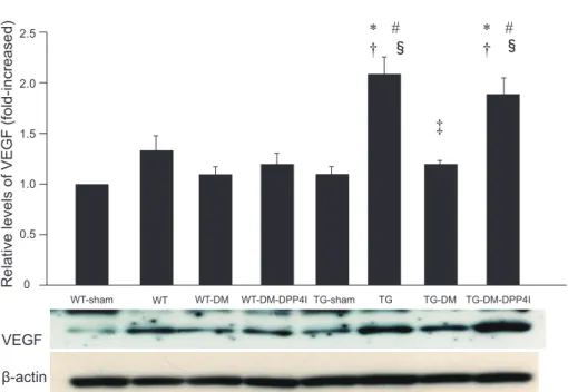
Evaluation of signaling promoting angiogenesis:
HMGB1 is known to promote tissue angiogenesis via VEGF.14,21) To investigate the mechanism of such promo- tion by DPP4 inhibition in diabetic TG mice, we analyzed the expression of VEGF in the MI border zone by West-
ern blotting (Figure 7). VEGF expression was promoted by the induction of MI in both the WT and TG mice in the MI border zone compared with sham-operated mice, although this increase was not statistically significant in the WT mice. The TG mice had significantly higher VEGF expression in the MI border zone than the WT mice (P < 0.05). VEGF expression in the MI border zone was significantly suppressed in the TG mice with diabetes and was ameliorated by inhibition of DPP4 activity (P <
Figure 7. VEGF expression in MI border zone. Representative figure shows Western blotting of VEGF protein in MI border zone at four days after induction of MI. The number of mice in each group was 6. Data are represented as relative values to the WT-sham group. *P < 0.05 versus WT-sham, #P < 0.05 versus WT,
†P < 0.05 versus TG-sham, ‡P < 0.05 versus TG, §P < 0.05 versus TG-DM.
0.5 1.5
1.0 2.5
Relative levels of VEGF (fold-increased) 0
WT-DM TG-DM
WT-sham WT-DM-DPP4I TG-sham TG-DM-DPP4I
* #†
VEGF ȕ-actin
WT TG
2.0
‡
㼲 * #㼲
†
0.05).
Discussion
This study demonstrated that the function of HMGB1 in promoting angiogenesis was reversed in the diabetic state. We additionally showed that DPP4 inhibition sup- pressed the degradation of HMGB1, resulting in the pro- motion of tissue angiogenesis and amelioration of cardiac function after MI. These findings indicate that HMGB1 was possibly one of the substrates of DPP4, and that the inhibition of DPP4 activity led to the preservation of car- diac function after MI via HMGB1.
HMGB1 is a non-histone DNA-binding protein con- sisting of 215 amino acids, and ubiquitously exists in every cell type.5-7,28)Intracellular HMGB1 plays important roles in regulating genome replication, transcription, and DNA repair.29,30) HMGB1 also has a crucial role in sur- vival because HMGB1-gene knockout mice die within the first day of life due to hypoglycemia.31)HMGB1 released from necrotic cells (extracellular HMGB1) acts as a cy- tokine involved in inflammation through the receptor for advanced glycation end products and toll-like receptors (TLR) 2 and 4.32,33) One of the local actions of extracellu- lar HMGB1 is stimulation of tissue repair by recruiting stem cells and promotion of their proliferation.11)With re- gard to the function of HMGB1 in tissue regeneration, Ki- tahara, et al. reported that HMGB1 restores cardiac func- tion through the activation of tissue angiogenesis after MI using male mice with a four-fold expression of HMGB1 in cardiac muscle cells (TG mice).12)Based on Kitahara,et al.’s report, we demonstrated that HMGB1-induced angio- genesis after MI arose from homing and differentiating endothelial progenitor cells from bone marrow, resulting
in promoting angiogenesis at the peri-infarct area.14)These reports showed that HMGB1 was released from necrotic myocardial cells into circulation, and plasma HMGB1 level after MI was much higher in TG mice than in WT mice. With regards to angiogenesis, HMGB1 promotes tis- sue angiogenesis by increasing the production of pro- angiogenic cytokines including VEGF, TNF-α, and IL-8 from endothelial cells and macrophages.34,35)
DPP4, a type of peptidase and one of the therapeutic targets for DM, reportedly degrades and inactivates not only incretin hormones but also other substances, includ- ing SDF-1, BNP, and substance P.19)Because HMGB1 has a component of partial DPP4 cleavage sites,20) it can also be degraded and inactivated by DPP4in vivo. Straino,et al.reported that HMGB1 levels of mice skin tissue were decreased in the diabetic state, and HMGB1 application to skin wounds promoted the tissue angiogenesis and wound healing.36) Furthermore, Marchetti, et al. proved that HMGB-1 was degraded by DPP4 inin vitro experiments and the inhibition of DPP4 enhanced tissue angiogenesis in a murine skin-wound model.20)
In the present study, we firstly demonstrated in vivo that cardio-protective effects, such as promotion of angio- genesis, suppression of cardiac remodeling, and enhance- ment of VEGF expression in the peri-infarct area, were re- versed in diabetic state and ameliorated by administration of DPP4 inhibitor in the TG mice. However, these reac- tions could not be observed in the WT mice. In WT-DM mice, slight decrease of plasma HMGB1 level was ob- served compared to non-DM WT mice, although this was not statistically significant (Figure 3). Additionally, in Fig- ure 6, the number of αSMA positive cells was lower in WT-DM mice than in non-DM WT mice, and that was partially recovered by the DPP4 inhibition, although these
Int Heart J
September 2017 CARDIOPROTECTION OF DPP4 INHIBITOR VIA HMGB1 785
differences were not statistically significant. Taken to- gether, similar trends with TG mice were observed in non- DM WT, WT-DM, and WT-DM-DPP4 inhibition groups, but these levels were low, and thus did not lead to de- crease in infarct size in WT mice. Sauve et al. reported that infarct size in genetic DPP4 deletion mice did not differ with that in WT mice,37)and their data were consis- tent with our present study. In addition, Shigeta, et al. re- ported that VEGF expression in cardiac muscle tissue was suppressed in the diabetic state with inhibition of DPP4 activity in a chronic heart failure model induced by tran- saortic constriction.38) In the current study, VEGF expres- sion was escalated in diabetic HMGB1-TG mice by DPP4 inhibition compared with that in diabetic HMGB-1 TG mice without inhibition of DPP4. This indicates that HMGB1 is a substrate of DPP4 in vivo, and is inactivated under diabetic conditions in which DPP4 activity in- creases, resulting in deterioration of cardiac function after MI in HMGB1-TG mice.
Although the DPP4 inhibition showed favorable ef- fects on heart failure in several basic researches, recent major clinical trials (SAVOR, EXAMINE, TECOS) sug- gested that DPP4 inhibitors were not associated with de- creases of major adverse cardiac events in DM patients with a history of established cardiovascular diseases or multiple cardiovascular risk factors.39-41) In addition, the secondary analysis of SAVOR trial indicated DPP4 inhibi- tor, saxagliptin, increased the hospitalization of worsening heart failure.42) However, these results did not deny the cardio-protective effects of DPP4 inhibition from some reasons. First, these trials were placebo-controlled trials carried out to confirm the safety of DPP4 inhibitors on patients with cardiovascular diseases, not designed to demonstrate beneficial effect of DPP4 inhibitor on heart failure. Moreover, it is also possible that observational pe- riods of these trials were too short to reveal the cardiac benefit of DPP4 inhibitors. Second, DPP4 inhibitors might have class effects on heart failure. Whereas subgroup analysis of SAVOR trial showed unfavorable cardiac effect of DPP4 inhibitor,42)EXAMINE and TECOS showed non- inferiority of DPP4 inhibitors on hospitalization of heart failure.43,44) In addition, subgroup analysis of EXAMINE trial showed DPP4 inhibitors decreased cardiovascular mortality compared with placebo in the highest BNP quar- tiles.43)Therefore, the impact of DPP4 inhibitor on cardio- vascular diseases has remained unclear, and further clini- cal studies should be conducted to clarify the effect of DPP4 inhibitors on cardiovascular diseases and heart fail- ure.
This study includes several limitations. First, since DPP4-inhibitors are generally used for treatment of type 2 DM, further studies are needed to verify cardio-protective effect of the DPP4 inhibition via HMGB1 in type 2 DM model mice. Second, we demonstrated cardio-protective effect of the DPP4 inhibition in only diabetic model in present study. Further study to examine the effect of the DPP4 inhibition in non-DM model is required. Third, the induction of experimental myocardial infarction might have influenced on DPP4 activity. However, our essential purpose of this study was to examine whether the DPP4 inhibition restores HMGB1-induced angiogenesis and tis-
sue repair in diabetic mice after MI. A recent article by Kubota and Takano, et al.45) has demonstrated that the treatment with DPP4 inhibitor increased the ratio of endo- thelial cell numbers to a cardiomyocyte, improved cardiac function and decreased the infarct size in C57BL/6 mice after MI, suggesting a key role of DPP4 activity after MI in non-diabetic condition. Thus, we should consider ef- fects of DPP4 activity on HMGB1 function in non- diabetic condition in the future study.
Conclusion
We demonstrated that the inhibition of DPP4 activity blocked HMGB-1 degradation, resulting in angiogenesis promotion and amelioration of cardiac function after myo- cardial infarction in diabetes.
Acknowledgment
We thank Ms. Tomiko Miura for her excellent techni- cal assistance.
Disclosures
Conflict of interest: This study was partly funded by Sanwa Kagaku Kenkyusho Co.Ltd., Aichi, Japan. The sponsor had no control over the interpretation, writing, or publication of this work.
References
1. Roger VL, Go AS, Lloyd-Jones DM, et al. Heart disease and stroke statistics―2012 update: a report from the American Heart Association. Circulation 2012; 125: e2-e220.
2. Miura T, Miki T. Limitation of myocardial infarct size in the clinical setting: current status and challenges in translating ani- mal experiments into clinical therapy. Basic Res Cardiol 2008;
103: 501-13. (Review)
3. Hausenloy DJ, Yellon DM. Targeting myocardial reperfusion in- jury―The Search Continues. N Engl J Med 2015; 373: 1073-5.
4. Heusch G. Molecular basis of cardioprotection: signal transduc- tion in ischemic pre-, post-, and remote conditioning. Circ Res 2015; 116: 674-99. (Review)
5. Thomas JO, Stott K. H1 and HMGB1: modulators of chromatin structure. Biochem Soc Trans 2012; 40: 341-6. (Review) 6. Aizawa S, Nishino H, Saito K, Kimura K, Shirakawa H, Yoshida
M. Stimulation of transcription in cultured cells by high mobil- ity group protein 1: essential role of the acidic carboxyl- terminal region. Biochemistry 1994; 33: 14690-5.
7. Thomas JO, Travers AA. HMG1 and 2, and related architec- tural’ DNA-binding proteins. Trends Biochem Sci 2001; 26:
167-74. (Review)
8. Yang H, Antoine DJ, Andersson U, Tracey KJ. The many faces of HMGB1: molecular structure-functional activity in inflamma- tion, apoptosis, and chemotaxis. J Leukoc Biol 2013; 93: 865- 73. (Review)
9. Yang H, Wang H, Czura CJ, Tracey KJ. The cytokine activity of HMGB1. J Leukoc Biol 2005; 78: 1-8. (Review)
10. Erlandsson Harris H, Andersson U. Mini-review: The nuclear protein HMGB1 as a proinflammatory mediator. Eur J Immunol 2004; 34: 1503-12. (Review)
11. Naglova H, Bucova M. HMGB1 and its physiological and pathological roles. Bratisl Lek Listy 2012; 113: 163-71. (Re- view)
12. Kitahara T, Takeishi Y, Harada M, et al. High mobility group box 1 restores cardiac function after myocardial infarction in transgenic mice. Cardiovasc Res 2008; 80: 40-6.
13. Kohno T, Anzai T, Naito K,et al. Role of high-mobility group box 1 protein in post-infarction healing process and left ven- tricular remodelling. Cardiovasc Res 2009; 81: 565-73.
14. Nakamura Y, Suzuki S, Shimizu T,et al. High Mobility Group Box 1 Promotes Angiogenesis from Bone Marrow-derived En- dothelial Progenitor Cells after Myocardial Infarction. J Athero- scler Thromb 2015; 22: 570-81.
15. Lamblin N, Fertin M, de Groote P, Bauters C. Cardiac remodel- ing and heart failure after a first anterior myocardial infarction in patients with diabetes mellitus. J Cardiovasc Med (Hager- stown) 2012; 13: 353-9.
16. Zhong J, Maiseyeu A, Davis SN, Rajagopalan S. DPP4 in cardi- ometabolic disease: recent insights from the laboratory and clinical trials of DPP4 inhibition. Circ Res 2015; 116: 1491- 504.
17. Manucci E, Pala L, Ciani S, et al. Hyperglycaemia increases dipeptidyl peptidase IV activity in diabetes mellitus. Diabetolo- gia 2005; 48: 1168-72.
18. Drucker DJ, Nauck MA. The incretin system: glucagon-like peptide-1 receptor agonists and dipeptidyl peptidase 4 inhibitors in type 2 diabetes. Lancet 2006; 368: 1696-705. (Review) 19. Koska J, Sands M, Burciu C, Reaven P. Cardiovascular effects
of dipeptidyl peptidase-4 inhibitors in patients with type 2 dia- betes. Diab Vasc Dis Res 2015; 12: 154-63. (Review)
20. Marchetii C, Di Carlo A, Facchiano F, et al. High mobility group box 1 is a novel substrate of dipeptidyl peptidase-IV. Dia- betologia 2012; 55: 236-44.
21. Biscetti F, Straface G, De Cristofaro R, et al. High-mobility group box-1 protein promotes angiogenesis after peripheral ischemia in diabetic mice through a VEGF-dependent mecha- nism. Diabetes 2010; 59: 1496-505.
22. Hirano T, Yamashita S, Takahashi M, Hashimoto H, Mori Y, Goto M. Anagliptin, a dipeptidyl peptidase-4 inhibitor, de- creases macrophage infiltration and suppresses atherosclerosis in aortic and coronary arteries in cholesterol-fed rabbits. Metabo- lism 2016; 65: 893-903.
23. Nakaya K, Kubota N, Takamoto I,et al. Dipeptidyl peptidase-4 inhibitor anagliptin ameliorates diabetes in mice with haploin- sufficiency of glucokinase on a high-fat diet. Metabolism 2013;
62: 939-51.
24. Ervinna N, Mita T, Yasunari E, et al. Anagliptin, a DPP-4 in- hibitor, suppresses proliferation of vascular smooth muscles and monocyte inflammatory reaction and attenuates atherosclerosis in male apo E-deficient mice. Endocrinology 2013; 154: 1260- 70.
25. Mulvihill EE, Varin EM, Ussher JR,et al. Inhibition of Dipepti- dyl Peptidase-4 Impairs Ventricular Function and Promotes Car- diac Fibrosis in High Fat-Fed Diabetic Mice. Diabetes 2016;
65: 742-54.
26. Lamont BJ, Drucker DJ. Differential antidiabetic efficacy of in- cretin agonists versus DPP-4 inhibition in high fat fed mice.
Diabetes 2008; 57: 190-8.
27. Miyata M, Suzuki S, Misaka T,et al. Senescence marker protein 30 has a cardio-protective role in doxorubicin-induced cardiac dysfunction. PLoS One 2013; 8: e79093.
28. Read CM, Cary PD, Crane-Robinson C, Driscoll PC, Norman
DG. Solution structure of a DNA-binding domain from HMG1.
Nucleic Acid Res 1993; 21: 3427-36.
29. Stros M. HMGB proteins: interactions with DNA and chroma- tin. Biochim Biophys Acta 2010; 1799: 101-13. (Review) 30. Bianchi ME, Beltrame M. Upwardly mobile proteins. Workshop:
the role of HMG proteins in chromatin structure, gene expres- sion and neoplasia. EMBO Rep 2000; 1: 109-14.
31. Calogero S, Grassi F, Aguzzi A,et al. The lack of chromosomal protein Hmg1 does not disrupt cell growth but causes lethal hy- poglycaemia in newborn mice. Nat Genet 1999; 22: 276-80.
32. Ellerman JE, Brown CK, de Vera M,et al. Masquerader: high mobility group box-1 and cancer. Clin Cancer Res 2007; 13:
2836-48. (Review)
33. Lotze MT, Tracey KJ. High-mobility group box 1 protein (HMGB1): nuclear weapon in the immune arsenal. Nat Rev Im- munol 2005; 5: 331-42. (Review)
34. van Beijnum JR, Nowak-Sliwinska P, van den Boezem E, Haut- vast P, Buurman WA, Griffioen AW. Tumor angiogenesis is en- forced by autocrine regulation of high-mobility group box 1.
Oncogene 2013; 32: 363-74.
35. van Beijnum JR, Dings RP, van der Linden E, et al. Gene ex- pression of tumor angiogenesis dissected: specific targeting of colon cancer angiogenic vasculature. Blood 2006; 108: 2339-48.
36. Straino S, Di Carlo A, Mangoni A,et al. High-mobility group box 1 protein in human and murine skin: involvement in wound healing. J Invest Dermatol 2008; 128: 1545-53.
37. Sauvé M, Ban K, Momen MA,et al. Genetic Deletion or phar- macological inhibition of dipeptidyl peptidase-4 improves car- diovascular outcomes after myocardial infarction in mice. Dia- betes 2010; 59: 1063-73.
38. Shigeta T, Aoyama M, Bando YK,et al. Dipeptidyl peptidase-4 modulates left ventricular dysfunction in chronic heart failure via angiogenesis-dependent and -independent actions. Circula- tion 2012; 126: 1838-51.
39. Scirica BM, Bhatt DL, Braunwald E,et al. Saxagliptin and car- diovascular outcome in patients with type 2 diabetes mellitus. N Eng J Med 2013; 369: 1317-26.
40. White WB, Cannon CP, Heller SR,et al. Alogliptin after acute coronary syndrome in patients with type 2 diabetes. N Eng J Med 2013; 369: 1327-35.
41. Green JB, Bethel MA, Armstrong PW,et al. Effect of sitagliptin on cardiovascular outcomes in type 2 diabetes. N Eng J Med 2015; 373: 232-42.
42. Scirica BM, Braunwald E, Raz I, et al. Heart failure, saxagliptin, and diabetes mellitus: observations from the SAVOR-TIMI 53 randomized trial. Circulation 2014; 130:
1579-88.
43. Zannad F, Cannon CP, Cushman WC, et al. Heart failure and mortality outcomes in patients with type 2 diabetes taking alo- gliptin versus placebo in EXAMINE: a multicentre, randomised, double-blind trial. Lancet 2015; 385: 2067-76.
44. McGuire DK, Vav de Werf F, Armstrong PW,et al. Association Between Sitagliptin Use and Heart Failure Hospitalization and Related Outcomes in Type 2 Diabetes Mellitus: Secondary Analysis of a Randomized Clinical Trial. JAMA Cardiol 2016;
1: 126-35.
45. Kubota A, Takano H, Wang H,et al. DPP-4 inhibition has bene- ficial effects on the heart after myocardial infarction. J Moll Cell Cardiol 2016; 91: 72-80.