Evolution of multigene families for lignin degradation in Pholiota microspora
( Pholiota microspora࡛࡛ࡢࣜࢢࢽࣥศゎࡢࡓࡵࡢከ㔜㑇ఏᏊࣇ࣑࣮ࣜࡢ㐍 )
SURASIT SUTTHIKHAMPA 2016
Contents
Chapter 1 General introduction ……… 1
Chapter 2 Only one major manganese peroxidase (MnP) is predominantly expressed for mycelial growth of Pholiota microspora on sawdust medium ………..10
Chapter 3 Relationship between fruiting body development and phenol oxidase gene expression in Pholiota microspora………... 33
Chapter 4 Conclusions and discussions ……… 54
Abstract ………. 57
Acknowledgements ………..…. 62
Literature cites ………..….63
List of publications ………..…. 75
1
Chapter 1 General introduction
Lignin is a complex aromatic polymers, amorphous and important in the formation of higher plant cell walls. It enables trees to grow taller and compete for sunshine. Wood and other vascular tissues generally are 20-30% lignin and forming a matrix that surrounds the orderly cellulose microfibrils. Lignin physically protects most of the world’s cellulose and hemicelluloses from enzymatic hydrolysis. In which fungi play the major role in lignin biodegradation (Kirk and Farrell, 1987).
According to differences in chemical and structural change in the plant cell wall during decomposition of wood by fungi. There were three distinguished types of lignin decay; white rot, brown rot and soft rot decay. Typically, white rot fungi simultaneously degrade the three major components of plant cell walls: lignin, hemicellulose and cellulose (Kirk and Highley, 1973). Their ability to metabolize large amounts of lignin is unique among microorganisms. Degradation is usually localized to cells colonized by fungal hyphae and substantial amounts of undecayed wood remains even after advanced decay has occurred. While brown rot and soft rot decay preferentially remove cell wall carbohydrate over lignin. (Kuhar et al., 2007).
The white rot fungi capable of extensive aerobic lignin biodegradation. This property is based on the white rot fungi capability to produce one or more extracellular lignin modifying enzyme (LME). The white rot fungi produce
2
extracellular enzymes that break down the woody cell wall. The lignin modifying enzymes, such as manganese peroxidase (MnP), lignin peroxidase (LiP) and laccase (Lcc) are directly involved in the degradation of lignin in their natural lignocellulosic substrates (Wesenberg et al., 2003). Some white rot fungi produce all three LMEs while others produce only one or two of them (Hatakka, 1994).
The wood-rotting basidiomycete Pholiota microspora (also known as P.
nameko or “nameko,” the common name in Japanese) (Neda, 2008). Nameko mushroom is one of the most popular edible mushroom and is widely cultivated in Japan. It is sold in local markets for food ingredient in Japan and is widely used as material in fungal research into aspects of sexual reproduction structure and mechanism (Babasaki et al., 2003; Aimi et al., 2005; Yi et al., 2010), DNA-mediated transformation system (Yi et al., 2009), tyrosinase protein structure and function (Kawamura-Konishi et al., 2007; 2011; Moe et al., 2015), inorganic phosphate deficiency (Joh et al., 2001; Tasaki et al., 2002; 2004; 2006), effect of polysaccharide (PNPS-1) of anti-flammatory, anti-tumor and anti-aging (Li et al., 2008; 2010; 2012;
2014; Qian et al., 2015; Zhang et al., 2014; Zheng et al., 2014; 2015), hypersensitivity pneumonitis (Nakazawa and Tochigi, 1989; Ishii et al., 1994; Inage et al., 1996) and production of enzyme lignin peroxidase, and manganese peroxidase (Xu et al., 2013).
In order to understand the lignin degradation system in P. microspora, we identify lignin degrading genes that belong to LME. Initially, the deduced amino
3
acid sequence of known MnP, LiP and laccases of different basidiomycetes such as Phanerochaete chrysosporium and Trametes versicolor from public databases were subjected to BLASTp searches of the P. microspora genome. LiP gene was not detected by this inquiry. It suggests that LiP absence in P. microspora, also, Xu et al. (2013) reported that the enzyme activity of LiP was not detected in their experiment. Therefore, the result obtained, P. microspora provides five MnP and nine laccase genes in their genome.
Either MnP or laccase genes were formed a multigene families that might be caused by gene duplication from ancient time. This phenomenon proposes that MnP and laccase genes were redundancy which one or more genes are performing the main function. On the other hand, one or more genes could has little or no effect on the biological phenotype. These suggested that genetic redundancy seems to be wide spread in genomes of higher organisms (Nowak et al., 1997) as well as in basidiomycetous fungi. Moreover, a study into redundancy among nine MnPs in Pleurotus ostreatus found that the mnp3 gene was the most strongly expressed; in an mnp3 knockout mutant, alternative transcription of mnp4 and 9 were observed on the sampling days, and although total MnP activity was reduced in Δmnp3 strains, the presence of minor MnPs was sufficient for maintaining lignin degradation capacity (Salame et al., 2013). Similar phenomenon was also observed in Laccaria bicolor, an ectomycorrhizal basidiomycete. L. bicolor has 11 laccase genes and showed the transcription profile by qRT-PCR of laccase genes in different stages of growth. Lb-
4
lcc3 and Lb-lcc8 were very abundant in ectomycorrhizas, Lb-lcc7 was in fruiting bodies, Lb-lcc9 and Lb-lcc10 were found in free-living mycelium grown on agar medium (Courty et al., 2009). Therefore, the evolution of multigene families among manganese peroxidase and laccase genes for lignin degradation in P. microspora were our major investigation.
1-1 Manganese peroxidase gene families in P. microspora
Manganese peroxidase (MnP), class II peroxidase, is the most common extracellular lignin-modifying peroxidase produced by lignolytic fungi. This heme- containing glycoprotein was first discovered in Phanerochete chrysoporium over 25 years ago and has been studied extensively. Secretory MnP is apparently limited to certain basidiomycetous fungi. Among lignolytic enzymes secreted by white rot fungi, MnP has been reported as the main enzyme involved in lignin depolymerisation (Hofrichter, 2002). Fungal peroxidases have been studied exclusively within the context of wood decomposition and MnP have been suggested to be involved in the degradation of humic compounds. For example, wood decomposers P. chrysoporium, T. versicolor, Phebia radiata and Pleurotus spp.
(Hatakka, 1994; Martinez, 2002).
Class II peroxidases belong to the plant peroxidase superfamily (Welinder, 1992), and are secreted as fungal heme peroxidases, including the lignin-modifying peroxidases lignin peroxidase (LiP), manganese peroxidase (MnP) and versatile
5
peroxidase (VP). There are known to be secreted by several homobasiomycetes (Hatakka, 1994). As mentioned above that LiP absence in P. microspora and VP was a combination between the catalytic properties of both LiPs and MnPs (Hatakka, 1994; Martinez, 2002; Passardi et al., 2007). Then, only MnP presents in P.
microspora, MnP oxidized Mn2+ to Mn3+, which then binds to an appropriate ligand and to polyphenolic substrates, which are oxidized and degraded. Variant MnP genes are reported to cluster in number in various fungi, i.e., there are three mnp genes in P. chrysoporium (Gettemy et al., 1998), two mnp genes in Trametes versicolor (Johansson et al., 2002), two mnp genes in Dichomitus squalens (Périé et al., 1996), nine mnp genes in P. ostreatus (Salame et al., 2013), and two unrelated genetic sequences for mnp-encoding genes in Phlebia radiate(Hildén et al., 2005). However, it is unclear whether all of these genes function in lignin degradation.
Five MnP nucleotide and deduced amino acid sequences in P. microspora will be analysis according to the intron-exon position and phylogenetic analysis. The cluster of P. microspora MnPs will be unveiled an evolutionary relationship within organism and among that of comparative basidiomycetous fungi. The duplication event was appeared in MnP family. Based on the evolutionary complexity and the developmental processes observed in higher organisms. The MnP gene family in P.
chrysoporium is actively function (Gettemy et al., 1998), but it was transcribed and expressed dissimilarly. This phenomenon was extensively observed among the mnp genes in P. ostreatus (Salame et al., 2013). Not only MnPs were unequal
6
transcription and expression, but different in protein sequences also presence in P.
radiata (Pr-mnp2 and Pr-mnp3). Pr-mnp2 and Pr-mnp3 were different in protein sequences but peroxidase function were similar (Hildén et al., 2005).
1-2 Peroxidase gene families
Laccase was first described by Yoshida (1883), and was characterized as a metal containing oxidase that have been reviewed by Mayer and Stables (2002). It is obvious that the laccases are very ancient enzymes from an evolutionary of view.
However, fungal laccase were discussed on the role in lignin degradation (Eggert et al., 1996; Youn et al., 1995). Laccases can degrade lignin in the absence of LiP and MnP. But laccase performed browning process in Lentinula edodes (Sakamoto et al., 2012). Moreover, laccase activity is strongly regulated and promoted fruiting body development in Agaricus bisporus and Schizophyllum commune (Wood, 1980; De Vries et al., 1986).
Laccase is belong to phenol oxidases (PO) that are enzymes containing copper atoms in the catalytic centre and are usually called multicopper oxidases. The catalytic activity of these enzymes is oxidation of diphenols to the corresponding quinones (Baldrian, 2006). Not only laccase is PO, but also includes tyrosinases (Durán et al., 2002). Laccases are multicopper enzymes that use molecular oxygen as a terminal electron acceptor and are capable of oxidizing various aromatic compounds such as substituted monophenols and polyphenols, aromatic amines, and
7
thiol compounds, with subsequent production of radicals (Selinheimo, 2008). Fungi generally produce several laccase isoenzymes encoded by complex multigene families (Giardina et al., 2010). The different transcriptional level can be different regulated mechanism and catalytic properties among isoenzymes. This occurrence suggests that different physiological functions related to fungal life cycle, such as morphogenesis, nutrition, fungal-plant/host interaction, stress defence and lignin degradation (Thurston, 1994). There were nine laccase genes in P. microspora according to BLASTp analysis. These genes families conducted us to analysis the possible physiological roles in native P. microspora cultivation.
Fungal PO, including tyrosinase, can catalyse melanin formation in fruiting bodies as well as browning of the fruiting body that occurs after harvest, as described in L. edodes (Sakamoto et al., 2012), but in A. bisporus, in which tyrosinase is responsible for melanogenesis during spore maturation (Hegnauer et al., 1985).
Intracellular and extracellular PO are produced in plants and fungi for a variety of purposes. PO from basidiomycetes fungi are connected with melanin production, lignin degradation, and morphogenic processes such as fruiting body formation (Sinsabaugh, 2010). Likewise, Schizophyllum commune can secrete high levels of laccase in dikaryotic strains, which are able to form fruiting bodies (De Vries et al., 1986). Furthermore, veratryl alcohol can stimulate laccase production during mycelial growth of P. ostreatus, increasing production of fruiting bodies, with fruiting occurring earlier in the medium containing veratryl alcohol than the medium
8
without (Suguimoto et al., 2001). However, the relationship between the roles of multicopper oxidases and veratryl alcohol in fruiting body development is unclear.
Therefore, these phenomena inducted us to study laccase genes in P. microspora.
1-3 The goal of this study
To estimate physiological roles of MnP and laccase multigene families in P.
microspora which were five and nine genes, respectively. We conducted quantitative RT-PCR (qRT-PCR) technique to study the transcriptional level of genes in P.
microspora. Firstly, to understand differential gene expression in different stages during mushroom development. Total RNA were extracted and quantified from mycelia grown in sawdust substrate, primordia and fruiting bodies. Secondly, to understand the substrate specificity for each gene. P. microspora was grown in M4 liquid medium containing various aromatic compounds, which were lignin related compounds. Total RNA were extracted and quantified from mycelia to investigate transcriptional level of MnP, laccase and related genes. Thirdly, nucleotide and deduced amino acid sequences were analysis. Number and position of introns were inspected. Phylogenetic trees were generated and analyzed of evolutionary relationship across basidiomycetous fungi.
Therefore, in Chapter 2, we will understand the possible physiological roles of MnPs in lignin degradation, since MnPs is a major lignolytic peroxidase in P.
microspora. Moreover, the possible roles of laccase among lignin degradation,
9
pigment synthesis and promote fruiting body development, will be explored in Chapter 3. Finally, the evolution of multigene families among MnPs and laccases for lignin degradation in P. micrpospora will be totally concluded and discussed in Chapter 4.
10
Chapter 2
Only one major manganese peroxidase (MnP) is predominantly expressed for mycelial growth of Pholiota microspora on sawdust medium
2-1 Abstract
In this study, we identified five manganese peroxidase genes (PnMnPs) in the Pholiota microspora haploid genome. Their amino acid sequences showed high similarity and were used to construct a phylogenetic tree. PnMnP5, 3, 2 and 4 were clustered tightly, but PnMnP1 was clustered relatively far from PnMnP5. qRT-PCR showed that PnMnP5 was the only MnP gene that was strongly transcribed, showing 15-fold higher expression than other PnMnPs in M4 liquid medium, while transcription of PnMnP5 in sawdust medium was 100 times higher than in M4 liquid medium. These results indicate that PnMnP5 plays a major role in the ligninolytic peroxidase reaction during mycelial growth in P. microspora. Based on a comparison of the position of introns, the phylogenetic relationships among PnMnPs and the predominant expression of PnMnP5, we believe that all PnMnPs are of the same origin and that they were amplified by duplication events in the ancient P.
microspora genome.
11
2-2 Introduction
Class II peroxidases belong to the plant peroxidase superfamily (Welinder, 1992), and are secreted as fungal heme peroxidases, including the lignin-modifying peroxidases lignin peroxidase (LiP), manganese peroxidase (MnP) and versatile peroxidase (VP). They show an exceptionally broad substrate spectrum that includes various organic and inorganic compounds (Hammel and Cullen, 2008). To date, MnPs are the most commonly occurring class II peroxidases and no other microorganisms have been reported to express and secrete MnP or contain mnp genes, which are limited to certain basidiomycetes families (Agaricales, Corticiales, Polyporales, Hymenochatales) (Hofrichter, 2002; Janusz et al., 2013). MnP was first discovered in Phanerochete chrysoporium over 25 years ago and has been studied extensively (Hofrichter, 2002; Martı́nez, 2002). Variant MnP genes are reported to cluster in number in various fungi, i.e., there are three mnp genes in P. chrysoporium (Gettemy et al., 1998), two mnp genes in Trametes versicolor (Johansson et al., 2002), two mnp genes in Dichomitus squalens (Périé et al., 1996), nine mnp genes in Pleurotus ostreatus (Salame et al., 2013), and two unrelated genetic sequences for mnp-encoding genes in Phlebia radiata (Hildén et al., 2005). However, it is unclear whether all of these genes function in lignin degradation.
The cluster of genes belonging to the MnP family appears to be the result of duplication, based on the evolutionary complexity and the developmental processes observed in higher organisms. The MnP gene family in P. chrysoporium is actively
12
functional (Gettemy et al., 1998), but not all of the genes are transcribed and expressed at all times. Differential expression is seen among the mnp genes in P.
ostreatus (Salame et al., 2013) and individual expression levels are affected by culture conditions (Gettemy et al., 1998). There are great dissimilarities in the mnp gene family with regard to exon-intron structure, with Corticiales, Polyporales and Hymenochaetales showing a lower number of introns (from 4 to 7 highly conserved short introns at similar position, except for Pr-mnp3) than Agaricales (up to 15 in P.
ostreatus, at positions corresponding to the sites of introns in LiP genes) (Hofrichter, 2002; Hildén et al., 2005).
In this study, we examined Pholiota microspora (P. nameko or “Nameko”, the common name in Japanese) culture under optimal conditions for fruit body formation, as MnP gene in this fungus has not previously been studied (Xu et al., 2013). P. microspora is traded in local markets for food in Japan and is widely used as a material in fungal research. We identify the five PnMnP genes and discuss the phylogenetic relationships among them, and transcription of PnMnPs in mycelium cultured in liquid medium containing various aromatic compounds and sawdust medium was investigated by qRT-PCR. Levels of transcription and phylogenetic relationships, including intron position, are also discussed.
13
2-3 Materials and Methods
2-3-1 Fungal strains and culture conditions
The monokaryotic strain of P. microspora NGW19-6 (A4, pdx1) with an auxotrophic mutant of pyridoxine, and NGW12-163 (A3, arg4) with an auxotrophic mutant of arginine (Masuda et al., 1995; Yi et al., 2009) were used. The dikaryotic strain was obtained by crossing NGW19-6 and NGW12-163 and referred to as NGW19-6/12-163 was then used for experiments.
I n o r d e r t o a n a l yz e the effects of different aromatic compounds on gene expression, the P. microspora NGW19-6/12-163 strain was grown on M4 agar at 25°C for 1 week, and then 10 mycelial agar blocks (3 × 3 mm) were transferred into 20 mL of M4 broth medium (Johansson et al., 2002) (components are presented in terms of g l-1: Glucose, 2.20 g; diammonium tartrate, 0.92 g; KH2PO4, 1.00 g;
NaH2PO4•2H2O, 0.26 g; MgSO4•7H2O, 0.50 g; Thiamine hydrochloride, 1.00 × 10-
4 g; CaCl2•2H2O 6.60 × 10⁻³g; FeSO4•7H2O, 5.00 × 10⁻³ g; MnSO4•H2O, 3.00 × 10⁻⁴ g; ZnSO4•7H2O, 5.00 × 10⁻⁴ g; CuSO4, 6.40 × 10⁻⁴ g; pH, 5.5; and 15 g agar for s o l i d M 4 me d i u m) in a 100-mL Erlenmeyer flask supplemented with aromatic compounds (final concentration): 0.01% lignosulfonate; 0.05 mM 2,5-xylidine; 3 mM veratryl alcohol; 0.1 mM guaiacol; 1 mM ferulic acid; 1 mM veratric acid; and 0.1 mM O-anisic acid. P. microspora NGW19-6/12-163 was then grown at 25°C for 10 days and the mycelia were harvested by filtration for RNA extraction.
14
The cultivation of fruit bodies of P. microspora was carried out on a sawdust substrate, which was prepared as follows. Beech sawdust was mixed with rice bran at a gravimetric ratio of 5:1 and was adjusted to 65% moisture content using tap water, and this medium was placed into a 100-mL Erlenmeyer flask, followed by autoclaving at 121°C for 60 min. After cooling the media in the air, five mycelial agar blocks (5 × 5 mm) containing NGW19-6/12-163 were inoculated and incubated at 25°C. When the mycelia had colonized the substrate (about 40 days after inoculation), the surface layer was scratched with a spatula, and then 50 mL of sterilized distilled water was poured into the flask. Water was removed after flasks were incubated at 15°C overnight, and then cultivation continued at 15°C until fruiting bodies developed. We defined the day after water removal as day 0. Sample for RNA extraction were taken from triplicate cultures at different stages of the mushroom developmental cycle: 30 days and 90 days.
2-3-2. Genomic DNA and total RNA preparation
Genomic DNA extraction from mycelia grown in the liquid medium was performed according to the method of Dellaporta et al. (1983). Harvested mycelia were frozen in liquid nitrogen, and ground to a fine powder in a mortar and pestle.
RNA was extracted using a MagExtractorTM Kit (Toyobo, Osaka, Japan) according to the manufacturer’s instructions. cDNA was synthesized using total RNA as a template with ReverTra Ace® qPCR RT Master Mix with gDNA Remover kit (Toyobo, Osaka, Japan). PCR was carried out using Takara Ex Taq® polymerase
15
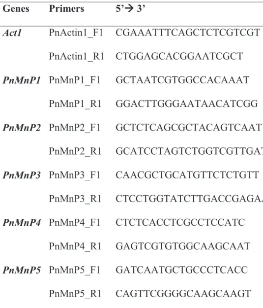
(Takara Bio, Japan). The oligonucleotide primers used in this study were listed in Table 2-1. Amplified fragments were subcloned into pMD20 T-vector (Takara Bio, Japan) and sequenced.
2-3-3. P. microspora genome and retrieved MnPs
Whole genomic sequences of monokaryon P. microspora NGW 19-6 were determined using the Illumina HiSeq 2000 paired-end technology, with the software (CASAVA ver.1.8.1) provided by Hokkaido System Science Co., Ltd. (Sapporo, Hokkaido, Japan), as described by Funo et al. (2014). This sequencing run yielded 30,935,254 high-quality filtered reads with 101-bp paired-end sequencing. The genome was assembled using Velvet assembler (hash length, 85 bp) (Zerbino and Birney, 2008). The final assembly contained 4,770 contigs with a total size 33,400,256 bp, and an N50 length of 72,431 bp. According to Higuchi, (2004), the deduced amino acid sequence of known peroxidase genes from public databases [Lip (Q01787) from Phanerochaete chrysosporium and MnP (Q6KB19) from T.
versicolor]. Five peroxidase genes were assigned as PnMnP1 to 5, and the coding sequences of intron-exon based on GT-AG rules (Breathnach et al., 1978; Wu and Krainer, 1999)) and open reading frames (ORFs) based on generic rules (start codon, ATG and stop codon TAA, TAG, or TGA) (Brown, 2010) were then predicted.
Nucleotide sequences of genomic DNA fragments of P. microspora MnP genes were deposited into the DNA Data Bank of Japan (DDBJ) under the following accession
16
numbers: MnP1 (LC068763); MnP2 (LC068764); MnP3 (LC068765); MnP4 (LC068766); and MnP5 (LC068767).
2-3-4 Quantitative RT-PCR (qRT-PCR) assays
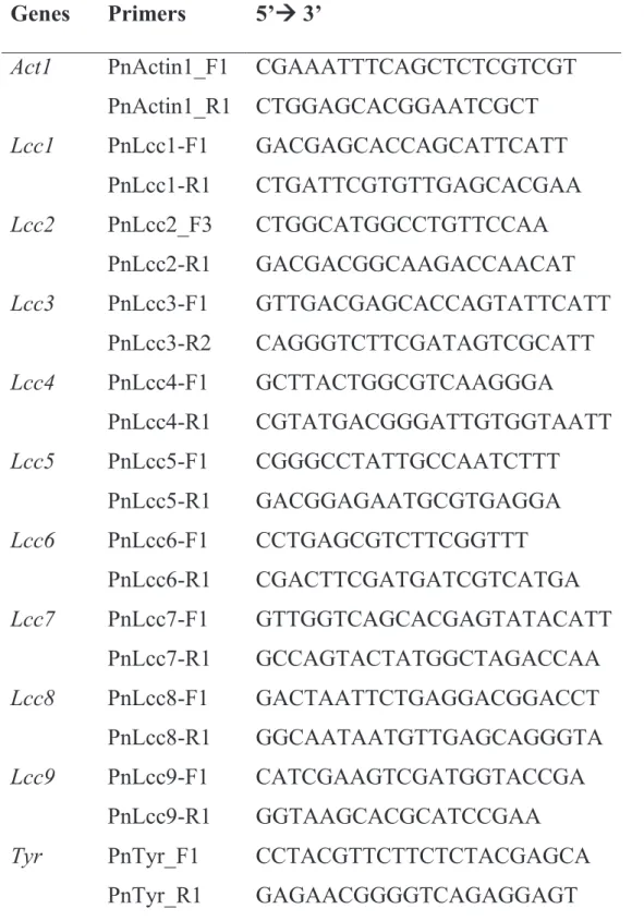
The actin gene (Act1) was used as the reference gene. Primer pairs for amplification of PnMnP1-5 and Act1 cDNAs were designed based on their cDNA sequences using Genetyx software. Amplification of genomic DNA was prevented by designing primers for exon-exon junctions. All primers were tested to ensure that they amplified a single band with no primer-dimers, as shown in Table 2-2. Plasmids inserted into the target gene (PnMnP1-5) and the housekeeping gene (Act1) were extracted as described by Birnboim (1983). Standard curves were constructed using four ten-fold dilutions of plasmid. Real-time PCR was performed using a KOD SYBR® qPCR Mix kit (Toyobo). Thermocycling was carried out using a PikoReal™
96 (Thermo Fisher Scientific) with an initial incubation for 1 min at 95°C, followed by 40 cycles of 95°C for 10 s, 60°C for 1 min. Each run was completed with a melting curve analysis to confirm the specificity of amplification and no primer dimers. Data analysis was performed in accordance with the manufacturer's instructions.
2-3-5 Analysis of sequences and phylogenetic tree
Nucleotide and protein sequences data were analyzed using Genetyx ver.
10.0.3 software (Genetyx, Tokyo, Japan). Protein sequence similarity was analyzed using the BLASTp algorithm (Altschul et al., 1997). Peroxidase genes were retrieved
17
from public domains (NCBI and UniProt). The phylogenetic tree was constructed by MEGA 6.06 software (Tamura et al., 2013) using the minimum evolution method with a bootstrap value of 2,000 replicates. Multiple alignment was performed using ClustalW (Larkin et al., 2007) with the Gonnet distance matrix (Hildén et al., 2005).
2-3-6 Statistical analysis
Mean value and standard deviation of the relative results in each treatments were calculated. Comparisons between control and treatment groups were made using Student’s t-test. Differences were regarded as statistically significant for P values under 5% (P<0.05).
2-4 Results
2-4-1 PnMnP sequence analysis and phylogenetic tree
Despite two kind of peroxidase proteins which were LiP and MnP, were subjected to tBLASTn searches of the P. microspora genome. Same five peroxidase genes were hit in each tBLASTn searches, and they were assigned as PnMnP1 to 5 from the similarity with MnPs from other fungi. Moreover, LiP activity was not detected in previous study in P. microspora (Xu et al., 2013). A phylogenetic tree based on the deduced amino acid sequences of five PnMnPs and other peroxidases from other basidiomycetous mushrooms extracted from the DNA/protein database
18
is shown in Fig. 2-1. There were four major clusters, assigned as clusters A, B, C and D, respectively. All five PnMnPs were clustered in D. Identity and similarity between PnMnP5 with PnMnP1, 2, 3 and 4 was as follows; PnMnP5 showed 83%
identity, 96% similarity to PnMnP3; 78% identity and 94% similarity to PnMnP2;
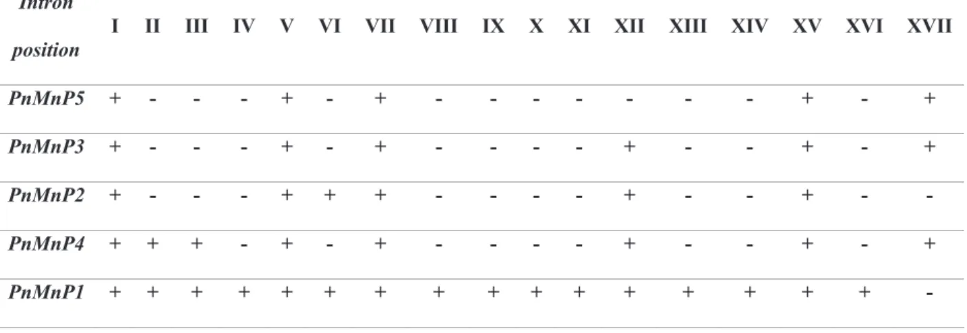
78% identity, 95% similarity to PnMnP4; and 68% identity, 91% similarity to PnMnP1. Therefore, the evolutional distance among PnMnPs (1, 2, 3 and 4) to PnMnP5 was in the rank order of 3, 2, 4 and 1. Moreover, identity and similarity between PnMnP5 with PnMnP1 was lower than for the other PnMnPs (2, 3 and 4), and the number of introns and their positions in PnMnP1 showed greater differences than for other PnMnPs. Therefore, to analyze whether the origin of PnMnP1 differed from that of other PnMnPs in the ancient P. microspora genome, the position and number of introns were analyzed.
Nucleotide sequences of ORFs of PnMnP cDNAs were similar sizes, ranging from 1,049 to 1,074 bp and their genomic DNA sequences contained 5 to 15 introns.
PnMnP cDNAs were coded from 354 to 372 amino acids proteins and their molecular weights were 36.68 to 39.11 kDa. PnMnP5 contains 5 introns, which is the lowest number of introns among PnMnPs, while PnMnP1 contains the highest molecular weight and the exons were interrupted by 15 introns, which is the maximum number among PnMnPs. Introns among PnMnPs were assigned as roman characters from I to XVII (Table 2-3 and Fig. 2-2). During evolution, intron insertion and excision in the genes located on the genome had occurred; therefore,
19
phylogenetic distance should correspond with the abundance and distribution of introns (Lynch, 2002). The inserted positions of introns I, V, VII, XV and XVII were conserved among PnMnPs, thus suggesting that the PnMnPs discovered in this study were of the same origin in the ancient genome of P. microspora. Moreover, PnMnP5 had the smallest number of introns, and is therefore believed to be the oldest ancestral gene. If the original PnMnP is PnMnP5, the hypothetical evolutionary steps would be as follows: i) PnMnP5 was duplicated as PnMnP3, and then intron XII was inserted (intron XII was conserved in PnMnP1, 2, 3 and 4); ii) PnMnP3 was duplicated as PnMnP2, and then intron VI was inserted and intron XVII was removed; iii) PnMnP3 was duplicated as PnMnP4, and introns II and III were inserted; iv) finally, PnMnP2 was duplicated as PnMnP1, and then introns II, III, VIII, IX, X, XI, XIII, XIV and XVI were inserted. This series of events is supported by phylogenetic distance based on amino acid sequences.
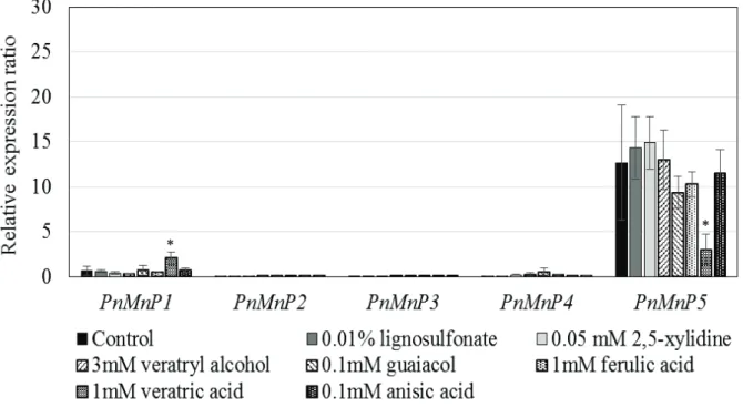
2-4-2 PnMnP5 gene expression in the presence of aromatic compound used for substrate for MnP
In order to investigate differences in the roles of PnMnPs, expression of PnMnPs in mycelia grown in M4 liquid medium supplemented with aromatic compound used for substrate for MnP was studied by quantitative RT-PCR analysis.
PnMnP1, 2, 3 and 4 transcripts were detected at very low levels, and no effect for supplemented aromatic compound was observed (Fig. 2-3). Surprisingly, however,
20
quantity of PnMnP5 mRNA in 10-day cultures with and without aromatic compound used for substrate for MnP were 10- to 15-fold larger than those of the other PnMnPs mRNA. In particular, PnMnP5 was not affected with 0.01% lignosulfonate, 0.05 mM 2,5-xylidine and 3 mM veratryl alcohol. These results suggest that: i) transcription of PnMnP5 is constitutive in M4 liquid medium, but the presence of aromatic compounds did not enhance PnMnP5 transcription; and ii) transcription of PnMnP1, 2, 3 and 4 is very low, suggesting that only PnMnP5 functions in lignin degradation in P. microspora.
2-4-3 PnMnP5 is the major manganese peroxidase in P. microspora
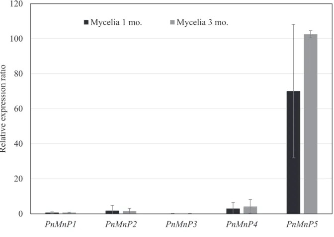
Transcription of PnMnPs in mycelia cultured on sawdust medium was then investigated. Mycelia were harvested from sawdust culture at 1 month after inoculation and at 2 months after kinkaki, which was performed at 1 month after inoculation, and total RNAs were extracted. Figure 2-4 shows expression levels of PnMnPs in the sawdust medium; and 100-fold higher transcription of PnMnP5, as compared with PnMnP1, 2, 3 and 4, was detected in total RNA from mycelia cultured for 1 and 3 months. Transcription of PnMnP5 in sawdust medium was also 100-fold higher than that of PnMnP5 in liquid M4 medium supplemented with aromatic compound used for substrate for MnP. Therefore, transcript of PnMnP5 with aromatic compounds such as veratryl alcohol was much lower than with sawdust. It is possible that aromatic compounds are not suitable for research into
21
lignin degradation by P. microspora, as they do not accurately reflect the response of mycelia to lignin. Moreover, transcription of PnMnP1, 2, 3 and 4 was also low in the sawdust medium. We thus concluded that PnMnP5 plays a major role in lignin degradation by P. microspora during mycelial growth on sawdust substrate.
2-5 Discussion
It has been well documented that nitrogen levels influence MnP production by white rot fungi (Johansson et al., 2002; Hamman et al., 1997). The results of this study show that PnMnP5 transcription in P. microspora is much higher than the other MnP genes presents in the genome. In addition, expression levels of PnMnP5 were not encouraged by various aromatic compound that used for substrate for MnP in modified liquid medium. But, when PnMnP5 transcription in M4 medium was compared with that in sawdust medium, we must consider that M4 liquid basal medium might not be suitable for MnP expression of P. microspora. Because contents of basal medium was not investigate in this study and aeration were much different between liquid culture and solid culture such as sawdust medium (Elisashvili et al., 2008; Camassola et al., 2013). The relatively poor production of the four PnMnPs in our experiments raised the issue of why transcription in the presence of lignin was unequal. In this respect, our results suggest the need for further consideration of assessing intragenic noncoding DNA in the context of evolutionary processes.
22
Our results showed that a well-used aromatic compound, veratryl alcohol, was unable to effectively induce the gene expression of PnMnP5 in P. microspora. This suggests that enhancement of MnP and LiP transcription in response to various aromatic compounds does not occur in white rot fungus as common feature (Cancel et al. 1993). PnMnP5 is expressed natively and is important for the ligninolytic system. A study into redundancy among nine MnPs in P. ostreatus found that the mnp3 gene was the most strongly expressed; in an mnp3 knockout mutant, alternative transcription of mnp4 and 9 were observed on the sampling days, and although total MnP activity was reduced in Δmnp3 strains, the presence of minor MnPs was sufficient for maintaining lignin degradation capacity (Salame et al., 2013). The accumulation of transcripts of mnp1, 2 and 3 genes in mycelia of P. chrysosporium cultured in Mn and nitrogen-limited medium indicated that mnp2 and mnp1 transcriptions are respectively predominant, but mnp3 transcription does not appear to be significantly regulated. These phenomena suggest that MnP genes are differentially regulated at the transcriptional level and respond to dissimilar environmental conditions (Gettemy et al., 1998). Moreover, the white rot basidiomycete Ganodema lucidum is grown and has the highest MnP activity on 10- day culture with poplar wood, but not in cultures grown with pine wood (D’Souza et al., 1999). Similar results have been reported for T. versicolor MnP. In addition, the transcription of P. chrysosporium genes was studied, and an important role for putative metal response elements (MREs) in transcriptional regulation was suggested, but they do not appear to regulate MnP in T. versicolor (Johansson et al.,
23
2002). Based on these phenomena, the differences in lignin degradation activity among white rot fungal species is the result of differential expression and transcription MnPs, not the number of genes in their genome.
With regard to genetic variability of gene families, MnP belongs to the class II peroxidases, which include lignin-modifying peroxidase, as well as LiP and VP.
LiP is catalytically the most powerful class II fungal peroxidase, having the ability to directly oxidize dimeric lignin model and nonphenolic substrates. LiP and MnP have been extensively studied with regard to substrate oxidation capacity, and LiP shares similarities with VP (Hofrichter, 2002). Our results suggest that PnMnP is an important gene for lignin degradation by P. microspora, as the LiP gene is absent from its genome. In addition, the lignin degradation capacity of P. microspora is relativity lower when compared with white rot basidiomyceteous fungi such as P.
chrysosporium, P. radiata and T. versicolor (Hatakka, 1994), which possess the multi-peroxidase gene family. Because of the high homology of amino acid sequences among the five PnMnPs, the genes appear to be part of the same family that was amplified by a duplication event during evolution. Therefore, P. microspora has only one type of enzyme.
Expression levels of PnMnP5 were highest; therefore, we believe it to be the original MnP in the P. microspora genome, as intragenic noncoding DNA interrupting the exons in the five PnMnPs is unequally present. The fewest introns are present in the most actively transcribed PnMnP5 gene. Intron excision and
24
insertion events promote mutation of PnMnPs, while duplication events promote disruption of promoter regions in PnMnPs. This may be why only PnMnP5 is strongly expressed and transcription of other PnMnPs is very low.
In this study, we examine the role of PnMnPs in P. microspora at the transcriptional level, and found that the number of genes is unimportant for lignin degradation. To clarify the relationship between lignin degradation capacity and related multi-gene families, such as laccase genes, in the fungal genome, we are planning further qRT-PCR studies.
25
Table 2-1. Primer sets for cDNA and plasmid construction.
Genes Primers 5’ÆÆ 3’
Act1 PnActin1_F1 CGAAATTTCAGCTCTCGTCGT PnActin1_R1 CTGGAGCACGGAATCGCT PnMnP1 PnMnP1_F1 GCTAATCGTGGCCACAAAT
PnMnP1_R1 GGACTTGGGAATAACATCGG PnMnP2 PnMnP2_F1 GCTCTCAGCGCTACAGTCAAT
PnMnP2_R1 GCATCCTAGTCTGGTCGTTGAT PnMnP3 PnMnP3_F1 CAACGCTGCATGTTCTCTGTT
PnMnP3_R1 CTCCTGGTATCTTGACCGAGAA PnMnP4 PnMnP4_F1 CTCTCACCTCGCCTCCATC
PnMnP4_R1 GAGTCGTGTGGCAAGCAAT PnMnP5 PnMnP5_F1 GATCAATGCTGCCCTCACC
PnMnP5_R1 CAGTTCGGGGCAAGCAAGT
26
Table 2-2. Primer sets for qPCR.
Genes Primers 5’ÆÆ 3’
Act1 PnActin1_CF1 GCTATGCTATGTCGCGCTTGAT PnActin1_R1 CTGGAGCACGGAATCGCT PnMnP1 PnMnP1_CF1 CTTCGCGCTCTCTCAAGACC
PnMnP1_R1 GGACTTGGGAATAACATCGG PnMnP2 PnMnP2_CF1 CTGTTCCCCGGAAATGGATC
PnMnP2_R1 GCATCCTAGTCTGGTCGTTGAT PnMnP3 PnMnP3_CF1 GCGATGGTCAACGACCAA
PnMnP3_R1 CTCCTGGTATCTTGACCGAGAA PnMnP4 PnMnP3_CF1 GGCAAGCCATGATCAACGA
PnMnP4_R1 GAGTCGTGTGGCAAGCAAT PnMnP5 PnMnP5_CF1 GTGGCAAGCTATGGTTAACGA
PnMnP5_R1 CAGTTCGGGGCAAGCAAGT
27
Table 2-3. Intron position according to the highest number of introns (PnMnP1).
Intron position is indicated by roman characters I to XVII from the N-terminal to the C-terminal. (+) indicates introns that are shared, while (-) indicates no intron in that position.
Intron
position I II III IV V VI VII VIII IX X XI XII XIII XIV XV XVI XVII PnMnP5 + - - - + - + - - - - - - - + - + PnMnP3 + - - - + - + - - - - + - - + - + PnMnP2 + - - - + + + - - - - + - - + - - PnMnP4 + + + - + - + - - - - + - - + - + PnMnP1 + + + + + + + + + + + + + + + + -
28
29
Fig. 2-1. Minimum evolution tree with 2000 bootstrap replications by Mega 6.06 based on ClustalW multiple sequence alignment using the Gonnet distance matrix and ignoring gaps in distance correlation, according to the method of Hildén et al.2005. PnMnP1-5 were clustered into the same ancestry as Hs-class II peroxidase, Ap-mnp1 and Lb-mnp, and was distant from groups A, B and C. (A) Pr-mnp3 was clustered with ligninase, lignin peroxidase (LiP) of Tv-Mn depentdent peroxidase and Tv-Pre-peroxidase, Po-mnp and Po-mnp3; (B) Pr-mnp2 was clustered with Ds- mnp1-2, Pc-mnp1-3 and Le-mnp2; and (C) no Pr-mnps were clustered in the clade with Ga-Ea-mnp1, Ga-MnP1, Ga-MnP2, Tv-mrp1,2 and Cc-peroxidase. Initials refer to fungal species with gene name and protein ID; Ab (Agaricus bisporus), Ap (Agrocybe praecox), Cc (Coprinopsis cinerea), Ds (Dichomitus squalens), Ga (Ganoderma applanatum), Hs (Hypholoma sublateritium), Lb (Laccaria bicolor), Le (Lentinula edodes), Pc (Phanerochaete chrysosporium), Pn (Pholiota nameko), Po (Pleurotus ostreatus), Pr (Phlebia radiata) and Tv (Trametes versicolor).
30
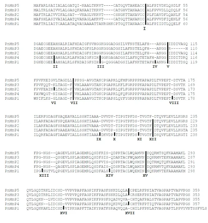
Fig. 2-2. Alignment of deduced amino acid sequences of PnMnP1-5 show intron- exon junctions. Sequences labeled from top to bottom are PnMnP5, 3, 2, 4 and 1, according to the phylogenetic tree (Fig. 2-1). Roman characters I to XVII refer to intron positions. Black vertical bars refer to interrupted intron positions and (-) indicates the gap.
31
Fig. 2-3. Relative expression ratio of PnMnP1-5 genes in P. microspora mycelial culture in amended M4 medium with aromatic compound used for substrate for MnP.
Total RNA was observed on day 10 by qRT-PCR in triplicate of samples, which are denoted by standard error bars. Asterisks indicate that the difference in expression level is significant between control basal medium and supplemented with aromatic compound used for substrate for MnP (t-test, p < 0.05).
*
*
32
Fig. 2-4 Relative expression ratio of PnMnP1-5 genes expression from mycelial growth of P. microspora in sawdust medium. Total RNA acquired by mycelial stages at 1 and 3 months. All samples were done in triplicates to denote in standard error bars.
0 20 40 60 80 100 120
PnMnP1 PnMnP2 PnMnP3 PnMnP4 PnMnP5
Relative expression ratio
Mycelia 1 mo. Mycelia 3 mo.
33
Chapter 3
Relationship between fruiting body development and phenol oxidase gene expression in Pholiota microspora
3-1 Abstract
We analysed nucleotide sequences of phenol oxidase genes in Pholiota microspora and identified three types of phenol oxidase: laccase (Lcc1-Lcc8), ferroxidase (Lcc9), and tyrosinase (Tyr). The expression of Lcc1 to Lcc9 and Tyr genes in P. microspora was examined by qRT-PCR. We quantified transcripts of these ten genes in mycelia, primordia, and fruiting bodies grown on sawdust substrate and in mycelia grown in M4 liquid medium supplemented with aromatic compounds. All Lcc genes were expressed at a very low level in mycelia grown on sawdust medium, but Lcc1 was transcribed at a level 8-fold higher in M4 liquid medium when supplemented with 3 mM veratryl alcohol. On the other hand, Lcc9 and tyrosinase were highly expressed in primordia and fruiting bodies. These results suggest that the content of melanin and related pigments in the fruiting body might be determined by complementary activity of two types of phenol oxidase, such as Lcc and Tyr, in P. microspora.
34
3-1 Introduction
Phenol oxidases (PO) are enzymes containing copper atoms in the catalytic centre and are usually called multicopper oxidases. The catalytic activity of these enzymes is oxidation of diphenols to the corresponding quinones (Baldrian, 2006).
PO includes tyrosinases and laccases (Durán et al., 2002). Tyrosinases oxidize p- monophenols and o-diphenols to the corresponding quinones, whereas laccases are capable of oxidizing various aromatic compounds such as substituted monophenols and polyphenols, aromatic amines, and thiol compounds, with subsequent production of radicals (Selinheimo, 2008). Intracellular and extracellular PO are produced in plants and fungi for a variety of purposes. PO from basidiomycetes fungi are connected with melanin production, lignin degradation, and morphogenic processes such as fruiting body formation (Sinsabaugh, 2010).
In fruiting bodies, fungal PO can catalyse melanin formation, as well as browning of the fruiting body that occurs after harvest, as described in Lentinula edodes (Sakamoto et al., 2012). In general, melanin involved in gill browning is considered to be synthesized from β-(3,4-dihydroxyphenyl)alanine (DOPA), derived from tyrosine. Oxidation of tyrosine is commonly catalysed by tyrosinase. The mechanisms of mushroom browning have been investigated extensively in Agaricus bisporus, in which tyrosinase is responsible for melanogenesis during spore maturation (Hegnauer et al., 1985); non-reproductive tissues have higher tyrosinase levels than fresh fruiting bodies (Burton, 1988). Pigmentation of mushrooms is
35
largely mediated by tyrosinase (Jolivet et al., 1998). Moreover, a correlation between melanin synthesis and intracellular laccase in Cryptococcus neoformans has been reported (Ikeda et al., 2002). Laccase activity also increases in fruiting bodies of L.
edodes after harvest. Laccase purified from L. edodes fruiting bodies after harvest can oxidize DOPA (Nagai et al., 2003). Therefore, PO are involved in melanin synthesis in fruiting bodies.
On the other hand, white-rot basidiomycetes produce extracellular PO to degrade lignin, and laccases are believed to be important for lignin degradation (Youn et al., 1995; Eggert et al., 1996). Almost all species of white-rot fungi reportedly produce laccase (Hatakka, 2001). In Pycnoporus cinnabarinus, which is capable of lignin degradation, laccase was described as the sole ligninolytic enzyme produced by this species (Eggert et al., 1996). Moreover, fruiting body development might be dependent on laccase activity in some fungi. In Schizophyllum commune, dikaryotic strains that are able to form fruiting bodies can secrete high levels of laccases, but monokaryotic strains do not produce any (De Vries et al., 1986). In A.
bisporus, laccase activity is strongly regulated during senescence of fruiting bodies;
therefore, fruiting bodies rapidly brown after harvest (Wood, 1980). These phenomena indicate that laccase may play an important role in the morphogenesis of mushrooms. Furthermore, veratryl alcohol can stimulate laccase production during mycelial growth of Pleurotus ostreatus, increasing production of fruiting bodies, with fruiting occurring earlier in the medium containing veratryl alcohol than the
36
medium without (Suguimoto et al., 2001). However, the relationship between the roles of multicopper oxidases and veratryl alcohol in fruiting body development is unclear.
The wood-rotting basidiomycete Pholiota microspora (=Pholiota nameko, also known as “nameko” in Japan) is sold in local markets as food in Japan and is widely used as material in fungal research into aspects of sexual reproduction (Aimi et al., 2005; Yi et al., 2010) and DNA-mediated transformation (Yi et al., 2009). To understand the role of laccase, we identified nine Lcc genes and evaluated their phylogenetic relationships, and then investigated the relationship between pigment production and lignin degradation.
3-3 Materials and Methods
3-3-1 Fungal strains and culture conditions
The monokaryotic strains P. microspora NGW19-6 (A4, pdx1), a pyridoxine auxotrophic mutant, and NGW12-163 (A3, arg4), an arginine auxotrophic mutant (Masuda et al., 1995; Yi et al., 2009) were used. A dikaryotic strain was obtained by crossing NGW19-6 and NGW12-163, referred to as NGW19-6/12-163, and used for experiments.
In order to analyse the effects of different aromatic compounds on gene expression, the P. microspora NGW19-6/12-163 strain was grown on M4 agar at
37
25°C for 1 week, and then 10 mycelial agar blocks (3 × 3 mm) were transferred into 20 mL of M4 broth medium (Johansson et al., 2002) (components are presented in terms of g l-1: glucose, 2.20 g; diammonium tartrate, 0.92 g; KH2PO4, 1.00 g;
NaH2PO4•2H2O, 0.26 g; MgSO4•7H2O, 0.50 g; thiamine hydrochloride, 1.00 × 10⁻⁴ g; CaCl2•2H2O 6.60 × 10⁻³g; FeSO4•7H2O, 5.00 × 10⁻³ g; MnSO4•H2O, 3.00 × 10⁻⁴ g; ZnSO4•7H2O, 5.00 × 10⁻⁴ g; CuSO4, 6.40 × 10⁻⁴ g; pH, 5.5; for solid M4 medium, 15 g agar was added) in a 100-mL Erlenmeyer flask supplemented with aromatic compounds (final concentration): 0.01% lignosulfonate; 0.05 mM 2,5-xylidine; 3 mM veratryl alcohol; 0.1 mM guaiacol; 1 mM ferulic acid; 1 mM veratric acid; 0.1 mM anisic acid; 1 mM gallic acid and 1 mM L-DOPA. P. microspora NGW19-6/12- 163 was then grown at 25°C for 20 days and the mycelia were harvested by filtration for RNA extraction.
The cultivation of fruiting bodies of P. microspora was carried out on a sawdust substrate, which was prepared as follows. Beech sawdust was mixed with rice bran at a gravimetric ratio of 5:1 and adjusted to 65% moisture content using tap water, and this medium was placed into a 100-mL Erlenmeyer flask, followed by autoclaving at 121°C for 60 min. After cooling the medium in the air, five mycelial agar blocks (5 × 5 mm) containing NGW19-6/12-163 were inoculated and incubated at 25°C. When the mycelia had colonized the substrate (about 40 days after inoculation), the surface layer was scratched with a spatula, and then 50 mL of sterilized distilled water was poured into the flask. Water was removed after flasks
38
were incubated at 15°C overnight, and then cultivation continued at 15°C until fruiting bodies developed. We defined the day after water removal as day 0. Samples for RNA extraction were taken from triplicate cultures at mycelial stages of the mushroom developmental cycle (30, 90 days), primordia, and fruiting body (1 cm).
3-3-2 Genomic DNA and total RNA preparation
Genomic DNA was extracted according to the method of Dellaporta et al. (1983).
RNA was extracted using a MagExtractor™ Kit (Toyobo, Osaka, Japan) according to the manufacturer’s instructions. cDNA was synthesized using total RNA as a template with ReverTra Ace® qPCR RT Master Mix with gDNA Remover kit (Toyobo, Osaka, Japan). PCR was carried out using a Takara Ex Taq® polymerase (Takara Bio, Japan). The oligonucleotide primers listed in Table 3-1. Amplified fragments were subcloned into pMD20 T-vector (Takara Bio, Japan) and sequenced.
3-3-3 P. microspora genome and retrieved genes
Whole genomic sequences of monokaryon P. microspora NGW 19-6 were determined using Illumina HiSeq 2000 paired-end technology with the software (CASAVA ver.1.8.1) provided by Hokkaido System Science Co., Ltd. (Sapporo, Hokkaido, Japan), as described by Funo et al. (2014). This sequencing run yielded 30,935,254 high-quality filtered reads with 101-bp paired-end sequencing. The genome was assembled using Velvet assembler (hash length, 85 bp) (Zerbino and Birney, 2008). The final assembly contained 4,770 contigs with a total size
39
33,400,256 bp and an N50 length of 72,431 bp. The deduced amino acid sequences of known laccase and tyrosinase genes from public databases were searched against the P. microspora genome using the BLASTp algorithm. The nine laccase genes and tyrosinase gene identified were named Lcc1-9 and Tyr. The coding sequences of intron-exon junctions were based on GT-AG rules (Breathach et al., 1978; Wu and Krainer, 1999) and homology of amino acid sequences to laccase proteins in the DNA Data Bank of Japan (DDBJ). The nucleotide sequences of genomic DNA fragments of P. microspora laccase genes were deposited in the DDBJ under the following accession numbers: Lcc1 (LC093451); Lcc2 (LC093452); Lcc3 (LC093453); Lcc4 (LC093454); Lcc5 (LC093455); Lcc6 (LC093456); Lcc7 (LC093457); Lcc8 (LC093458); and Lcc9 (LC093459). Subcellular localization was predicted using the PSORTII (Nakai and Kanehisa, 1992) (http://psort.hgc.jp/form2.html), and SOSUI (Hirokawa et al., 1998) (http://harrier.nagahama-i-bio.ac.jp/sosui/sosui_submit.html) online tools.
3-3-4 Quantitative RT-PCR (qRT-PCR) assays
The actin gene (Act1) was used as a reference gene. Primer pairs for amplification
of Lcc1-9, Tyr, and Act1 cDNAs were designed based on their cDNA sequences using Genetyx software. Amplification of genomic DNA was prevented by designing primers for exon-exon junctions. All primers were tested to ensure that they amplified a single band with no primer-dimers, as shown in Table 3-2. Plasmids with inserted of the target gene (Lcc1-9, Tyr) and the housekeeping gene (Act1) were
40
extracted as described by Birnboim (1983). Standard curves were constructed using four ten-fold dilutions of plasmid. Real-time PCR was performed using a KOD SYBR® qPCR Mix kit (Toyobo). Thermocycling was carried out using a PikoReal™
96 system (Thermo Fisher Scientific) with an initial incubation for 1 min at 95°C, followed by 40 cycles of 95°C for 10 s, 60°C for 1 min. Each run was completed with a melting curve analysis to confirm the specificity of amplification and absence of primer-dimers. Data analysis was performed in accordance with the manufacturer's instructions.
3-3-5 Analysis of sequences and phylogenetic tree
Nucleotide and protein sequence data were analysed using Genetyx ver. 10.0.3 software (Genetyx, Tokyo, Japan). Protein sequence similarity was analysed using the BLASTp algorithm (Altschul et al., 1997). Laccase genes were retrieved from public domains (NCBI and UniProt). A phylogenetic tree was constructed by MEGA 6.06 software (Tamura et al., 2013) using the neighbour joining method with a bootstrap value of 1,000 replicates. Multiple alignment was performed using ClustalW (Larkin et al., 2007).
41
3-4 Results
3-4-1 Role of Lcc1 and Lcc9
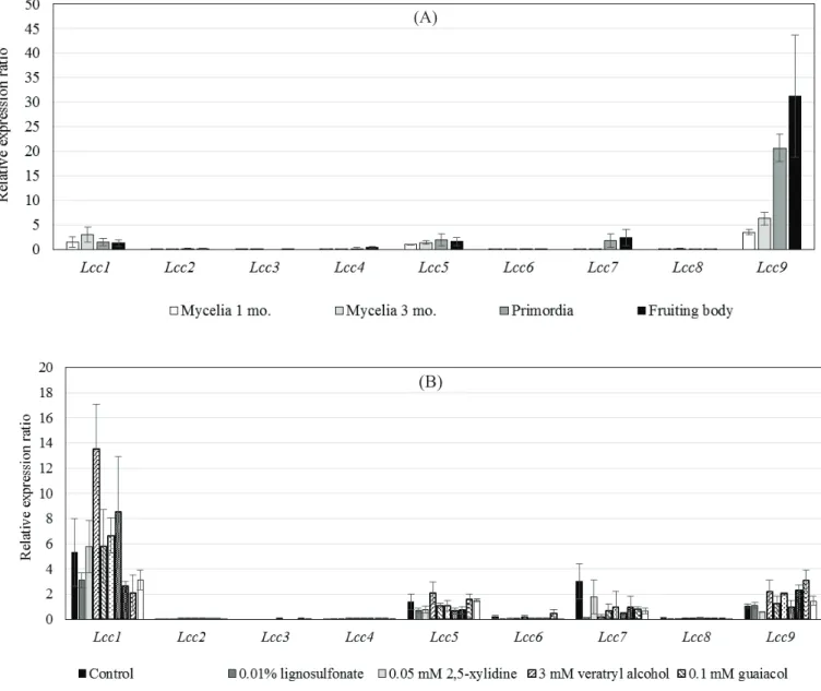
Transcription of the nine laccase genes (Lcc1-9) in mycelia and fruiting bodies grown on sawdust medium was investigated. Mycelia were harvested from sawdust medium 1 month after inoculation and 2 months after kinkaki. Primordia and fruiting bodies were also harvested and total RNA was extracted from the tissue and the mycelium. Fig. 3-1(A) shows expression levels of the Lccs (1-9).
Significant expression was detected in only Lcc1 and Lcc9. On the other hand, no or only slight expression of the other Lccs (2-8) was detected. Lcc9 showed the highest expression of all Lccs in the fruiting body. Lcc9 expression in primordia was also relatively high. On sawdust medium, the expression of Lcc9 in primordia was 10-fold higher and in fruiting bodies 15-fold higher than in mycelia.
In order to investigate the roles of Lcc1 and Lcc9, expression of Lcc1 and Lcc9 in mycelia grown in M4 liquid medium supplemented with aromatic compounds was studied by quantitative RT-PCR [Fig. 3-1(B)]. Maximum expression of Lcc1 was observed in mycelium grown in M4 liquid medium supplemented with 3 mM veratryl alcohol, and expression was 8-fold higher than that of Lcc1 in mycelia grown in M4 liquid basal medium. Other tested aromatic compounds did not affect Lcc1 expression. In contrast, Lcc9, which was transcribed at the highest level in fruiting bodies, did not show any significant response in the presence of aromatic compounds.
42
Therefore, the role of Lcc1 may be lignin degradation, and Lcc9 may be related to morphogenesis involving colouration of the fruiting body.
3-4-2 Expression of Tyr also involved in fruiting body
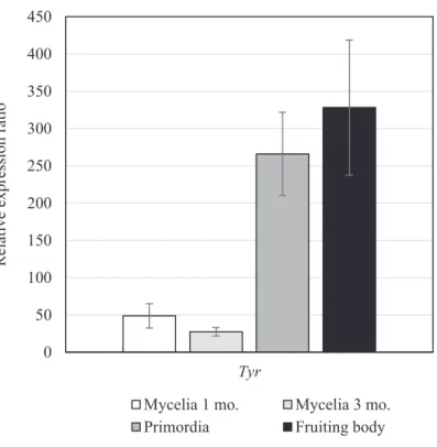
Laccase and tyrosinase are PO and are closely related to production of melanin pigment in the fruiting body. Thus, in order to confirm the relationship between PO expression and fruiting body development, transcription of Tyr in mycelia, primordia, and fruiting bodies that were cultured on sawdust medium were investigated.
Expression of Tyr in primordia was 250-fold higher and in fruiting bodies 300-fold higher than that of Tyr in mycelia (Fig. 3-2). These results indicated that tyrosinase is required for processes during fruiting body development in P. microspora such as pigment production.
3-4-3. Phylogenetic relationship among Lccs and origin of the Lcc genes
From a phylogenetic tree based on the deduced amino acid sequences of P.
microspora Lccs and laccases from other basidiomycetous mushrooms extracted from the DNA and protein databases, Lcc1-7, Lcc8, and Lcc9 fit into three major clusters (Fig. 3-3). The first cluster, containing Lcc1-7, fell into the same clade.
Based on their phylogenetic relationships and the structure of the genes including intron position, Lcc2-7 may have been amplified from Lcc1. Therefore, the origin of
43
the Lcc1 group was Lcc1. These laccases are clustered tightly with other fungi in the same family, Hypholoma sublateritium and Stropharia aeruginosa, whereas laccase gene families of other basidiomycetes are tightly clustered within the same genus.
The second cluster, containing Lcc8, included laccase protein sequences from the anamorphic fungi Thanatephorus cucumeris (Rhizoctonia solani) and Flammulina velutipes. Transcription of Lcc8 was not detected in mycelia, primordia, or fruiting bodies grown on sawdust substrate or in mycelia grown in M4 liquid medium supplemented with aromatic compounds. These results suggested that Lcc8 has a different function than lignin degradation or morphogenesis or has lost its function, although its origin differed from Lcc1 and Lcc9. The third cluster, Lcc9, was grouped with ferroxidase. The role of ferroxidase in iron uptake has been analysed extensively in Saccharomyces cerevisiae (De Silva et al., 1995), but remains unclear in basidiomycetes, although it has strong activity to iron (Larrondo et al., 2003). Thus, three types of laccases might have originally been present in the P. microspora genome. The PSORTII and SOSUI program estimated that the Lcc1, 3, 4, 5 and 8 were extracellular proteins, Lcc2 and 7 were cytoplasmic proteins, and Lcc6 and 9 were transmembrane proteins. Moreover, Tyrosinase was estimated as cytoplasmic protein. Therefore, development of the colour might be occur in cytoplasm or periplasmic spaces of the cell.
44
3-4-4 Intron structure and origin of Lccs
To analyse whether the origin of Lcc1 differed from that of Lcc8 and Lcc9 in the ancient P. microspora genome, the position and number of introns were analysed.
Nucleotide sequence of Lccs ranged from 2118 bp to 2643 bp. ORFs of Lcc8 and 9 were the largest. The nucleotide sequence of Lccs belonging to the Lcc1 group (Lcc1- 7) ranged from 1551 to 1575 bp. Fig. 3-4 shows the structure of the Lcc genes. There were 18 introns in Lcc8; 13 in Lcc1, 2, 5, and 7; 12 in Lcc3 and Lcc6; 10 in Lcc4;
and 9 in Lcc9. Introns I to XIII were inserted in the same positions for Lcc1-7, which were clustered in the same group in the phylogenetic tree, with the exception of introns VII, VIII, and XII, which were absent in Lcc4 and intron XIII, which was absent in Lcc3. Based on these results, the position of the introns corresponded with phylogenetic and evolutionary relationships; therefore, the ancient P. microspora genome originally had three genes, a Lcc gene belonging to the Lcc1 group, Lcc8, and Lcc9.
All P. microspora laccase coding sequences were used as search queries against the DDBJ using the Blastx algorithm. The percentage of protein sequence identity of Lcc1 to Lcc9 was 83% to multicopper oxidase in H. sublateritium (KJA22755.1), 80% and 81% to two laccases in S. aeruginosa (AFE48786.2 and AFE48786.2), 61% to multicopper oxidase in H. sublateritium (KJA22010.1), 74%
to a laccase-like protein in Galerina marginata (KDR80952.1), 79% to multicopper oxidase in H. sublateritium (KJA22017.), 71% to multiple oxidase in H.
45
sublateritium (KJA22017.1), 66% to multicopper oxidase in Sphaerobolus stellatus (KIJ32231.1), and 83% to multicopper oxidase in H. sublateritium (KJA26904.1).
Consensus motifs (L1, L2, L3, and L4) among laccases (Larrondo et al., 2003;
Kumar et al., 2003) were conserved in all P. microspora Lccs. Therefore, Lcc1-8 were identified as laccases and Lcc9 as a homologue of ferroxidase.
3-5 Discussion
In this study, high Lcc9 and Tyr expression in primordia and fruiting bodies were shown, so the colour of the fruiting body in P. microspora may be determined by the combined activity of these two enzymes because they are closely related to oxidation of phenolic compounds and melanin production. Lcc9 is a ferroxidase that can reduce Fe3+ to Fe2+, and the substrate specificity of ferroxidase for Fe3+ was higher than for phenolic compounds in Phanerochaete chrysosporium (Larrondo et al., 2003). In order to minimize production of active oxygen in redox cycling in fungi, the coupling of Fe3+ reduction with a reductant is required (De Luca and Wood, 2000;
Kosman, 2003). Therefore, this highly suggests that Lcc9 and Tyr are related to not only colouring, but also active oxygen scavenging. Furthermore, in L. edodes, laccases are believed to have a role in morphogenesis of fruiting bodies (Zhao and Kwan, 1999). Likewise, in P. ostreatus, the presence of phenolic compounds such as veratryl alcohol in the culture medium promotes fruiting body formation and shortens the culture period16). These observations support our results that metabolism
46
of phenolic compound by PO was connected with fruiting body formation. Moreover, small lignin degradation products might be one of the initiation signals for fruiting body development.
Our results showed that expression of all Lccs was poor in mycelia grown on sawdust substrate. Therefore, laccases do not directly degrade lignin during growth on sawdust substrate. However, P. microspora contains manganese peroxidase, which is highly expressed in mycelia on sawdust medium, for degrading lignin (Sutthikhampa et al., 2015). Lcc1 was induced by aromatic compounds and Lcc2-7 were expressed at a basal level during mycelial growth on sawdust substrate. Based on the evolutionary distance of the first cluster, position of the introns, and expression profile, Lcc1-7 might have been generated by repeated duplication from a single Lcc, and Lcc1 might be the origin of Lcc2-7. Moreover, enzyme activities in P. microspora are affected by their level of expression rather than the number of genes, because only two of the nine genes were actively transcribed.
In this study, we investigated phenol oxidase expression in P. microspora, finding that Lcc9 and Tyr are closely related to fruiting body formation, though their role remains unresolved. In subsequent experiments, therefore, we will further study their role by knockout of Lcc9 to determine the relationship between this gene and fruiting body formation, including pigment production, in P. microspora.
47
Table 3-1. Primer sets for cDNA and plasmid construction.
Genes Primers 5’ÆÆ 3’
Act1 PnActin1_F1 CGAAATTTCAGCTCTCGTCGT PnActin1_R1 CTGGAGCACGGAATCGCT Lcc1 PnLcc1-F1 GACGAGCACCAGCATTCATT
PnLcc1-R1 CTGATTCGTGTTGAGCACGAA Lcc2 PnLcc2_F3 CTGGCATGGCCTGTTCCAA
PnLcc2-R1 GACGACGGCAAGACCAACAT Lcc3 PnLcc3-F1 GTTGACGAGCACCAGTATTCATT
PnLcc3-R2 CAGGGTCTTCGATAGTCGCATT Lcc4 PnLcc4-F1 GCTTACTGGCGTCAAGGGA
PnLcc4-R1 CGTATGACGGGATTGTGGTAATT Lcc5 PnLcc5-F1 CGGGCCTATTGCCAATCTTT
PnLcc5-R1 GACGGAGAATGCGTGAGGA Lcc6 PnLcc6-F1 CCTGAGCGTCTTCGGTTT
PnLcc6-R1 CGACTTCGATGATCGTCATGA Lcc7 PnLcc7-F1 GTTGGTCAGCACGAGTATACATT
PnLcc7-R1 GCCAGTACTATGGCTAGACCAA Lcc8 PnLcc8-F1 GACTAATTCTGAGGACGGACCT PnLcc8-R1 GGCAATAATGTTGAGCAGGGTA Lcc9 PnLcc9-F1 CATCGAAGTCGATGGTACCGA
PnLcc9-R1 GGTAAGCACGCATCCGAA
Tyr PnTyr_F1 CCTACGTTCTTCTCTACGAGCA
PnTyr_R1 GAGAACGGGGTCAGAGGAGT
48
Table 3-2. Primer sets for qPCR.
Genes Primers 5’ÆÆ 3’
Act1 PnActin1_CF1 GCTATGCTATGTCGCGCTTGAT PnActin1_R1 CTGGAGCACGGAATCGCT Lcc1 PnLcc1-F1 GACGAGCACCAGCATTCATT
PnLcc1-CR1 CCGTCACAGTATTGCGTCGAAT Lcc2 PnLcc2_CF1 CTATTCACTTGCACGGCCACA
PnLcc2-R1 GACGACGGCAAGACCAACAT Lcc3 PnLcc3-F1 GTTGACGAGCACCAGTATTCATT
PnLcc3_CR1 GAGACCGTCGCAATACTGTGTAGA Lcc4 PnLcc4-F1 GCTTACTGGCGTCAAGGGA
PnLcc4-CR1 CCGTCACAATACTGAGTGGAAT Lcc5 PnLcc5-F1 CGGGCCTATTGCCAATCTTT
PnLcc5-CR1 GCCAATGAATGCTCGTGCT Lcc6 PnLcc6-F1 CCTGAGCGTCTTCGGTTT
PnLcc6-CR1 CATTGAGATTGAAGGTATCGCCCT Lcc7 PnLcc7-CF1 GATCACCTGGTTTCCCGCA
PnLcc7-R1 GCCAGTACTATGGCTAGACCAA Lcc8 PnLcc8-F1 GACTAATTCTGAGGACGGACCT PnLcc8-CR1 GGGTCAACGGGGTCATAAACA Lcc9 PnLcc9-CF1 GTCTGGTTCCTTCATTGCCA
PnLcc9-R1 GGTAAGCACGCATCCGAA Tyr PnTyr_CF1 GATCCTGCTGTCGCTGCTT
PnTyr_R1 GAGAACGGGGTCAGAGGAGT
49
Fig. 3-1 Relative expression ratio of Lcc1-9 genes during development of P.
microspora in sawdust medium. Total RNA was extracted from mycelia cultivated for 1 and 3 months (mo.), primordia, and fruiting bodies (A). Mycelia cultured in M4 medium with aromatic compounds (B) were observed by qRT-PCR. All samples were assessed in triplicate, with variation denoted by standard error bars.
(A)
(B)
50
Fig. 3-2 Relative expression ratio of Tyr gene during development of P. microspora in sawdust medium. Total RNA was extracted from mycelia cultivated for 1 and 3 months (mo.), primordia and fruiting bodies. All samples were assessed in triplicate, with variation denoted by standard error bars.
0 50 100 150 200 250 300 350 400 450
Tyr
Relative expression ratio
Mycelia 1 mo. Mycelia 3 mo.
Primordia Fruiting body
51
52
Fig. 3-3 Phylogenetic tree of laccases. The tree was constructed by the neighbour joining method with 1000 bootstrap replications of Lcc1-9 and laccases from public databases according to gene family: Emericella nidulans, Flammulina velutipes, Hypholoma sublateritium, Laccaria bicolor, Lentinula edodes, Pleurotus ostreatus, Saccharomyces cerevisiae, Sphaerobolus stellatus, Stropharia aeruginosa, and Thanatephorus cucumeris. Taxa contain organism and gene name, then protein ID.
53
Fig. 3-4 Intron positions within laccase genes Lcc1 to Lcc9 of P. microspora define three gene types. Horizontal lines indicate the laccase genes, and vertical bars indicate intron positions. Dotted lines indicate introns that interrupt the coding sequence of the different genes at exactly the same codon position for Lcc1-7. The Roman characters I to XIII indicate intron positions. Lcc8 and Lcc9 do not have introns at the same positions.
54
Chapter 4
Conclusions and discussions
From the evolutionary perspective, genetic redundancy of MnP and laccase gene families might be occurred by gene duplication since ancient time. From the results suggested that among five MnP, only one MnP predominant transcript in mycelia grown on sawdust substrate and relatively constitutive transcription even grown in liquid medium with or without aromatic compounds. Moreover, due to nucleotide and protein sequences analysis, we believe that all P. microspora MnPs are of the same origin and that they were amplified by duplication events in the ancient P. microspora genome. Furthermore, analysis of nine laccases in P.
microspora, the result obtained that Lcc1-9 were low level of expression in mycelia grown on sawdust medium, also in liquid medium containing aromatic compounds.
It suggested that P. microspora laccases were not involved in lignin degradation. But Lcc9 and tyrosinase was highly expressed in primordia and fruiting bodies. The results suggest that the content of melanin and related pigment in the fruiting body might be decided by harmony of the two kinds of phenol oxidase activity such as Lcc and Tyr in P. microspora.
In the Chapter 2, P. microspora MnP5 transcription in P. microspora is much higher than the other MnP genes presents in the genome. This result may occurred by the concentration of nitrogen that presence in M4 liquid medium. Since, the