hypertrophy, a history of syncope, left ventricular outflow tract obstruction (LVOTO), non-sustained ventricular tachycardia, and left atrial dilatation.4–6
For patients with multiple risks, documenting VAEs (i.e., non-sustained [NSVT] and sustained ventricular tachycardia [VT]) leads to ICD implantation in most patients.7 How- ever, VAEs are often asymptomatic and comparatively rare (reported rates are as high as 15–30%), with repeated Holter electrocardiography and stress tests failing to reveal high-risk patients who should have an ICD implanted to avoid SCD.8
Fragmented QRS (fQRS) is defined as multiple fine spikes on normal resting electrocardiograms (ECGs).
Reflecting regional myocardial damage, an association between fQRS and the occurrence of VAEs during myocar- dial infarction and Brugada syndrome has been reported.9 It has also been reported that regional myocardial damage,
H
ypertrophic cardiomyopathy (HCM) is caused by numerous mutations of genes encoding sarcomere proteins, with widely different phenotypic expres- sion.1–3 The main causes of death in HCM are left ven- tricular dysfunction and ventricular arrhythmic events (VAEs). In recent reports, improved diagnosis and treat- ment resulted in a reduction of the mortality rate to <1%per year, which is comparable to that of the general popu- lation. However, some high-risk groups experience progres- sive deterioration of cardiac function and fatal arrhythmic events.
Among HCM patients, the rate of sudden cardiac death (SCD) from arrhythmic events has been reported to be 0.5%
per year, and SCD is preventable with the proper use of implantable cardiac defibrillators (ICDs).3 Conventionally, decisions regarding ICD implantation are guided by many risk factors, including family history, massive left ventricular
Received October 27, 2019; revised manuscript received January 9, 2020; accepted January 15, 2020; J-STAGE Advance Publication released online February 8, 2020 Time for primary review: 24 days
Department of Cardiovascular Medicine (S.O., K. Nakamura, H.M., N.T., K. Nakagawa, M.Y., A.W., N.N., T.M., H.I.), Department of Cardiovascular Therapeutics (H.M., N.N.), Okayama University Graduate School of Medicine, Dentistry and Pharmaceutical Sciences, Okayama, Japan
Mailing address: Kazufumi Nakamura, MD, PhD, Department of Cardiovascular Medicine, Okayama University Graduate School of Medicine, Dentistry and Pharmaceutical Sciences, 2-5-1 Shikata, Okayama 700-8558, Japan. E-mail: [email protected].
ac.jp
ISSN-1346-9843 All rights are reserved to the Japanese Circulation Society. For permissions, please e-mail: [email protected]
New Appearance of Fragmented QRS as a Predictor of Ventricular Arrhythmic Events in Patients
With Hypertrophic Cardiomyopathy
Soichiro Ogura, MD; Kazufumi Nakamura, MD, PhD; Hiroshi Morita, MD, PhD;
Norihisa Toh, MD, PhD; Koji Nakagawa, MD, PhD; Masashi Yoshida, MD, PhD;
Atsuyuki Watanabe, MD, PhD; Nobuhiro Nishii, MD, PhD;
Toru Miyoshi, MD, PhD; Hiroshi Ito, MD, PhD
Background: Multiple spikes in the QRS complex (fragmented QRS [fQRS]) on 12-lead electrocardiography have been associated with ventricular arrhythmic events (VAEs) in patients with hypertrophic cardiomyopathy (HCM). The aim of this study was to assess the association between new appearances of fQRS and cardiac events in patients with HCM.
Methods and Results: The association between baseline fQRS and cardiac events, namely VAEs, heart failure-related hospitalization, and all-cause death, was evaluated retrospectively in 146 HCM patients (46 patients with fQRS, 100 without fQRS). The median follow-up was 5.3 years. Cardiac events occurred in 29 patients with baseline fQRS and 32 patients without baseline fQRS (63% vs.
32%; P<0.001). VAEs occurred in a significantly larger percentage of patients with than without baseline fQRS (54% vs. 23%, respectively; P<0.001). Of the 100 patients without baseline fQRS, 33 had a new appearance of fQRS during the 4.6-year follow-up, whereas 67 did not. VAEs occurred more frequently in the 33 patients with the appearance of fQRS than in those without (42% vs.
13%, respectively; P=0.001). Multivariable analysis showed that the new appearance of fQRS documented before VAEs was associated with VAEs (hazard ratio 4.29, 95% confidence interval 1.81–10.2; P=0.001).
Conclusions: The new appearance of fQRS was associated with an increased risk of VAEs in HCM patients.
Key Words: Electrocardiography; Fragmented QRS; Hospitalization; Hypertrophic cardiomyopathy; Ventricular arrhythmia
appearance of fQRS in HCM patients has never been reported.
The aim of the present study was to examine the asso- ciation between baseline fQRS and cardiac events in HCM patients. In addition, we investigated the association between the new appearance of fQRS and VAEs in patients without baseline fQRS.
Methods Study Population
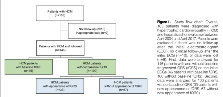
The study population comprised 165 consecutive patients who were suspected of having HCM and who had been referred to Okayama University Hospital between April 2004 and April 2017. Patients without follow-up after their initial ECG (n=10) or with inadequate clinical data (n=9) were excluded from the study, leaving 146 patients enrolled in this study. Data for 100 patients without baseline fQRS were also used to analyze the new appearance of fQRS (Figure 1).
HCM was diagnosed according to the 1995 criteria of the World Health Organization/International Society and Federation of Cardiology Task Force.18 The study protocol was approved by the Institutional Ethics Committee on Human Research of Okayama University (Approval no.
1809-029). Because of the anonymous nature of the data, the requirement for informed consent was waived. The first author takes complete responsibility for the integrity of the data and the accuracy of the data analysis.
ECG Recording and Definition of fQRS
Standard 12-lead electrocardiography with a 0- to 150-Hz filter was used to record ECGs for all patients. ECGs acquired at admission were analyzed as the initial ECGs.
The digital ECG without a filter was evaluated at 400% of the initial size on a personal computer monitor; each parameter and fragmentation of the QRS complex were measured as described previously;9 a typical example of the ECGs obtained is shown in Figure 2. Abnormal fragmen- tation within the QRS complex was defined as more than 2 positive spikes within the QRS complex in 2 contiguous leads. Analysis of fragmentation needed 3 consecutive beats demonstrated by late gadolinium enhancement (LGE) during
cardiac magnetic resonance imaging (CMR), is associated with the presence of fQRS in HCM patients.10
An association between baseline fQRS and the occur- rence of VAEs has also been reported in HCM patients,11–15 and a recent meta-analysis showed a significant increase in arrhythmic events in HCM patients with baseline fQRS.16 However, in some HCM patients without baseline fQRS, fQRS newly appears during follow-up, and little is known about the significance of this newly appeared fQRS. In 1 case report, an HCM patient with new appearance of fQRS was reported to have VAEs 7 years after the baseline ECG,17 but an association between cardiac events and the
Figure 1. Study flow chart. Overall, 165 patients were diagnosed with hypertrophic cardiomyopathy (HCM) and hospitalized for evaluation between April 2004 and April 2017. Patients were excluded if there was no follow-up after the initial electrocardiogram (ECG), no clinical follow-up after the initial ECG (n=10), or data were lost (n=9). First, data were analyzed for 146 patients with and without baseline fragmented QRS (fQRS) on the initial ECGs (46 patients with baseline fQRS, 100 without baseline fQRS). Second, data were analyzed for 100 patients without baseline fQRS (33 patients with new appearance of fQRS, 67 without new appearance of fQRS).
Figure 2. Typical example of a fragmented QRS complex.
(A) Standard 12-lead electrocardiogram (ECG) with a filter.
(B) Standard 12-lead ECG without a filter. (C) ECG at the beginning of the study. (D) ECG 4 years after the first ECG.
disc summation method. Cardiac catheterization, including coronary angiography and myocardial biopsy, was per- formed using the Seldinger technique, as described previ- ously.19 In all, 64 patients underwent CMR. A high-intensity area in the myocardium appearing as gadolinium enhance- ment was considered positive for LGE. Plasma B-type natriuretic peptide (BNP) concentrations were measured using a radioimmunoassay specific for human BNP.
Endpoints
Endpoints were defined as cardiac events, namely VAEs, or more. The minimum size of the QRS fragmentation was
as small as 0.02 μV. The ECGs of patients without fQRS were retrospectively analyzed and the new appearance of fQRS was investigated (Figure 2). ECGs were reviewed by 3 cardiologists (S.O., K. Nakamura and H.M.) in a blinded manner.
Other Clinical Examinations
All other clinical examinations were performed at the time of diagnosis. All patients underwent echocardiography and the left ventricular ejection fraction was calculated using the
Yes (n=46) No (n=100)
Age (years) 61±16 58±16 0.39
Male sex 31 (67) 63 (63) 0.60
Body mass index (kg/m2) 23±3.8 23±4.1 0.30
Family history 13 (28) 17 (17) 0.12
Syncope history 6 (13) 25 (25) 0.10
Follow-up duration (years) 6.1±4.6 5.0±4.3 0.15
Medications
ACEI or ARB 25 (54) 42 (42) 0.16
β-blockers 28 (61) 56 (56) 0.58
MRA 9 (20) 8 (8.0) 0.043
Amiodarone 6 (13) 5 (5.0) 0.088
Diuretics 16 (35) 23 (23) 0.13
Plasma BNP (pg/mL) 377 [13–1,905] 297 [5–2,126] 0.21
Electrocardiogram
SR 31 (67) 85 (85) 0.014
AF or AFL 5 (11) 11 (11) 0.98
AVB 1 (2.2) 0 (0) 0.14
RBBB 15 (33) 7 (7.0) <0.001
LBBB 6 (13) 0 (0) <0.001
PR (ms) 187±46 171±33 0.78
QRS (ms) 124±36 101±18 <0.001
QTc (ms) 462±38 442±32 <0.001
Fragmentations (n/100 ms) 3.2±0.78 1.8±1.2 <0.001
Late-potential positive 7 (44) 17 (41) 0.88
Echocardiography
LA diameter (mm) 44±5.5 42±8.1 0.091
Ejection fraction (%) 57 [14–84] 63 [21–93] 0.015
LVMI (g/m2) 135±42 140±55 0.59
Massive LVH (LVMI >100 g/m2) 39 (85) 73 (73) 0.12
E/e’ 17±7.8 17±7.6 0.88
LVOTO (pressure gradient >10 mmHg) 5 (11) 10 (10) 0.89
Apical aneurysm 6 (14) 9 (9.0) 0.47
Previous procedure
Pacemaker implantation 7 (15) 2 (2.0) 0.002
ICD implantation 5 (11) 7 (7.0) 0.43
Septal ablation 1 (2.2) 0 (0) 0.14
Myectomy 2 (4.3) 0 (0) 0.036
Risk of SCD at 5 years 2.15 [0.49–18] 1.89 [0.69–14] 0.96
Data are expressed as the mean ± SD, median [interquartile range], or n (%). ACEI, angiotensin-converting enzyme inhibitor; AF, atrial fibrillation; AFL, atrial flutter; ARB, angiotensin II type 1 receptor antagonist; AVB, atrioventricular block; BNP, B-type natriuretic peptide; E/e’, ratio between early mitral inflow velocity and mitral annular early diastolic velocity; fQRS, fragmented QRS; ICD, implantable cardioverter-defibrillator; LA, left atrial; LBBB, left bundle block;
LVH, left ventricular hypertrophy; LVMI, left ventricular mass index; LVOTO, left ventricular outflow tract obstruction;
MRA, mineralocorticoid receptor antagonist; RBBB, right bundle branch block; SCD, sudden cardiac death; SR, sinus rhythm.
ventricular hypertrophy, and fQRS. Hazard ratios (HRs) are presented with 95% confidence intervals (CIs). Statistical analyses were performed using SPSS version 24 (IBM Inc., Armonk, NY, USA) and P<0.05 was considered significant.
Results
Baseline Characteristics of Patients With and Without Baseline fQRS
The clinical characteristics of patients with and without baseline fQRS are given in Table 1. Data for 146 patients (64% men; mean age 60±16 years, range 14–80 years) were analyzed. Patients’ physical characteristics, medical history, and medications were similar between the 2 groups. How- ever, some differences were noted between the 2 groups.
First, pacemaker implantation was performed more often in patients with baseline fQRS. Second, the percentage of patients with right bundle branch block (RBBB) and left bundle branch block (LBBB) was much higher in the group with fQRS, therefore the duration of both QRS and QTc was longer in patients with fQRS (QRS: 124 vs. 101 ms [P<0.001]; QTc: 462 vs. 442 ms [P<0.001]). However, the number of fragmentations per QRS duration was higher in patients with than without baseline fQRS (3.2±0.8 vs.
1.8±1.2 n/100 ms, respectively; P<0.001). The ejection fraction determined with echocardiography was lower in heart failure-related hospitalization, and all-cause deaths,
with VAEs defined as NSVT, VT and ventricular fibrilla- tion. NSVT was defined as spontaneous ventricular tachycardia at a rate of >120 beats/min that did not last
>30 s. VT was defined as spontaneous VT at a rate of
>120 beats/min that lasted >30 s. Patients were followed from the date of the initial ECG until the first documented date of cardiac events or the end of follow-up. Follow-up information was obtained from patients’ medical records, contact with the patients’ physicians, or interrogation of the ICDs. In patients without baseline fQRS, the association between the new appearance of fQRS and cardiac events was also evaluated.
Statistical Analyses
Continuous variables are presented as the mean ± SD or as the median with interquartile range (IQR). Categorical variables are presented as frequencies and percentages. The significance of differences was analyzed using Student’s t-test for continuous variables and the χ2 test for categorical variables. The event-free survival rate was estimated using a Kaplan-Meier analysis, with the significance of differences analyzed using a log-rank test. Predictors of cardiac events were analyzed using Cox proportional hazards analysis and logistic regression analysis. Variables for the univariate analysis were family history, syncope history, massive left
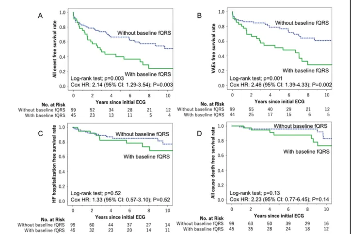
Figure 3. Kaplan-Meier curves for the selected end points: (A) primary end point of ventricular arrhythmic events (VAEs); (B) heart failure (HF)-related hospitalization; (C) all-cause death; and (D) all cardiac events. CI, confidence interval; ECG, electrocardiogram;
fQRS, fragmented QRS; HR, hazard ratio.
implantation were performed more often in patients with new appearance of fQRS than in those without (pacemaker implantation: 6.1% vs. 0% [P=0.042]; ICD implantation:
18% vs. 1.5% [P=0.002]). No patients in either group underwent septal ablation or myectomy.
Plasma BNP concentrations did not differ between patients with and without new appearance of fQRS (340 vs. 275 pg/mL, respectively; P=0.43). Ejection fraction determined by echocardiography was within the normal range in both groups, but lower in patients with new appearance of fQRS (59% vs. 65%, respectively; P=0.019).
The percentage of patients with LVOTO and massive left ventricular hypertrophy did not differ between those with and without new fQRS (LVOTO: 5% vs. 13%, respectively [P=0.60]; massive left ventricular hypertrophy: 70% vs.
75%, respectively [P=0.55]).
With regard to ECG parameters, the percentage of patients with RBBB was higher in the group with than with- out new appearance of fQRS (18% vs. 1.5%, respectively;
P=0.068), but LBBB was not seen in either group. Thus, QRS duration was slightly longer in patients with than without new appearance of fQRS (109 vs. 98 ms, respec- tively; P=0.029), but QTc duration did not differ between the 2 groups (445 vs. 441 ms, respectively; P=0.58). The number of fragmentations per QRS duration in the initial ECG did not differ between the group with and that without new appearance of fQRS (1.8±1.0 vs. 1.6±1.0 n/100 ms, respectively; P=0.61). In addition, we evaluated the vari- ability and reproducibility of fQRS through repeated recording of ECGs. The number of fragmentations was counted in patients without fQRS at baseline and during short-term follow-up. Fifty-one patients without baseline fQRS had repeated follow-up ECGs within 3 months, and the median duration until repeated ECG was 25 days (IQR 2–84 days). The numbers of fragmentations did not differ at the 2 time points (1.8±1.0 vs. 1.8±0.9, respectively; P=0.57).
Except for minor differences in the percentage of patients with RBBB, QTc duration, and ejection fraction, the characteristics in the 2 groups at follow-up were similar to those seen at baseline.
Association Between VAEs and New Appearance of fQRS Among the 100 patients without baseline fQRS, the new appearance of fQRS was seen in 33. During the 4.6-year follow-up, VAEs and heart failure-related hospitalization were seen more frequently in the 33 patients with new patients with than without fQRS, but was within the normal
range in both groups (57% and 62%, respectively; P=0.015).
There were no significant differences in plasma BNP concentrations between the 2 groups (377 vs. 297 pg/mL in patients with and without fQRS, respectively; P=0.21).
Univariable Analysis
Cardiac events occurred in 29 patients with fQRS and in 32 without fQRS (63% vs. 32%, respectively; P=0.0001;
Supplementary Table). In terms of cardiac events, VAEs (NSVT, VT, ventricular fibrillation) and all-cause death occurred in a significantly larger percentage of patients with than without fQRS (Supplementary Table). There was no significant difference in the frequency of hospitalization for heart failure between the 2 groups (Supplementary Table).
Kaplan-Meier Analysis
Kaplan-Meier analysis showed a significant difference in the frequency of all events and VAEs between patients with and without fQRS (log-rank P=0.001 and 0.001, respectively; Figure 3). Kaplan-Meier analysis did not show significant differences in the occurrences of hospitalization for heart failure and all-cause death between the 2 groups (log-rank P=0.52 and 0.14, respectively).
Multivariable Analysis
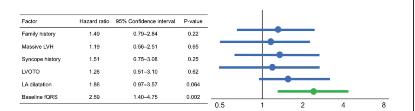
Multivariable analysis showed that baseline fQRS was associated with VAEs (HR 2.59; 95% CI 1.41–4.75;
P=0.002), but not with hospitalization for heart failure (HR 0.95; 95% CI: 0.37–2.41; P=0.92; Figure 4).
LGE With CMR
LGE with CMR was observed more frequently in patients with than without baseline fQRS (17/19 [90%] vs. 29/45 [64%], respectively; P=0.042).
Baseline Characteristics of Patients With and Without New Appearance of fQRS
The clinical characteristics of patients with and without new appearance of fQRS are given in Table 2. Data were analyzed for 100 patients (63% men; mean age 61±16 years, range 17–80 years). Patients’ physical characteristics, medical history, medications, and follow-up duration were similar in the 2 groups. The follow-up period was 4.6±4.3 years and did not differ between the 2 groups.
Regarding previous procedures, pacemaker and ICD
Figure 4. Forest plot of the Cox proportional hazard model analysis. The left ventricular mass index was >100 g/m2. fQRS, fragmented QRS; LA, left atrial; LVH, left ventricular hypertrophy; LVOTO, left ventricular outflow tract obstruction.
Discussion
In this study we investigated associations between baseline fQRS and cardiac events, as well as the new appearance of fQRS and cardiac events in HCM patients. The results showed that baseline fQRS was associated with the occur- rence of VAEs and all-cause death, but not with heart failure-related hospitalization. Baseline fQRS also corre- sponded well with CMR findings with LGE. In patients without fQRS, the new appearance of fQRS during the follow-up period was associated with VAEs and heart failure-related hospitalization.
Although previous studies have shown that fQRS in Brugada syndrome and myocardial infarction are associated with VAEs,9 the association between cardiac events and fQRS in HCM patients has not been clarified. A recent appearance of fQRS (n=14) than in those without (n=9;
VAEs: 42% vs. 13%, respectively [P=0.001]; heart failure- related hospitalization: 24% vs. 7.5%, respectively [P=0.019]).
However, all-cause death did not differ between the 2 groups (9.1% vs. 4.5% in those with vs. without new fQRS, respectively; P=0.36).
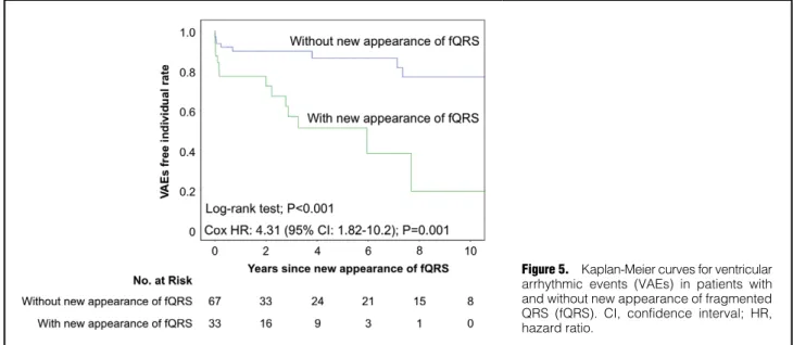
Time-dependent associations were evaluated the between new appearance of fQRS documented before VAEs and the occurrence of VAEs. Kaplan-Meier analysis showed a significant difference in the frequency of VAEs between patients with and without new appearance of fQRS (log- rank P<0.001; Figure 5). Multivariable analysis showed that new appearance of fQRS documented before VAEs was associated with VAEs (HR 4.29; 95% CI 1.81–10.2;
P=0.001; Table 3).
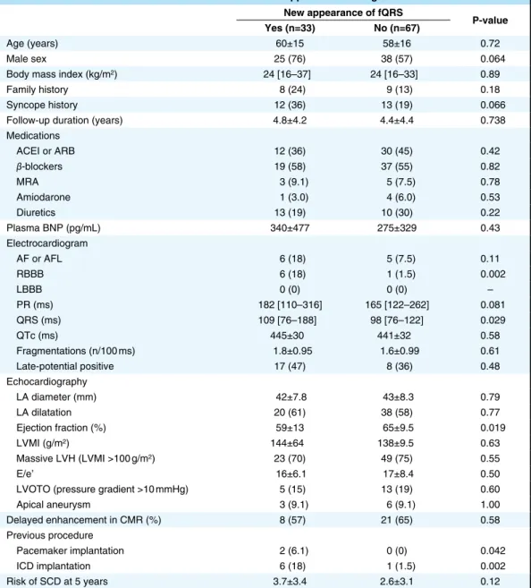
Table 2. Characteristics of Patients With and Without New Appearance of Fragmented QRS New appearance of fQRS
P-value Yes (n=33) No (n=67)
Age (years) 60±15 58±16 0.72
Male sex 25 (76) 38 (57) 0.064
Body mass index (kg/m2) 24 [16–37] 24 [16–33] 0.89
Family history 8 (24) 9 (13) 0.18
Syncope history 12 (36) 13 (19) 0.066
Follow-up duration (years) 4.8±4.2 4.4±4.4 0.738
Medications
ACEI or ARB 12 (36) 30 (45) 0.42
β-blockers 19 (58) 37 (55) 0.82
MRA 3 (9.1) 5 (7.5) 0.78
Amiodarone 1 (3.0) 4 (6.0) 0.53
Diuretics 13 (19) 10 (30) 0.22
Plasma BNP (pg/mL) 340±477 275±329 0.43
Electrocardiogram
AF or AFL 6 (18) 5 (7.5) 0.11
RBBB 6 (18) 1 (1.5) 0.002
LBBB 0 (0) 0 (0) –
PR (ms) 182 [110–316] 165 [122–262] 0.081
QRS (ms) 109 [76–188] 98 [76–122] 0.029
QTc (ms) 445±30 441±32 0.58
Fragmentations (n/100 ms) 1.8±0.95 1.6±0.99 0.61
Late-potential positive 17 (47) 8 (36) 0.48
Echocardiography
LA diameter (mm) 42±7.8 43±8.3 0.79
LA dilatation 20 (61) 38 (58) 0.77
Ejection fraction (%) 59±13 65±9.5 0.019
LVMI (g/m2) 144±64 138±9.5 0.63
Massive LVH (LVMI >100 g/m2) 23 (70) 49 (75) 0.55
E/e’ 16±6.1 17±8.4 0.50
LVOTO (pressure gradient >10 mmHg) 5 (15) 13 (19) 0.60
Apical aneurysm 3 (9.1) 6 (9.1) 1.00
Delayed enhancement in CMR (%) 8 (57) 21 (65) 0.58
Previous procedure
Pacemaker implantation 2 (6.1) 0 (0) 0.042
ICD implantation 6 (18) 1 (1.5) 0.002
Risk of SCD at 5 years 3.7±3.4 2.6±3.1 0.12
Data are expressed as the mean ± SD, median [interquartile range], or n (%). CMR, cardiac magnetic resonance (imaging); E/e’, ratio of transmitral Doppler early filling velocity to tissue Doppler early diastolic mitral annular velocity.
Other abbreviations as in Table 1.
ance of fQRS was associated with VAEs and hospitalization for heart failure. The incidence of VAEs in patients with new appearance of fQRS was as high as that in patients with baseline fQRS (i.e., on the initial ECG; 42% vs. 52%, respectively). If the appearance of fQRS indicates progres- sion of myocardial injury and increased risk of VAE and SCD, it may be possible to determine whether ICD implan- tation is required by evaluating normal follow-up ECGs.
Regarding heart failure, the new appearance of fQRS was associated with heart failure-related hospitalization (24%
vs. 7.5% in those with and without fQRS, respectively;
P=0.019), although baseline fQRS was not associated with heart failure-related hospitalization (20% vs. 13%, respec- tively; P=0.30) despite the lower ejection fraction. This may be explained by the size, distribution, and rate of progression of regional scars relative to the entire ventricular myocardium. Chronic scars representing baseline fQRS may be too small and too widely distributed to affect global left ventricular function to result in heart failure-related hospitalization. However, secondary to progression, the emergence of minor regional scars representing the new appearance of fQRS could have caused the heart failure events.
meta-analysis showed that baseline fQRS significantly increased the risk of major arrhythmic events in HCM patients.16 An observational study showed that a myocar- dial scar visualized by CMR with LGE predicted major adverse events in HCM patients,10 but the cost and avail- ability of CMR prevents repetitive testing during long-term follow-up. Thus, more convenient and safer clinical measures to assess cardiac event risks in HCM patients are needed. fQRS is expected to be a useful predictor of VAEs because fQRS can be obtained from a standard normal resting ECG; however, problems remain for patients without baseline fQRS. In some HCM patients without baseline fQRS, we saw the new appearance of fQRS during follow-up. An association between the new appearance of fQRS and adverse events has been reported in adult congenital heart disease,20 and 1 case report discussed an HCM patient without baseline fQRS who developed VAEs with the new appearance of fQRS 7 years after the initial ECG.17 To use fQRS as a risk predictor for HCM patients, the association between the new appearance of fQRS and VAEs must be clarified.
The new appearance of fQRS occurred in 33% of patients without fQRS during the 4.6-year follow-up. The appear-
Figure 5. Kaplan-Meier curves for ventricular arrhythmic events (VAEs) in patients with and without new appearance of fragmented QRS (fQRS). CI, confidence interval; HR, hazard ratio.
Table 3. ORs for the Occurrence of VAEs
Univariate Multivariate
OR (95% CI) P-value OR (95% CI) P-value
Age (over 65 years) 1.18 (0.59–2.37) 0.65
Male sex 0.89 (0.43–1.81) 0.74
Family history 2.52 (1.11–5.72) 0.03 1.08 (0.40–2.94) 0.88
Massive LVH 1.48 (0.63–3.49) 0.36
Syncope history 1.39 (0.61–3.16) 0.44
LVOTO 0.68 (0.25–1.86) 0.45
LA dilatation 2.02 (0.95–4.28) 0.65
New appearance of fQRS 4.75 (1.77–12.7) 0.001 4.29 (1.81–10.2) 0.001 All variables with a significant association in univariate analysis were included in the multivariate analysis. The odds ratio (OR), 95% confidence interval (CI), and P-value are given for each independent variable. VAEs, ventricular arrhythmic events. Other abbreviations as in Table 1.
Cardiac Death Prevention in Hypertrophic Cardiomyopathy (EVIDENCE-HCM). Circulation 2018; 137: 1015 – 1023.
5. Elliott P, McKenna WJ. Hypertrophic cardiomyopathy. Lancet 2004; 363: 1881 – 1891.
6. Elliott PM, Poloniecki J, Dickie S, Sharma S, Monserrat L, Varnava A, et al. Sudden death in hypertrophic cardiomyopathy:
Identification of high risk patients. J Am Coll Cardiol 2000; 36:
2212 – 2218.
7. Wang W, Lian Z, Rowin EJ, Maron BJ, Maron MS, Link MS.
Prognostic implications of nonsustained ventricular tachycardia in high-risk patients with hypertrophic cardiomyopathy. Circ Arrhythm Electrophysiol 2017; 10: e004604.
8. Adabag AS, Casey SA, Kuskowski MA, Zenovich AG, Maron BJ.
Spectrum and prognostic significance of arrhythmias on ambula- tory Holter electrocardiogram in hypertrophic cardiomyopathy.
J Am Coll Cardiol 2005; 45: 697 – 704.
9. Morita H, Watanabe A, Morimoto Y, Kawada S, Tachibana M, Nakagawa K, et al. Distribution and prognostic significance of fragmented QRS in patients with Brugada syndrome. Circ Arrhythm Electrophysiol 2017; 10: e004765.
10. Ratheendran AC, Subramanian M, Bhanu DK, Prabhu MA, Kannan R, Natarajan KU, et al. Fragmented QRS on electro- cardiography as a predictor of myocardial scar in patients with hypertrophic cardiomyopathy. Acta Cardiol, doi:10.1080/
00015385.2018.1547355.
11. Debonnaire P, Katsanos S, Joyce E, Van Den Brink OV, Atsma DE, Schalij MJ, et al. QRS fragmentation and QTc duration relate to malignant ventricular tachyarrhythmias and sudden cardiac death in patients with hypertrophic cardiomyopathy. J Cardiovasc Electrophysiol 2015; 26: 547 – 555.
12. Femenia F, Arce M, Van Grieken J, Trucco E, Mont L, Abello M, et al. Fragmented QRS as a predictor of arrhythmic events in patients with hypertrophic obstructive cardiomyopathy. J Interv Card Electrophysiol 2013; 38: 159 – 165.
13. Kang KW, Janardhan AH, Jung KT, Lee HS, Lee MH, Hwang HJ.
Fragmented QRS as a candidate marker for high-risk assessment in hypertrophic cardiomyopathy. Heart Rhythm 2014; 11: 1433 – 1440.
14. Nomura A, Konno T, Fujita T, Tanaka Y, Nagata Y, Tsuda T, et al. Fragmented QRS predicts heart failure progression in patients with hypertrophic cardiomyopathy. Circ J 2015; 79:
136 – 143.
15. Ozyilmaz S, Akgul O, Uyarel H, Pusuroglu H, Karayakali M, Gul M, et al. Assessment of the association between the presence of fragmented QRS and the predicted risk score of sudden cardiac death at 5 years in patients with hypertrophic cardiomyopathy.
Anatol J Cardiol 2017; 18: 54 – 61.
16. Rattanawong P, Riangwiwat T, Kanitsoraphan C, Chongsathidkiet P, Kanjanahattakij N, Vutthikraivit W, et al. Baseline fragmented QRS increases the risk of major arrhythmic events in hypertrophic cardiomyopathy: Systematic review and meta-analysis. Ann Noninvasive Electrocardiol 2018; 23: e12533.
17. Femenia F, Arce M, Arrieta M, Baranchuk A. Surface fragmented QRS in a patient with hypertrophic cardiomyopathy and malignant arrhythmias: Is there an association? J Cardiovasc Dis Res 2012;
3: 32 – 35.
18. Richardson P, McKenna W, Bristow M, Maisch B, Mautner B, O’Connell J, et al. Report of the 1995 World Health Organization/
International Society and Federation of Cardiology Task Force on the Definition and Classification of cardiomyopathies. Circulation 1996; 93: 841 – 842.
19. Nakamura K, Kusano KF, Matsubara H, Nakamura Y, Miura A, Nishii N, et al. Relationship between oxidative stress and systolic dysfunction in patients with hypertrophic cardiomyopathy.
J Card Fail 2005; 11: 117 – 123.
20. Helsen F, Vandenberk B, De Meester P, Van De Bruaene A, Gabriels C, Troost E, et al. Appearance of QRS fragmentation late after Mustard/Senning repair is associated with adverse outcome. Heart 2017; 103: 1036 – 1042.
Supplementary Files Please find supplementary file(s);
http://dx.doi.org/10.1253/circj.CJ-19-0968 Study Limitations
Several limitations of this study should be considered.
First, this was a single-center retrospective study that involved only 146 Japanese patients with HCM. Further investigation in a larger population is needed to definitively determine the association between VAEs and fQRS in HCM patients. Second, confounding factors may have existed between patients with and without fQRS in our cohort. There were differences in physiological functional test results and medications between the 2 groups, especially regarding ejection fraction and ECG parameters. Third, variability in the interpretation of fQRS is possible, but we used special precautions in this study to ensure that the ECG interpretations by the 3 reviewers were consistent with the fQRS criteria. Fourth, we did not show a clear mechanism for the relationship between fQRS and SCD risk. This study showed that VAEs in HCM patients were associated with both the presence and appearance of fQRS, but not with SCD. Nevertheless, we believe that identifying fQRS in a single normal ECG could be a useful clue to preventing SCD in HCM patients. Fifth, we did not analyze QRS fragmentations with differential or double differential values in this study. Because QRS fragmentations were smaller changes in the ECG, analysis with differential or double differential values could have helped us evaluate fQRS more scientifically. Further studies are needed to clarify this point.
Conclusions
The new appearance of fQRS in patients without baseline fQRS was a predictor of VAEs and myocardial injury in HCM patients.
Acknowledgements
The authors thank Miyuki Fujiwara and Masayo Ohmori for their excellent technical assistance and Nancy Schatken and Jane Charbonneau from the Edanz Group (www.edanzediting.com/ac) for editing a draft of this manuscript.
Sources of Funding This study did not receive any specific funding.
Conflict of Interests
None of the authors has any financial disclosures that are relevant to this article. H.I. is a member of the Editorial Board of Circulation Journal.
References
1. Miura K, Nakagawa H, Morikawa Y, Sasayama S, Matsumori A, Hasegawa K, et al. Epidemiology of idiopathic cardiomyopathy in Japan: Results from a nationwide survey. Heart 2002; 87:
126 – 130.
2. McKenna W, Deanfield J, Faruqui A, England D, Oakley C, Goodwin J. Prognosis in hypertrophic cardiomyopathy: Role of age and clinical, electrocardiographic and hemodynamic features.
Am J Cardiol 1981; 47: 532 – 538.
3. Elliott PM, Gimeno JR, Thaman R, Shah J, Ward D, Dickie S, et al. Historical trends in reported survival rates in patients with hypertrophic cardiomyopathy. Heart 2006; 92: 785 – 791.
4. O’Mahony C, Jichi F, Ommen SR, Christiaans I, Arbustini E, Garcia-Pavia P, et al. International external validation study of the 2014 European Society of Cardiology Guidelines on Sudden