Acta Med. Nagasaki 24:63-73
Regeneration in Cirrhosis of the Liver Following Partial Hepatectomy
Kenichiro SHIGEOKA
2nd Department of Surgery Nagasaki University School of Medicine
Received for publication, June 27, 1979
Distribution of regenerative hepatocytes was observed by autoradiographic study in at liver after partial hepatectomy in order to investigate the relationship between hepatic blood flow around the pseudolobule and regeneration in cirrhosis of the liver. In cirrhotic liver, pseudolobules were mostly supplied by hepatic arterial flow as most of portal blood flow were bypassed around the pseudolobule resulting in marked decrease of sinusoidal blood flow. Regenerative rate of liver remnant was not increased in cirrhotic hepatectomized group during the experimental period, while it was increased to 105.0±21.2% at 48 hours in normal hepatectomized group after operation (P<0. 01). Unelevated regenerative rate in cirrhotic liver may have been induced by marked decrease of sinusoidal portal blood
flow caused by intrahepatic portavenous shunt. As judged by H3-thymidine incorporation, DNA synthesis was increased to a maximum of 8335.1±1668.8 dpm/mg/min. at 36 hours in normal liver, and of 6538.9±2898.5 dpm/mg/min. at 48 hours in cirrhotic liver after partial hepatectomy (P<0.01). Average labeled hepatocyte number was also significantly increased to a peak of 48.0±9.1 at 36 hours in lobules of normal liver (P<0.001) and
of 12.9±4.8 at 48 hours in pseudolobules of cirrhotic liver after partial hepatectomy (P<
0.01). Dissociation of unelevated regenerative rate of liver remnant from increase of DNA synthesis in cirrhotic hepatectomized group may indicate that cell volume stimulating factor is entirely different from cell number stimulating factor, and each factor plays a role in- dividually. Direction of spreading of regeneration in normal liver was from peripheral zone to central zone, while spreading of regeneration in cirrhotic liver did not have specific direction in the pseudolobule. Direction of spreading of regeneration in normal hepatecto- mized group may substantiate the relation between direction of intralobular blood flow and
liver regeneration. Nonspecific distribution in cirrhotic liver which has no direction of regeneration, indicates that regeneration does not always need the portal blood flow and can be supported by hepatic arterial flow which is expected to contain the portal blood
factor after systemic circulation.
重岡 健 一郎
INTRODUCTION
Factors which control the liver regeneration following partial hepatectomy have been studied by many workers. Hepatic blood volume (16) and humoral factors (2)(4)(8)(24) (29) were experimentally pointed out as extracellular factors which regulate the liver re- generation in normal liver. However, such regulatory factors in cirrhotic liver has not been well investigated, except some reports regarding capacity of regeneration. (5) (10) (11) (15) (17) (23) (25) (27) . Liver cell carcinomas are frequently associated with cirrhosis of the liver (7) (12) (18) and have been resected successfully due to advances of diagnostic and surgical technics and postoperative management. Therefore, it seems important to study the regulatory mechanism in relation to specific environment such as vascular system in cirrhotic liver. In the present report, distribution of regenerative hepatocytes in pseudolo- bules was examined by autoradiographic study in cirrhotic rats in order to clarify the relationship between intralobular regeneration and intrahepatic vascular system around the pseudolobule.
MATERIALS AND METHODS
(1) Experimental Animals
Male Wistar albino rats, weighing 230-260 gm were used. Cirrhosis of the liver was produced by i. m. injection of 1 ml per 100 gm body weight of 20 per cent CCL4- olive oil twice a week for 3 months. Sixty-three animals which showed obvious cirrhosis of the liver microscopically which consisted of fibrous connective tissue and pseudolobule, were operated on at 8 days after discontinuation of oil injection. Normal 48 rats, weighing 250-350 gm, were used as control.
(2) Operation
Following fasting for 5 hours, either left lateral lobectomy of the liver or sham operation in which the liver was exposed and abdomen was closed without hepatectomy, was performed under ether anesthesia. After operation, the animals were offered food and water freely up to the time when they were sacrificed. Each rat was given intraperi- toneally 6-methyl-H3-thymidine (specific activity 5 Ci/mmol) in dose of 1 pCi/gm body weight at 1 hour before sacrifice. Experimental animals were divided into four groups ; normal hepatectomized, normal sham, cirrhotic hepatectomized, cirrhotic sham. The animals were sacrificed at 12, 24, 36 and 48 hours and 4 and 7 days after operation, respectively. Remaining liver was excised from each rat and the weight was recorded.
In sham operation groups, left leteral lobe was removed and weighed. Small liver samples from each rat were weighed in wet condition and immersed in 1 ml of tissue solubilizer (Soluene 350).
Aside from the above experiment, investigation of the distribution of portal vein and hepatic artery in both normal and cirrhotic liver by dye injection technic was per- formed. About 7 ml of India ink was infused into portal vein after ligation of hepatic
artery, and into hepatic artery by direct heart injection after ligation of portal vein, using wings-equiped needle under ether anesthesia. After fixation by 10% neutral formalin,
liver specimens were embedded in paraffin, cut 25p thick and mounted.
(3) H3-thymidine Incorporation into DNA
Nine ml of Instagel was used scintillator and incorporation of isotope was measured in Aloka LSC 602 liquid scintillation spectrometer. Value of the activity was corrected by external standard method.
(4) Autoradiographic Study
After fixation in 10% neutral formalin, liver specimens were embedded in paraffin and cut 4p thick. These sections were deparaffined, dipped in the photographic emulsion (Sakura NR-M2), dried and stored in darkness at 4°C for 5 weeks. Then, each slide was developed and stained with hematoxylin and eosin by Harris' method.
Labeled hepatocyte number was counted in the lobules of normal liver and in the pseudolobules of cirrhotic liver. To determine the distribution of labeled hepatocytes, such cells were marked on the paper sketching the lobule in normal liver or the pseudolobule in cirrhotic liver. Circles, the diameter of which equaled one-third and two-third of the distance between portal area and central vein of lobule, and between perilobular area and central point of pseudolobule, were drawn on the map, with the use of an ocular reticule under a microscope. Thus, lobules and pseudolobules were divided into three areas, namely peripheral, middle and central zones. Labeled hepatocytes in all three areas were totaled and the numbers in each zone were expressed as per cent of the total number.
(5) Regenerative Rate of Liver Remnant
Average percentage of weight of left lateral lobe to whole liver in normal and cirrho- tic sham groups, were 29.7 per cent, and 36.0 per cent respectively. Regenerative rate was estimated by percentage of weight of liver remnant to calculated whole liver weight.
Change of body weight during experimental period was not taken into consideration.
(6) Statistical Analysis
The statistical comparison for significance was carried out utilizing the Student t test, and a P value of less than 0.05 was considered to be significant.
RESULTS
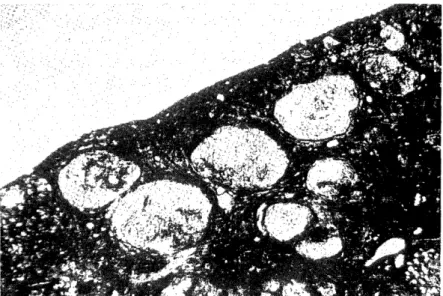
(1) Distribution of portal vein and hepatic artery shown by dye injection technic (Fig.1) In normal liver, the india ink was diffusely distributed into the lobule, whichever route, arterial or portal venous, was selected. However, in cirrhotic liver, the india ink which was infused into portal vein, was distributed only slightly into the pseudolobules, but abundantly at the perilobular spaces. While, the dye which was infused into the hepatic artery, was distributed into the pseudolobules diffusely.
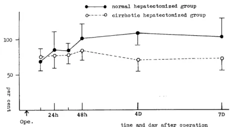
(2) Regenerative rate of liver remnant (Fig. 2)
Regenerative rate was increased in normal hepatectomized group to 105.0:i:21.2%0 at 48 hours after partial hepatectomy, while it was not increased in cirrhotic hepatectomized group for as long as seven days after operation. Regenerative rate in normal hepatectomized
Distribution of portal vein
Distribution of hepatic artery
Fig. 1 Distribution of portal vein and hepatic artery shown by dye injection technic India ink which was infused into portal vein, was distributed only slightly into
the pseudolobules, but abundantly at the perilobular spaces. While, the dye which was infused into the hepatic artery, was distributed into the pseudolobules
diffusely.
Fig. 2 Regenerative rate of liver remnant
Regenerative rate was increased in normal hepatectomized group to 105.0±21.2%
at 48 hours after partial hepatectomy, while it was not increased in cirrhotic hepatectomized group for as long as seven days after operation. Regenerative
rate in normal hepatectomized group was significantly higher than that in cirrhotic hepatectomized group (P<0.01). Mean ±S. E. (vertical lines), normal liver
group : n=4 at 12hrs, n=5 at 24hrs, n=5 at 36hrs, n=5 at 48hrs, n=4 at 4
days, n=5 at 7days, cirrhotic group : n=6 at 12hrs, n=7 at 24hrs, n=5 at 36hrs,
n=7 at 48hrs, n=5 at 4days, n=5 at 7days.
group was significantly higher than that in cirrhotic hepatectomized group (P<0.01).
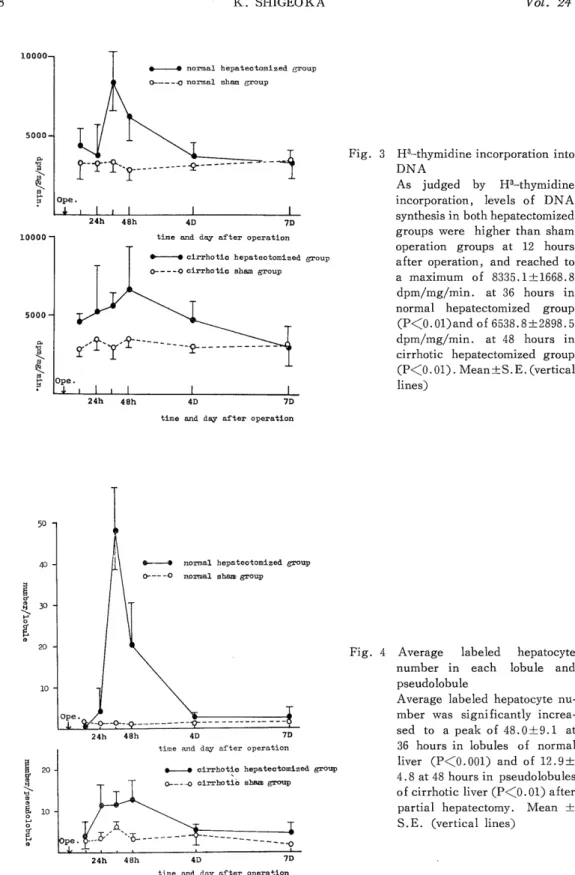
(3) H3-thymidine incorporation into DNA (Fig. 3)
As shown by Fig. 3, levels of DNA synthesis in both hepatectomized groups were higher than sham operation groups at 12 hours after operation, and reached to a maximum of 8335.1--L1668.8 dpm/mg/min. at 36 hours in normal hepatectomized group (P<0.01) and of 8538.9±2898.5 dpm/mg/min. at 48 hours (P<0.01) in cirrhotic hepatectomized group. The peak DNA synthetic response in cirrhotic hepatectomized group was lower than normal hepatectomized group.
(4) Autoradiographic study (Fig. 4, 5 and 6)
Average labeled hepatocyte number was significantly increased to a peak of 48.0±
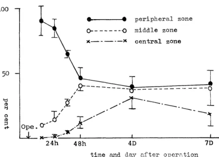
9.1 at 36 hours in lobules of normal liver compared with the normal sham group (P<0.001) and of 12.9±4.8 at 48 hours in pseudolobules of cirrhotic liver compared with the cirrhotic sham group (P<0.01) after partial hepatectomy similar to the result of H3-thymidine in- corporation. In normal hepatectomized group (Fig. 5), there was prominent localization of labeled hepatocytes in peripheral zone for 36 hours after operation, and from 48 hours such localization disappeared as the results of decrease of labeled hepatocytes in peripheral zone and increase in middle and central zones.
In cirrhotic hepatectomized group (Fig. 6), percentage of the labeled hepatocytes in each zone of the pseudolobules was the most prominent in peripheral zone, the least in central zone and in-between in middle zone. The rates of these localization in three zones
Fig. 3 H3-thymidine incorporation into DNA
As judged by H3-thymidine
incorporation, levels of DNA synthesis in both hepatectomized
groups were higher than sham operation groups at 12 hours
after operation, and reached to
a maximum of 8335.1±1668.8
dpm/mg/min. at 36 hours in normal hepatectomized group
(P<O. 01) and of 6538.8±2898.5
dpm/mg/min. at 48 hours in cirrhotic hepatectomized group
(P<O. 01). Mean ±S. E. (vertical
lines)
Fig. 4 Average labeled hepatocyte number in each lobule and
pseudolobule
Average labeled hepatocyte nu- mber was significantly increa-
sed to a peak of 48.0±9.1 at
36 hours in lobules of normal
liver (P<0.001) and of 12.9±
4.8 at 48 hours in pseudolobules
of cirrhotic liver (P<0.01) after partial hepatectomy. Mean ±
S.E. (vertical lines)
Fig. 5 The time course of distribution of labeled hepatocytes in lobules in normal hepa- tectomized group
There was prominent localization of labeled hepatocytes in peripheral zone for 36 hours after operation, and from 48 hours such localization disappeared as the
results of decrease of labeled hepatocytes in peripheral zone and increase in
middle and central zones.
Mean±S.E. (vertical lines), n=4 at 12hrs, n=5 at 24hrs, n=5 at 36hrs, n=5 at 48hrs, n=4 at 4days, n=5 at 7days.
Fig. 6 The time course of distribution of labeled hepatocytes in pseudolobules in cirrhotic hepatectomized group
Percentage of the labeled hepatocytes in each zone of the pseudolobules was the most prominent in peripheral zone, the least in central zone and in-between in
middle zone. The rates of these localization in three zones were almost stationary
during the experimental period.
Mean±S.E. (vertical lines), n=6 at 12hrs, n=7 at 24hrs, n=5 at 36hrs, n=7 at 48hrs, n=5 at 4days, n=5 at 7days
were almost stationary during the experimental period. The results which the percentage of labeled hepatocytes in three zones were not increased in spite of increase of average
labeled hepatocytes in pseudolobules after partial hepatectomy in cirrhotic liver as shown in Fig. 4, implicate that rates of increased labeled hepatocytes in three zones were almost same.
DISCUSSION
The control of liver regeneration may be considered to be done by intracellular and extracellular regulation(31). Intracellular regulation is thought to exist and to be a control mechanism within the cell which initiate and mediate the change in nucleic acids and in protein synthesis associated with cell division(9).
Extracellular regulation relates to conditions arising outside of a liver cell which promote cell division and cell enlargement, and hepatic blood volume(6) (16) and humoral factor are proposed as extracellular factors. As humoral factor, for instance, insulin(3) (30), glucagon(3) (24) (34) and portal blood factor (P . B. F) (8) have been reported. In this study, in order to investigate the relationship between hepatic blood flow and regene- ration in cirrhotic liver, distribution of regenerative hepatocytes was observed by autoradio- graphic method in both normal and cirrhotic livers in rats after partial hepatectomy.
As shown by Fig. 4, 5 and 6, there was increase of labeled hepatocyte after partial hepatectomy in both normal and cirrhotic livers. Direction of spreading of regeneration in normal liver was from peripheral zone to central zone, while in cirrhotic liver such direction of spreading was not seen. The rates of labeled hepatocyte in three zones were almost constant throughout the experiment, in spite of demonstrating an increase of average labeled hepatocytes after partial hepatectomy. These results indicate that rates of increased labeled hepatocytes in three zones in cirrhotic liver were the same.
Rabes(28) concluded from the proliferative differences in time and intensity within lobule in his autoradiographic study in normal rat liver after partial hepatectomy that microenvironment related to distribution of blood flow in liver lobules, would determine a topography of optimal condition for cell proliferation. Sigel(32) reported that localization of new hepatocyte formation could not be attributed to an inherently greater capacity of peripheral liver cell to divide but was probably related to their preferential exposure to blood constituent changes from his experiment which labeled hepatocytes were more prevalent about the central vein in the "reverse" blood flow graft.
In liver cirrhosis, portal blood flow circuits the pseudolobules via intrahepatic shunt which was reported to be specific finding of vascular change of liver cirrhosis with reduction of vascular bed (1) (13) (14) (19) (20) (26). Koo(13) and Kretz(14) reported that regene- rative nodules in liver cirrhosis were mainly supplied by hepatic arterial flow. As shown by Fig. 1, portal blood flow perfuses mainly perilobular area of the pseudolobules resulting in marked decrease of sinusoidal portal blood flow, and pseudolobules are mostly supplied by hepatic arterial flow.
Weinbren(35) showed that portal blood diversion away from a remnant of liver tissue after partial hepatectomy did not impair regeneration. Fisher(8) reported that there was a portal blood factor (P.B.F.) which was capable of stimulating hepatic cells and was neither destroyed nor inhibited in systemic blood. Bucher(2) and Price(24) showed that hepatic artery alone was capable of supporting liver cell proliferation. Thus, the same increasing rates of labeled hepatocytes in three zones in cirrhotic liver after partial hepatecto- my as shown by Fig. 4, may substantiate that liver regeneration is not always affected by contact with portal blood flow, and can be stimulated by hepatic arterial flow which is expected to contain the portal blood factor after systemic circulation. Because, if the portal blood flow only could stimulate the liver regeneration, the increasing rate of labeled hepatocyte in the peripheral zone should be higher than the other two.
There was no elevation of regenerative rate though there was increase of DNA synthesis in cirrhotic liver after partial hepatectomy. Fisher(8) reported that volume of the portal blood flow was related to hypertrophy and humoral factor was related to hyper- plasia. Malamud(22) suggested that cellular hypertrophy and cellular hyperplasia were independent each other. Dissociation of unelevated regenerative rate of liver remnant from increase of DNA synthesis may indicate that cell volume stimulating factor is not identical with cell number stimulating factor, and each factor plays a role individually. Unelevated regenerative rate of liver remnant in cirrhotic liver may be induced by marked decrease of sinusoidal portal blood flow which is caused by intrahepatic portavenous shunt. Volume of sinusoidal blood flow in cirrhotic liver may affect liver regeneration and restoration of postoperative liver function in critical stage after partial hepatectomy. Then, clinically it may be useful for estimating the indication of hepatic resection of liver cell carcinoma associated with liver cirrhosis to calculate the intrahepatic shunting rate before operation.
Furthermore, it may be recommended to perform periarterial neurectomy of the hepatic artery which was reported that it would increase the hepatic arterial blood flow(21), bring a good effect to postoperative course(36) and promate liver regeneration after hepatic surgery(33).
ACKNOWLEDGEMENT
Grateful acknowledgement is made to Prof. R. Tsuchiya for his constant guidance and encouragement. The auther wish also to express his gratitude to Assist. Prof. T.
Itoh and Dr. R. Nishimura for their kind advices.
REFERENCES
1) ALPERT L. I. et al ; Anatomic and Functional Circulatory Change in Experimental Cirrhosis. Gastroenterology 48 ; 579, 1966
2) BuCHER N. L. R. and MIRIAN N.; Regeneration of Liver in Rats in the Abscence of Portal Splanchnic Organs and a Portal Blood Supply, Cancer Res. 33 : 3189, 1973
3) BUCHER N. L. R. and SWAFF'IELD M. N. ; Synergistic Action of Glucagon and Insulin in Regulation of Hepatic Regeneration. Adv. Enz. Deg. 13 ; 281, 1975 4) CHRISTENSEN B. G. and JACOBSON E. ; Studies on Liver Regeneration. Acta Med.
Scand. Suppl. 234 : 103, 1949-1950
5) CAMERON G. R. and KARUNARATNE W. A. E. ; Carbon-Tetrachloride Cirrhosis in Relation to liver Regeneration. J. Path. Bact. 42(1) : 1, 1936
6) CHILD C. G., BARR D., HOLSWADE G. R. and HARRISON C. S. ; Liver Regeneration
following Portacaval Transposition in Dogs. Ann. Surg. 138(4) : 600, 1953 7) EDMONDSON H. A. and STEINER P. E. ; Primary Carcinoma of the Liver. A Study
of 100 Cases among 48900 Necropsis. Cancer 7(3) ; 462, 1954
8) FISHER B., LSZHCH P. and FISHER E. R. ; Evaluation of a Humoral Factor in Liver Regeneration utilizing Liver Transplant. Cancer Res. 31 : 322, 1971
9) FUJIOKA M., KOGA M. and LIEBERMAN I. Metabolism of Ribonucleic Acid after Partial Hepatectomy. J. Biol. Chem. 238(10) : 3401, 1963
10) HANEY A., PEACOCK J. E. E. and MADDEN J. W. ; Liver Regeneration and Hepatic Collagen Deposition in Rats with Dimethylnitrosamin-induced Cirrhosis. Ann. Surg.
175(6) : 863, 1972
11) ISLAMI A. H., PACK G. T. and HUBBARD J. C. ; Regenerative Hyperplasia of the Cirrhotic Liver following Partial Hepatectomy. Cancer 11(4) : 663, 1958
12) KOHNO A., MIZUMOTO R. and HoNJO I. ; Changes after Major Resection of Experi- mental Cirrhotic Liver. Amer. J. Surg. 134 : 248, 1977
13) Koo A., LIANG I. Y. S. and CHENG K. K.; Intrahepatic Microvascular Changes in CC14-induced Cirrhotic Liver in the Rat. AJeBak 54 : 277, 1976
14) KRETZ R. ; Cirrhosis of the Liver. Internat, Clin. 3 : 289, 1905
15) LIN T. Y. et al; Metabolic Function and Regeneration of Cirrhotic and Noncirrhotic
Livers after Hepatic Lobectomy in Man. Ann. Surg. 162(6) : 959, 1965
16) MANN F. C. ; Portal Circulation and Restoration of Liver after Partial Removal.
Surgery 8 : 225, 1940
17) MANN F. C. et al ; Experimental Pathology of Liver. Arch. Path. 12(5) : 787, 1931
18) MACDONALD R.A. et al ; Primary Carcinoma of the Liver. A Clinicopathologic Study of One hundred Eight Cases. Arch. Ind. Med. 99 : 266, 1957
19) MITRA S. K. ; Hepatic Vascular Change in Human and Experimental Cirrhosis. J.
Path. Bact. 92 : 405, 1966
20) MANN J. D., WAKIN K. G. and BAGGENSTOSS A. H. ; Alteration the Vasculature
of the Diseased Liver. Gastroenterology 25 : 540, 1953
21) MAKINO Y.; Effect of Hepatic Periarterial Neurectomy upon Hepatic Blood Flow and Regeneration of the Liver after Partial Hepatectomy in the Dog. Arch. Fur.
Japa. Chiru. 37 : 485, 1968
22) MALAMUD D., GONZALEG E. M., CHIN H. and MALT R. A. ; Inhibition of Cell Proliferation by Azathioprine. Cancer Res. 32 : 1226, 1972
23) OLSON R.: Partial Hepatectomy in Experimental Carbon-Tetrachloride induced Liver Fibrosis. Acta Chir. Scand. 366(1) : 1, 1966
24) PRICE T. B., TAKASHIGE K., MAX M. H. and VOORHEES A. B. ; Glucagon as the Portal Factor modifying Hepatic Regeneration. Surgery 72(1) : 74, 1972
25) PECHET G. and MACDONALD R.A. ; Repair of Nutritional Cirrhosis. Autoradiographic
and Histological Study after Partial Hepatectomy. Cancer 14(5) : 963, 1961
26) POPPER H., ELIAs H. and PETTY DAVID E. ; Vascular Pattern of the Cirrhotic Liver.
Am. J. Clin. Path. 22 : 717, 1952
27) RABINOvICi N. and WIENER E. ; Liver Regeneration after Partial Hepatectomy in Carbon-Tetrachloride induced Cirrhosis in the Rat. Gastroenterology 40 : 410, 1961 28) RABES H. and TUCZEK H.V. ; Quantitative Autoradiographic Study of Heterogeneity
of Liver Cell Proliferation After Partial Hepatectomy. Virchows Arch. Abt. B Zellpath 6 : 302, 1970
29) STARZEL T. E., PORTER K. A., KASHIWAGI N. and PUTNAM C. W. ; Portal Hepatotrophic Factors, Diabetes Mellitus and Acute Liver Atrophy, Hypertrophy and
Regeneration. Surg. Gynecol. Obstet. 141(6) : 843, 1975
30) STARZEL T. E. et al; The Origin, Hormonal Nature, and Action of Hepatotrophic
Substances in Portal Venous Blood. Surg. Gynecol. Obstet. 137 : 179, 1973
31) SIGEL B.; Current Research Review. The Extracellular Regulation of Liver Regenera- tion. J. Surg. Res. 9(6) : 387, 1969
32) SIGEL B., BALDIA L. B., BRIGHTMAN S. A., DUNN M. R. and PRICE R. I. M. ; Effects of Blood Flow Reversal in Liver Autotransplants upon the Site of Hepatocyte
Regeneration. J. Clin. Invest. 47 : 1231, 1968
33) TsuCHIYA R. ; Hepatectomy, Vascular Ligation and Liver Regeneration. Nippon Igakukai Soukai Kaishi. No. 19, 1200-1201 (in Japanese)
34) WHITTEMORE A. D., KASUYA M., VOORHEES H. B. and PRICE J. B. ; Hepatic Regeneration in the Abscence of Portal Visceras. Surgery 77(3) : 419, 1975
35) WEINBREN K. ; The Portal Blood Supply and Regeneration of the Rat Liver. Brit.
J. Exp. Path. 36 : 583, 1955
36) WEXLER M. et al; Regeneration and Maintenance of Integrity of Canine Liver. The Role of Arterial, Portal and Systemic Venous Blood. Arch. Surg. 101 : 267, 1970