Acta Med. Nagasaki 58: 131−134−
Introduction
Intrahepatic cholangiocarcinoma (ICC) is a comparative- ly rare cancer and one of the most difficult cancers to treat.
Although surgical resection is the best therapeutic strategy for this malignant disorder, the long-term outcome after surgical intervention remains unfavorable because of fre- quent local and/or regional lymph node recurrences [1, 2].
The 5-year survival rate for patients with curative resection is 20-40% [1-5]. Heterochronous metastasis to the adrenal glands from ICC is uncommon, and to the best of our knowl- edge no resected cases have been reported. We report herein a case of adrenalectomy for heterochronous metastasis from ICC.
Case Report
A 61-year-old Japanese man who had been followed-up for ICC after curative resection at our hospital was diag- nosed with a nodule in the left adrenal gland. At the age of 58 years, he had undergone right hepatic lobectomy with regional lymph node resection for nodular-type ICC in he- patic segment 6. The histopathologic findings of the liver tumor had showed poorly differentiated adenocarcinoma without metastatic lymph node (T2N0M0 StageⅡ ). The pa- tientʼs post-operative course had been uneventful. The pa- tient had received post-operative adjuvant chemotherapy with gemcitabine for 6 months. Three years after the initial surgery, abdominal computed tomography (CT) revealed a nodule in the left adrenal gland (Fig. 1). In addition, posi- tron emission tomography/computed tomography (PET/CT) showed significant fluorodeoxyglucose (FDG) uptake at the
MS#AMN 07142
Case Report
Heterochronous Adrenal Metastasis from Intrahepatic Cholangiocarcinoma:
Report of a Case
Yasuhiro M
aruya1, Tamotsu K
uroKi1, Tomohiko A
dachi1, Tatsuya o
KaMoto1, Kuniko a
be2, Takashi K
aneMatsu1, and Susumu e
guchi11 Department of Surgery, Nagasaki University Graduate School of Biomedical Sciences, 1-7-1 Sakamoto, Nagasaki 852-8501, Japan
2 Department of Pathology, Nagasaki University Graduate School of Biomedical Sciences, 1-7-1 Sakamoto, Nagasaki 852-8501, Japan
We herein report a case of laparoscopic adrenalectomy for solitary adrenal metastasis from intrahepatic cholangiocarci- noma (ICC). Although the treatment of extrahepatic metastasis from primary liver tumors is essentially palliative, solitary metastasis from such tumors offers the possibility of a cure by surgical resection. The adrenal gland is an uncommon site for metastasis from ICC. A patient who had received a right hepatic lobectomy with lymph node resection for ICC later developed a heterochronous adrenal metastasis, and thus underwent laparoscopic adrenalectomy 30 months after the liver resection. To the best of our knowledge, this is the first case of adrenalectomy for heterochronous metastasis from ICC.
ACTA MEDICA NAGASAKIENSIA 58: 131−134, 2014 Key words: intrahepatic cholangiocarcinoma, adrenal metastasis, laparoscopic adrenalectomy
Address correspondence: Tamotsu Kuroki, Department of Surgery, Nagasaki University, Graduate School of Biomedical Sciences, 1-7-1 Sakamoto, Nagasaki 852-8501, Japan.
Tel: +81-95-8197316, Fax: +81-95-8197319
Received October 9, 2013; Accepted November 26, 2013
132 Yasuhiro Maruya et al.: adrenal metastasis, ICC
left adrenal gland [standardized uptake rate (SUV) MAX early phase=5.6, late phase=8.1], with suspicion of a malig- nant tumor (Fig. 2). Routine hematologic and biochemical examinations were normal. Serum levels of carcinoembry- onic antigen and carbohydrate antigen 19-9 were within normal limits. Adrenal hormone, including vanillylman- delic acid in the urine, 4 renin, aldosterone, and adrenocor- ticotropic hormone, were all within normal limits. Serum and urine laboratory data showed that the mass was non- functioning. Reevaluation 4 months later with contrast-en- hanced CT demonstrated a progressively enlarging solitary nodule in the left adrenal gland. However, no swollen lymph nodes, local recurrences, or distant metastases were noted.
Therefore, we diagnosed the nodule in the left adrenal gland as metastasis from ICC. Thirty months after the initial op- eration, the patient underwent a laparosopic left adrenalec- tomy operation by a laparoscopic left transperitoneal ap- proach, in order to establish a definitive diagnosis. Surgery was performed with the patient in the flank position under CO
2pneumoperitoneum at 12 cmH
2O. A 12 mm port was inserted at the umbilicus, and another 12 mm port was in- serted at a location of about 3 cm to the left of, and above, the umbilicus. Two 5 mm ports were also inserted: one at the lateral border of the rectus abdominis mid and the other under the left costal margin. The distal pancreas and the spleen were mobilized and retracted by a snake retractor (Mediflex
®) to expose the inferior left adrenal gland. The procedure required extensive adhesiolysis, but otherwise, dissection of the tumor proceeded without intraoperative 5 complications. The left adrenal vein and artery were identi- fied and divided by a LigaSure (Valleylab, Boulder, CO) device. The specimen was extracted through the umbilical
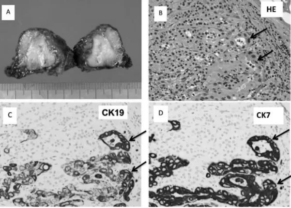
trocar site. The patient was discharged uneventfully on the 10th postoperative day without any complications. The cut surface of the surgical specimen showed a solid mass lesion measuring 3.0 cm× 3.5 cm (Fig. 3A). Histologically, the le- sion consisted of atypical epithelial cell proliferation with focal duct formation (Fig. 3B). The pathologic findings were similar to those of the primary lesion of cholangiocarcino- ma (data not shown).
Immunohistochemically, the tumor cells were positive for cytokeratin 19 (CK19) and cytokeratin 7 (CK7) (Fig. 3C, D). CK19 and CK7 are normally expressed in the lining of the gastro-entero-pancreatic and hepato-biliary tracts. Thus, the tumor in the left adrenal gland appeared compatible with metastatic cholangiocarcinoma. The patient received postoperative chemotherapy with seven cycles of S-1. Ten months after the second operation, para-aortic lymph node metastasis recurred, along with left kidney metastases and multiple liver metastases. The patient received palliative care but died of liver failure 15 months after the second op- eration.
Discussion
The adrenal gland is not a common site for metastases from ICC. Zheng et al. [6] reported that the majority of met- astatic lesions in the adrenal gland occurred after early de- tection of a primary tumor of which the lung was the most common primary tumor site, followed by kidney, liver, breast, melanoma, and others. The histological types of ad- renal metastasis of patients who underwent adrenalectomy were estimated as follows: small cell lung carcinoma, non-
Figure 1. Three years after the initial surgery, CT revealed a nod-ule, measuring 3.0 cm ×3.8 cm, in the left adrenal gland.
Figure 2. PET/CT showed significant FDG uptake at the left ad- renal gland [SUV MAX early phase = 5.6, late phase=8.1] and suspected malignant tumor.
133 Yasuhiro Maruya et al.: adrenal metastasis, ICC
small cell lung carcinoma, kidney clear cell carcinoma, he- patocellular carcinoma, melanoma, breast cancer, and un- known carcinoma. The worldwide literature to date on adrenalectomy for metastasis from ICC is limited to case reports. To our knowledge, a few cases of synchronous ad- renal metastasis from ICC have been described [7]. On the other hand, our case is the first report of adrenalectomy for heterochronous metastasis from ICC. Several reports have suggested that selected patients with isolated adrenal metas- tasis from non-small cell lung carcinoma can achieve long- term survival following adrenalectomy and definitive treat- ment of the primary tumor [8-10]. For non-small cell lung carcinoma patients, it was also found that patients with syn- chronous metastasis who underwent adrenalectomy had a shorter median overall survival than those with heterochro- nous metastasis for an isolated adrenal metastasis [10]. Cho- langiocarcinomas have often metastasized to distant organs, and
18F-FDG PET is valuable for discovering unsuspected distant metastases of cholangiocarcinoma [11-13]. There is no standardized SUV cutoff point for discriminating be- tween benign disease and cholangiocarcinoma, but there
are several studies about the usefulness of PET/CT for cho- langiocarcinoma. Ruys et al. [14] reported that, for hilar cholangiocarcinoma, patients with distant metastases had significantly higher SUVs for the primary tumor. Reinhardt et al. [15] reported that 3.6 is the ideal SUV cutoff point for discriminating between benign disease and cholangiocarci- noma. In our case, SUV MAX in the early phase was 5.6 and that in the late phase was 8.1, so this SUV point sug- gested that the left adrenal gland was a malignant tumor.
Because surgical resection is the only curative treatment for ICC,
18F-FDG PET may be useful for the appropriate man- agement of metastasis from ICC [16, 17].
Surgical resection of the liver is the only curative treat- ment for ICC patients; its 5-year survival rate is around 30
%, with a median overall survival of 2-3 years [1-5]. There is little support from evidence-based evaluation for the ef- ficacy of chemotherapy for ICC patients. However, recent advances facilitate the use of chemotherapy to achieve a re- sponse rate of around 30% and a median survival of more than one year for ICC patients. Key drugs currently avail- able for the therapy are gemcitabine, fluoropyrimidines, and
Figure 3A. The cut surface of the surgical specimen showed a solid mass lesion measuring 3.0 cm×3.5 cm.Figure 3B. Histologically, there were normal adrenocortical cells on the left upper side and atypical epithelial cell proliferation with focal duct formation (arrow) on the right lower side. The pathologic findings were simi- lar to those of the primary lesion of cholangiocarcinoma (hematoxylin and eosin staining, 14 ×100.) Arrows indicate tumor cells. Figure 3C, D. Immunohistochemically, the tumor cells were positive for cytokeratin 19 (CK19) (C) and cytokeratin7 (CK7) (D). Adrenocortical cells were negative for CK19 (C) and CK7 (D). (x100) Arrows indicate tumor cells.
134 Yasuhiro Maruya et al.: adrenal metastasis, ICC
References
1. Tajima Y, Kuroki T, Fukuda K, Tsuneoka N, Furui J, Kanematsu T. An intraductal papillary component is associated with prolonged survival after hepatic resection for intrahepatic cholangiocarcinoma. Br J Surg.
2004 Jan ;91(1):99-104
2. Nakagohri T, Kinoshita T, Konishi M ,Takahashi S, Gotohda N. Surgi- cal outcome and prognostic factors in intrahepatic cholangiocarcino- ma. World J Surg.2008 Dec;32(12):2675-80
3. Madariaga JR, Iwatsuki S, Todo S, Lee RG, Irish W, Starzl TE. Liver resection for hilar and peripheral cholangiocarcinoma: a study of 62 cases. Ann Surg.1998 Jan;227(1):70-9
4. Weber SM, Jarnagin WR, Klimstra D, DeMatteo RP, Fong Y, Blumqart LH. Intrahepatic cholangiocarcinoma: resectability, recurrence pat- tern, and outcomes. J Am Coll Surg. 2001 Oct; 193(4):384-91 5. Ohtsuka M , Ito H, Kimura F, Shimizu H, Togawa A, Yoshidome H,
Miyazaki M. Results of surgical treatment for intrahepatic cholangio- carcinoma and clinicopathological factors influencing survival. Br J Surg. 2002 Dec; 89(1):1525-31
6. Zheng QY, Zhang GH, Zhang Y, Guo YL. Adrenalectomy may in- crease survival of patients with adrenal metastases. Oncology Letters.
2012 Apr ; 3(4):917-20
7. Durgatosh P, Kai-Chah T. Surgical resection of adrenal metastasis from primary liver tumor: report of two cases. Hepatobiliary Pan- creat Dis Int. 2008 Aug ;7(4):440-2
8. Salah S, Tanvetyanon T, Abbasi S. Metastatectomy for extra-adrenal non- small cell lung cancer solitary metastases: Systematic review and analysis of reported cases. Lung Cancer. 2012 Jan;75(1):9-14
9. Raz DJ, Lanuti M, Gaissert HC, Wright CD, Mathisen DJ, Wain JC.
Outcomes of patients with isolated adrenal metastasis from non-small cell lung carcinoma. Ann Thorac Surg. 2011 Nov; 92(5):1788-92 10. Tanvetyanon T, Robinson LA, Schell MJ, Strong VE, Kapoor R, Coit
DG, Bepier G. Outcomes of adrenalectomy for isolated synchronous versus metachronous adrenal metastasis in non-small-cell lung can- cer: a systematic review and pooled analysis. J Clin Oncol.2008 Mar;
26(7):1142-7
11. Park SK, Kim YS, Kim SG, Jang JY, Moon JH, Lee MS et al. Detec- tion of distant metastasis to skeletal muscle by 18F-FDG PET in a case of intrahepatic cholangiocarcinoma. The Korean Journal of Hepatol- ogy. 2010 Sep; 16(3):325-8
12. Seo S, Hatano E, Higashi T, Hara T, Tada M, Tamaki N et al. Fluo- rine-18 fluorodeoxyglucose positron emission tomography predicts tumor differentiation, P-glycoprotein expression, and outcome after resection in hepatocellular carcinoma. Clin Cancer Res.2007 Jan ; 13(2):427-33
13. Kluge R, Schmidt F, Caca K, Barthel H, Hesse S, Georgi P et al. Posi- tron emission tomography with [(18)F]fluoro-2-deoxy-D-glucose for diagnosis and staging of bile duct cancer. Hepatology. 2001 May;
33(5):1029-35
14. Ruys AT, Bennink RJ, van Westreenen HL, Engelbrecht MR, Busch OR, Gouma DJ et al. FDG-positron emission tomography/computed tomography and standardized uptake value in the primary diagnosis of hilar cholangiocarcinoma. HPB. 2011 Apr;13(4):256-62
15. Reinhardt MJ, Strunk H, Gerhardt T, Roedel R, Jaeqer U, Bucerius J et al. Detection of Klatskinʼs tumor in bile duct structures using de- layed 18F-FDG PET/CT: preliminary results for 22 patient studies. J Nucl Med.2005 Jul; 46(7):1158-63
16. Anderson CD, Rice MH, Pinson CW, Chapman WC, Chari RS, Del- beke D. Fluorodeoxyglucose PET imaging in the evaluation of gall- bladder carcinoma and cholangiocarcinoma. J Gastrointest Surg.
2004 Jan; 8(1): 90-7
17. Breitenstein S, Apestequi C, Clavien PA. Positron emission tomogra- phy (PET) for cholangiocarcinoma. HPB.2008 Apr; 10(2):120-1 18. Okusaka T, Ishii H, Funakoshi A, Yamao K, Ohkawa S, Saito S et al.
Phase II study of single-agent gemcitabine in patients with advanced biliary tract cancer. Cancer Chemother Pharmacol. 2006 May;
57(5):647-53
19. Furuse J, Okusaka T, Boku N, Ohkawa S, Sawaki A, Masumoto T et al. S-1 monotherapy as first-line treatment in patients with advanced biliary tract cancer: a multicenter phase II study. Cancer Chemother Pharmacol.2008 Oct; 62(5):849-55
![Figure 2. PET/CT showed significant FDG uptake at the left ad- ad-renal gland [SUV MAX early phase = 5.6, late phase=8.1] and suspected malignant tumor.](https://thumb-ap.123doks.com/thumbv2/123deta/10138309.1972188/2.892.460.821.142.396/figure-showed-significant-uptake-renal-gland-suspected-malignant.webp)