◆
症例報告
◆
左蝶形骨縁髄膜腫術後に反対側に発生した
右海綿静脈洞部 dAVF の 1 例
瀬川 将史
1, 2)小野 秀明
1)青野 峻也
1)三谷 知広
1, 2)庄島 正明
3)谷島 健生
1)田村 晃
1)齋藤 勇
1)要旨:【緒言】硬膜動静脈瘻(dural arteriovenous fistula: dAVF)は硬膜内での異常な動静脈シャントで
あり,稀な疾患である.蝶形骨縁髄膜腫術後に対側海綿静脈洞部に発生した症候性の dAVF を経験
したので文献的考察を加えて報告する.【症例】69 歳,女性.ふらつき精査にて見つかった左蝶形骨
縁髄膜腫が経過の中で増大傾向を認め,開頭腫瘍摘出術を施行した.周術期合併症なく経過した
が,術半年後より右拍動性耳鳴を自覚,さらに半年後に複視が出現し,精査の DSA で海綿静脈洞部
dAVF
と診断した.経静脈的に塞栓術を施行し,シャントの完全閉塞が得られた.術後 1 年の段階で
症状の再発なく,髄膜腫,dAVF 共に再発なく良好に経過している.【結論・考察】髄膜腫摘出後に遠
隔に dAVF が生じた症例を経験し,硬膜面動静脈血流や頭蓋内血行動態の変化など複合的な発生機
序を考えた.文献的考察を加えて報告する.
Key words: dural arteriovenous fistula, meningioma, cavernous sinus
1)富士脳障害研究所附属病院脳神経外科 2)東京大学医学部附属病院脳神経外科 3)埼玉医科大学総合医療センター脳神経外科 責任著者:〒418-0021 静岡県富士宮市杉田 270-12 富士脳障害研究所附属病院脳神経外科 小野秀明 E-mail: [email protected] (2019 年 12 月 4 日受付,2020 年 2 月 17 日受理) doi: 10.3995/jstroke.10785
はじめに
硬膜動静脈瘻(dural arteriovenous fistula: dAVF)は硬膜
内での異常な動静脈シャントであり,発生頻度は頭蓋内
動静脈奇形の 10∼15%程度と比較的稀な疾患である.
成因ははっきりとわかっていないが,静脈洞血栓症,外
傷,開頭手術などが関連している症例が報告されてい
る
1–7).今回我々は蝶形骨縁髄膜腫術後に対側海綿静脈
洞をシャントポイントとして発生した症候性の dAVF を
経験したので,文献的考察を加えて報告する.
症 例
患者:69 歳,女性
既往歴:特記すべきことなし.
現病歴:6 年前にふらつき精査にて見つかった左蝶形
骨縁に主座を置く腫瘤が最大径 38 mm に増大し,精査
加療目的に入院となった.
神経学的所見:明らかな異常所見なし.
神経放射線学的所見:MRI 造影 T1 強調画像にて,左
蝶形骨縁に付着し均一に造影される最大径 38 mm の腫
瘍を認め,dural tail sign を呈していた(Fig. 1A).Digital
subtraction angiography
(DSA)では,左眼動脈を介した後
篩骨動脈と左中硬膜動脈が栄養血管として同定された.
その他,対側の総頸動脈,椎骨動脈撮影で血管異常を認
めなかった.また,静脈相ではシルビウス静脈の描出は
不良であった(Fig. 2A).以上より,髄膜腫を疑い手術
を行った.
腫瘍摘出手術所見:左前頭側頭開頭で腫瘍摘出術を施
行した.腫瘍は硬膜に付着しており,髄膜腫に矛盾しな
い所見であった.蝶形骨縁内側で肉眼的に正常硬膜が確
認できるところまで腫瘍の摘出と浸潤硬膜の焼灼を行っ
た(Simpson 分類 Grade III,Fig. 1B).腫瘍によって術側
の sylvian vein は圧排されており,腫瘍の摘出に伴って
良好な静脈の拡張が確認された(Fig. 2B).永久病理結果
は microcystic meningioma であった.
腫瘍摘出術後経過:術後 3 カ月での MRI で髄膜腫の
再発なく,MR angiography(MRA)にて明らかな異常を
認めなかった.術半年後より右拍動性耳鳴を自覚,他院
耳鼻科にて精査行うも明らかな異常を指摘されなかっ
脳卒中 43: 142–147, 2021
J-STAGE 早期公開 2020年7月20日
た.さらに半年後,右注視時に複視が出現し,精査加療
目的に入院となった.
血管内治療前神経学的所見:意識清明,右外転神経麻
痺,右拍動性耳鳴あり.明らかな眼球突出や充血はなし.
血管内治療前神経放射線学的所見:MRI T2 強調画像
で右眼静脈の拡張所見あり,MRA で右海綿静脈洞部の
描出を認めた(Fig. 1C, D).DSA で両側の dorsal
menin-geal artery,accessory meninmenin-geal artery から動脈相での右
海綿静脈洞の描出を認め,右眼静脈に導出されており,
海綿静脈洞部 dAVF と診断した(Fig. 3A–D).この際,
腫瘍摘出術前に描出不良であったシルビウス静脈が認め
られるようになった(Fig. 3E).
血管内治療手術所見:経静脈的に塞栓術を施行する方
針とした.右内頸静脈から下錐体静脈洞経由で海綿静脈
洞にマイクロカテーテルを誘導した.術中診断でシャン
トポイントは海綿静脈洞後部であると考えられたため,
同部位にマイクロカテーテルの誘導を試みるも困難で
あった.流出静脈の近位部の塞栓術を行う方針として,
Fig. 1 Pre/post-operative, follow up MRI
Preoperative T1-weighted axial image with gadolinium enhancement showed a solid mass at the left sphenoid ridge (A), and postoperative MRI (B). T2-weighted axial image 12 months after tumor resection showed dilatation of the right opthalmic vein (C), and MR angiography showed high signal intensity in right cavernous sinus (D).
Fig. 2 Preoperative DSA and intraoperative finding
Left sylvian vein was not identified on angiogram before tumor removal (A). Sylvian vein was compressed by the tumor and became dilated after tumor removal (B, white arrow).
眼静脈を離脱式コイルにより packing し,流出量を減ら
した.その段階で右シルビウス静脈への逆流の所見あ
り,こちらもコイルによる閉塞を追加した.さらに瘻孔
の存在が示唆される右海綿静脈洞部後部にもコイリング
を行い,これによりシャントの完全閉塞が得られた
(Fig. 3F).
血管内治療後経過:術直後より右外転神経麻痺と拍動
性の耳鳴は消失した.術後 1 年の段階で症状の再発な
く,髄膜腫,dAVF 共に再発なく良好に経過している.
症例報告にあたり患者の同意を得ている.
考 察
dAVF は硬膜動脈を主な feeding artery とし,硬膜静脈
洞あるいは脳表静脈に流出する動静脈シャントである.
その多くは後天性と考えられており,静脈洞血栓症,脳
梗塞,開頭手術,頭部外傷,感染症などに続いて起こる
ものが報告されている
1–7).
開頭術後の dAVF の報告例は,三浦らの報告以降散見
されており,われわれが渉猟した限りでは 19 文献 23 症
例(男性 11 名,女性 12 名,平均年齢 55 歳)が報告され
ている
1–4, 8–20)(Table 1).術前の原疾患としては,脳血管
障害が 10 例,機能的脳神経外科疾患が 4 例,頭蓋内腫
瘍が 8 例,感染症が 1 例であり,自験例のような髄膜腫
は 4 例であった.初回手術から dAVF の診断までの期間
に関しては,中央値が 8.5 カ月(最短で 2 週間,最長で
48
カ月)であり,自験例では術後 1 年,耳鳴の症状出現
を発症と考えると術後 6 カ月であり,中央値に近いもの
であった.発生部位としては,横静脈洞−S 状静脈洞が
最も多く,自験例のように海綿静脈洞部の dAVF は過去
に 1 例の報告があるのみであった
17).この報告では右内
頸動脈−後交通動脈分岐部瘤に対しての開頭クリッピン
グ術後に同側の dAVF が出現しており,今回のような開
頭手術と対側にシャントポイントを有する dAVF の報告
は本症例が初めてである.
本症例のように髄膜腫術後に発生した dAVF は 4 例の
報告が存在し
11, 15, 19, 20),その発生機序としては下記のよ
うに考察されている.先天的な小動静脈吻合路が髄膜腫
摘出に伴う特異な頭蓋内外の血流動態,頭蓋内圧の変動
などによって後天的に開放され,次第に動静脈瘻へ成長
したもの
11),また,髄膜腫摘出に伴う硬膜面の血行動態
の変化の中で,静脈還流圧の上昇を来した部分に血管内
皮細胞増殖因子の発現が誘導され,dAVF の新生が促さ
れるとのもの
20),などである.
自験例に関しては,髄膜腫手術側と反対側の海綿静脈
洞後部にシャントポイントを有しており,腫瘍摘出の操
作そのものが直接的に関与した可能性は低く,髄膜腫手
術による頭蓋内静脈灌流の状態変化が dAVF 発症の一つ
の原因と考えられる.今回の症例の所見として,腫瘍摘
出術前の DSA では髄膜腫のある左側のシルビウス静脈
の描出が悪く,術中所見ではこの静脈は腫瘍により強く
圧迫されていたが,摘出後は静脈への圧迫は解除され,
dAVF
治療時の DSA でもシルビウス静脈は認められて
いることにより,髄膜腫摘出により左シルビウス静脈を
介した海綿静脈洞への血流が変化し,海綿静脈洞周囲の
血行動態が変化したことが,dAVF が生じた一つの原因
となった可能性が考えられる(Fig. 4).また,髄膜腫術
前の DSA では左後篩骨動脈・左中硬膜動脈から腫瘍へ
の血流が認められていたが,腫瘍の摘出でこれらの血流
が遮断されたことにより硬膜面の血行動態も変化してお
り,これも dAVF の発生に寄与した可能性が考えられる.
Fig. 3 DSA before and after the endovascular treatment DAVF seen on right internal (A, B) and external (C, D) carotid artery angiogram (A, C: AP view, B, D: lateral view) 1 year after tumor re-section was disappeared after endovascular surgery (F: right carotid artery angiogram). Left sylvian vein was identified at endovascular surgery (E, black arrow).
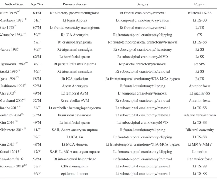
Table 1 Reported cases of dural arteriovenous fistula developed after surgery
Author/Year Age/Sex Primary disease Surgery Region
Miura 197511) 60/M Rt olfactory groove meningioma Rt frontal craniotomy/removal Bilateral TS-SS
Mizukawa 197812) 61/F Lt brain abscess Lt temporal craniotomy/evacuation Lt TS-SS
Bito 197815) 67/M Lt frontal convexity meningioma Rt frontal craniotomy/removal Lt TS
Watanabe 198417) 59/F Rt ICA Aneurysm Rt frontotemporal craniotomy/clipping CS
57/F Rt craniopharyngioma Rt frontotemporoparietal craniotomy/removal Lt TS-SS
Nabors 1987 70/F Rt trigeminal neuralgia Rt suboccipital craniotomy/rhyzotomy Rt SS
62/M Lt hemifacial spasm Rt suboccipital craniotomy/MVD Lt SS
Ugrinovski 198919) 46/F Rt parietal falx meningioma Rt parietal craniotomy/removal Rt SPS
Sasaki 199516) 46/F Rt trigeminal neuralgia Rt suboccipital craniotomy/removal Rt SS
Igase 199610) 58/M Rt ICA occlusion Rt frontotemporal craniotomy/STA-MCA bypass Rt TS
Hashimoto 19989) 52/M Acom Aneurysm Bifrontal craniotomy/clipping Anterior fossa
Ahn 20038) 49/M Lt temporal AVM Lt temporal craniotomy/removal Lt jugular-SS
Murakami 20054) 52/M Rt cerebellar AVM Rt suboccipital craniotomy/removal Anterior fossa
Tanabe 20133) 64/F Lt cerebellar hemangiopericytoma Lt suboccipital craniotomy/removal Lt TS-SS
Sadahiro 201418) 37/M brain stem cavernoma Lt suboccipital craniotomy/removal inferior vermian vein
Kim 201413) 49/M Lt hemifacial spasm Lt suboccipital craniotomy/MVD Lt TS-SS
Nishimoto 20142) 41/F SAH, Acom aneurysm rupture Bifrontal craniotomy/clipping Bilateral convexity
69/F Lt ICA An Lt frontotemporal craniotomy/clipping Lt TS-SS
Kim 201514) 48/M Lt MCA stenosis Lt frontotemporal craniotomy/STA-MCA bypass Lt MMA-MMV
Yamaki 20151) 47/F SAH, Lt MCA aneurysm rupture Lt frontotemporal craniotomy/clipping Lt pterion
Kawahara 2016 52/M Rt intracerebral hemorrhage Lt frontotemporal craniotomy/removal Rt anterior fossa
Yokoyama 201920) 63/F CPA meningioma Lt suboccipital craniotomy/removal Lt TS-SS
56/F epidermoid tumor Lt suboccipital craniotomy/removal Lt TS-SS
Acom: anterior communicating artery, CPA: cerebellopontine angle, CS: cavernous sinus, ICA: internal carotid artery, Lt: left, MCA: middle cere-bral artery, MMA: middle meningeal artery, MMV: middle meningeal vein, MVD: microvascular decompression, Rt: right, SAH: subarachnoid hemorrhage, SPS: superior petrosal sinus, SS: sigmoid sinus, STA: superficial temporal artery, TS: transverse sinus
Fig. 4 Scheme of the blood flow around cavernous sinus
Blood flow into cavernous sinus through left sylvian vein changed after tumor removal, which might have contributed to the formation of dAVF.
手術部位の対側に生じた dAVF は手術により発生した
とするには確証に乏しいが,dAVF の発生には上記のよ
うな複数の機序が関与している可能性が考えられ,さら
なる症例の集積が待たれる.また,開頭術後の画像フォ
ローでは,頭蓋内外の血管評価を含めた,注意深い経過
観察が重要と考える.
結 語
蝶形骨縁髄膜腫術後に対側の海綿静脈洞部に発生した
dAVF
の 1 例を経験した.腫瘍摘出に伴う硬膜面動静脈
の血流変化や,腫瘍により圧排された静脈が腫瘍摘出に
よって圧迫解除されることにより頭蓋内血行動態変化が
生じ,動静脈シャント,硬膜動静脈瘻の発生に寄与する
と考えた.開頭術後の画像検査においては,脳血管系の
評価も併せて行うことが望ましいと思われた.
著者は日本脳卒中学会への COI 自己申告を完了して
おり,本論文の発表に関して,開示すべき COI はない.
参考文献 1) 山木 哲,近藤 礼,毛利 渉ら:クリッピング術後に 生じた蝶形骨翼部硬膜動静脈瘻の 1 手術例.脳外速報 25: 88–93, 2015 2) 西本陽央,高橋 潔,林 悟ら:開頭術後、開頭範囲 から離れた部位に新たに発生した硬膜動静脈瘻の 2 例. 脳神外ジャーナル 23: 667–671, 2014 3) 田邉 淳,石川達哉,師井淳太ら:外側後頭下開頭術後 に発生した硬膜動静脈瘻の 1 例:開頭術後に発生する機 序の検討.Neurol Surg, 41: 711–716, 2013 4) 村上謙介,松本乾児,沼上佳寛ら:後頭蓋窩開頭術後に 出現した二次性前頭蓋窩硬膜動静脈瘻の 1 例.Neurol Surg 33: 1101–1105, 20055) Kusaka N, Sugiu K, Katsumata A, et al: The importance of ve-nous hypertension in the formation of dural arterioveve-nous fistu-las: a case report of multiple fistulas remote from sinus throm-bosis. Neuroradiology 43: 980–984, 2001
6) 桑山直也,久保道也,遠藤俊郎ら:わが国における硬膜 動静脈瘻の治療の現状(〈特集〉脳脊髄動静脈奇形の診断・ 治療の進歩).脳神外ジャーナル 20: 12–19, 2011
7) Yu J, Guo Y, Wu Z, et al: Traumatic arteriovenous fistula be-tween the extracranial middle meningeal artery and the ptery-goid plexus: A case report and literature review. Interv Neurora-diol 23: 90–96, 2017
8) Ahn JY, Kim OJ, Joo YJ, et al: Dural arteriovenous tion occurring after craniotomy for pial arteriovenous malforma-tion. J Clin Neurosci 10: 134–136, 2003
9) Hashimoto H, Iida J, Masui K, et al: Dural arteriovenous mal-formation of the anterior cranial fossa occurring after bifrontal craniotomy. Surg Neurol 49: 47–50, 1998
10) Igase K, Oka Y, Kumon Y, et al: [A case of dural AVM detected after STA-MCA anastomosis]. Neurol Surg 24: 81–85, 1996 (in Japanese) 11) 三浦直久,門田紘輝,小川信子ら:髄膜腫摘出後にみら れた外頸動脈および内頸動脈─静脈洞間 A-V Fistula の 1 例.Neurol Surg, 3: 265–269, 1975 12) 水川典彦,角南典生,則兼 博ら:後頭蓋窩硬膜動静脈 奇形の 1 症例.Neurol Surg 6: 295–302, 1978
13) Kim SH, Chang WS, Jung HH, et al: Delayed dural arteriove-nous fistula after microvascular decompression for hemifacial spasm. J Korean Neurosurg Soc 56: 168–170, 2014
14) Kim SW, Chae KS, Shim JH, et al: Iatrogenic dural arteriove-nous fistula after superficial temporal artery to middle cerebral artery anastomosis: a case report. Korean J Neurotrauma 11: 151–153, 2015
15) 尾藤昭二,大西俊輝,滝本 昇ら:髄膜腫摘出術後にみ られた硬膜動静脈瘻の 1 例.Neurol Surg 6: 397–400, 1978 16) Sasaki T, Hoya K, Kinone K, et al: Postsurgical development of
dural arteriovenous malformations after transpetrosal and trans-tentorial operations: case report. Neurosurgery 37: 820–824; discussion 824–825, 1995
17) Watanabe A, Takahara Y, Ibuchi Y, et al: Two cases of dural arte-riovenous malformation occurring after intracranial surgery. Neuroradiology 26: 375–380, 1984
18) Sadahiro H, Ishihara H, Goto H, et al: Postoperative dural arte-riovenous fistula in a patient with Cowden disease: a case report. J Stroke Cerebrovasc Dis 23: 572–575, 2014
19) Ugrinovski J, Vrcakovski M, Lozance K: Dural arteriovenous malformation secondary to meningioma removal. Br J Neuro-surg 3: 603–607, 1989
20) Yokoyama S, Nakagawa I, Kotsugi M, et al: Dural arteriovenous fistula arising after intracranial surgery in posterior fossa of nondominant sinus: two cases and literature review. Asian J Neurosurg 14: 602–606, 2019
Abstract
Development of cavernous sinus dural arteriovenous fistula at contralateral site
after removal of meningioma: a case report and literature review
Masafumi Segawa, M.D.,
1, 2)Hideaki Ono, M.D., Ph.D.,
1)Toshiya Aono, M.D.,
1)Tomohiro Mitani, M.D.,
1, 2)Masaaki Shojima, M.D., Ph.D.,
3)Takeo Tanishima, M.D., Ph.D.,
1)Akira Tamura, M.D., Ph.D.,
1)and Isamu Saito, M.D., Ph.D.
1)1)Department of Neurosurgery, Fuji Brain Institute and Hospital 2)
Department of Neurosurgery, The University of Tokyo Hospital
3)
Department of Neurosurgery, Saitama Medical Center
Background: Dural arteriovenous fistula (dAVF) is an abnormal arteriovenous shunt in the dura mater and is a relatively rare disease. We experienced a case of symptomatic dAVF that developed at contralateral cavernous sinus after operation of the sphenoid ridge meningioma. Herein, we report our case along with the relevant literature. Case report: A 69-year-old woman underwent a surgical removal of left sphenoid ridge meningioma, which gradually in-creased. The operation was conducted via left frontotemporal craniotomy. Although there were no perioperative com-plications, she became aware of right pulsatile tinnitus six months later, and double vision developed one year after the operation. A close examination was conducted, and digital subtraction angiography revealed the dural shunt at right cavernous sinus in the arterial phase. We diagnosed cavernous sinus dAVF, and performed coiling intravenously, resulting in complete occlusion of the shunt. The symptoms of the patient disappeared soon after the operation, she has had an uneventful course after discharge without recurrence of symptoms, as well as tumors and dAVF. Conclu-sion: We experienced a case in which dAVF developed after the excision of meningioma, and considered multiple mechanisms of development of dAVF in this case such as changes in blood flow of dural arteries and veins, and intra-cranial hemodynamics.
Key words: dural arteriovenous fistula, meningioma, cavernous sinus