博士論文(要約)
論文題目
A comprehensive understanding of
modulators regulating tau aggregation
(
Tau タンパク質の凝集に関与する因子の包括的理解)
The microtubule associated protein tau is implicated in a broad range of neurodegenerative diseases collectively known as tauopathies, characterized by the accumulation of hyperphosphorylated and ubiquitinated insoluble aggregates of tau. To date, numerous studies have aimed to identify cellular pathways regulating the aggregation of tau. However, while several genetic screens utilizing Drosophila models of tau have been performed, no genome-wide screens using mammalian cells for modulators of tau aggregation have been reported. This is caused in part because no simple cell culture models of tau aggregation were available, since tau is a highly soluble, natively disordered protein, and overexpression alone does not induce aggregation of tau, as is the case for the poly-glutamine repeat expansion proteins. Recently, Sanders et al. reported a tau aggregation model cell line based on the microtubule binding repeat domain of tau (tau RD) that indefinitely propagates aggregates even after cell division (Neuron, 2014). Therefore, we performed a genome-wide siRNA screen for modulators of tau RD aggregation through utilizing this cell culture model.
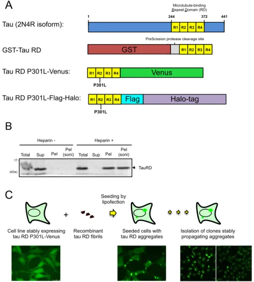
We first independently reproduced the tau RD aggregation-propagating model cell line. HEK293A cells stably expressing tau RD fused to Venus was established (Fig. 1A). To increase the aggregation of tau RD, we introduced the P301L mutation, which is a causative mutation of frontotemporal dementia and is known to increase the aggregation propensity of tau. We next generated recombinant tau RD fibrils in vitro through incubation of purified tau RD with heparin (Fig. 1B). These recombinant tau RD fibrils were transduced into HEK293A cells stably expressing tau RD P301L-Venus through lipofection to induce aggregation of tau RD. Fibril-transduced cells containing tau RD aggregates were sparsely diluted, and monoclonal cell lines stably propagating tau RD aggregates, hereafter referred to as aggregate-positive cells, were obtained (Fig. 1C).
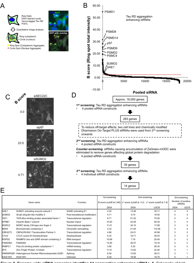
We used these aggregate-positive cells to perform high-content genome-wide siRNA screening based on quantification of the total intensity of cytoplasmic tau RD aggregates (Fig. 2A). For the 1st screening, we used a genome-wide siRNA library targeting approximately 18,000 genes, with 4 pooled siRNA constructs per gene, and selected 283 genes as significantly enhancing tau RD aggregation when knocked down (Fig. 2B). Representative images from the 1st screening are shown in Fig. 2C. To reduce off-target effects, from the 2nd screening, we used two different cell types and chemically modified siRNAs (Fig 2D). For the 2nd screening, we validated results of the 1st screening using HEK293A and U2OS tau RD aggregate-positive cell lines, again with 4 pooled siRNA constructs. Genes that were positive in both cell lines were taken as hits. Among these hit genes, we reasoned that some genes
would affect general protein degradation. To eliminate such genes, we performed a counter screen using HEK293A and U2OS cells stably expressing ZsGreen-mODC, a well-characterized proteasome model substrate consisting of the fluorescent protein ZsGreen fused to a mouse ornithine decarboxylase degron. Genes that caused accumulation of ZsGreen-mODC when knocked down likely affected proteasome activity, and were therefore removed from 2nd screening hits, resulting in 35 aggregation suppressor candidate genes. Finally, to further remove off-target effects, each of the 4 pooled siRNAs were tested individually in both HEK293A and U2OS tau RD aggregate-positive cell lines for the 3rd screening. Genes for which more than 3 of 4 individual siRNAs tested enhanced tau aggregation in either HEK293A or U2OS cells were chosen, resulting in 14 candidate aggregation suppressor genes.
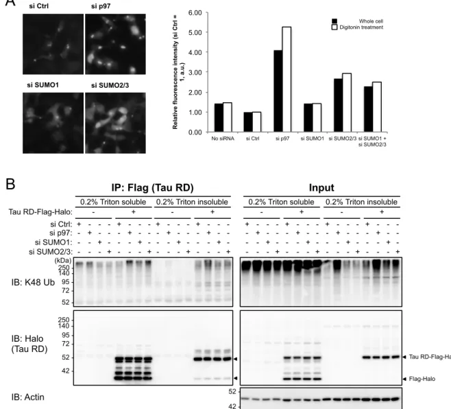
Among these 14 genes, three genes were positive for 4 of 4 siRNA constructs tested in both cell lines (Fig. 2E). Of these, two were components of the SUMO modification (SUMOylation) pathway: SUMO2, and SAE1, a subunit of the SUMO activating E1 enzyme heterodimer. We therefore decided to investigate the role of SUMOylation on the aggregation of tau RD. Human cells express three isoforms of SUMO, of which SUMO2 and SUMO3 are nearly identical and often regarded as one protein, SUMO2/3, while SUMO1 is only about 50% identical to SUMO2/3. Therefore, we first tested the specificity of SUMO1 and SUMO2/3 modification through using 4 siRNA constructs against SUMO1, SUMO2, and SUMO3. We confirmed that only SUMO2 knockdown caused accumulation of tau RD aggregates in both HEK293A and U2OS cells. We further validated SUMO1 versus SUMO2/3 knockdown specificity on tau RD aggregation by flow cytometry following digitonin treatment to remove soluble tau RD, and confirmed that while knockdown of SUMO2/3 resulted in about a three-fold increase compared to non-targeting control siRNA, knockdown of SUMO1 had almost no effect on tau RD aggregates (Fig. 3A).
To facilitate immunoprecipitation (IP) of tau RD, we next established a HEK293T cell line stably expressing tau RD P301L-Flag-Halo (Fig. 1A). Using an aggregate-positive form of this cell line, SUMO1 and SUMO2/3 were knocked down, and the ubiquitination of tau RD-Flag-Halo was analyzed by Flag-IP followed by blotting for tau RD and K48-linked ubiquitin (Fig. 3B). This revealed that knockdown of SUMO1 and SUMO2/3 both increased the ubiquitination levels of soluble tau RD, with SUMO2/3 having a larger effect. In contrast, both SUMO1 and SUMO2/3 knockdown did not significantly affect the ubiquitination of insoluble tau RD.
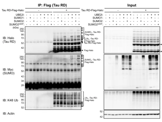
We next investigated if tau RD was directly SUMOylated by overexpressing Myc-tagged SUMO1 or SUMO2 in a HEK293T tau RD aggregate-positive cell line, followed by Flag-IP of tau RD. Immunoblot analysis of immunoprecipitated tau RD from total cell lysate revealed additional higher molecular weight bands above that expected for tau RD (Fig. 4A). Comparison with immunoblots to detect SUMO and K48-linked ubiquitin revealed these bands to be due to both ubiquitination and SUMOylation of tau RD. Consistent with the effect of SUMO1 and SUMO2/3 knockdown increasing the ubiquitination of tau RD, we found that when UBE2I was overexpressed, there was a decrease in ubiquitination of tau RD. We then performed the same experiment using a HEK293T tau RD aggregate-negative cell line, and found that tau RD was also SUMOylated, and to a higher extent than in the aggregate-positive cell line (Fig. 4B).
Next, since the majority of tau RD expressed in aggregate-negative cell lines are soluble, we reasoned that SUMOylation may target soluble tau RD. We tested this through IP of tau RD from both the 0.2% Triton X-100 soluble and insoluble fractions of a tau RD aggregate-positive cell line. We found that soluble tau RD was mostly SUMOylated, while insoluble tau RD was mainly ubiquitinated (Fig. 5A and 5B).
Taken together, our results suggest a mechanism where SUMOylation competes with ubiquitination to prevent the aggregation of tau RD. While we do not currently have sufficient data to present a more detailed model, past studies have shown that SUMOylation can increase the solubility and inhibit the aggregation of misfolded proteins. Furthermore, SUMOylation has been shown to compete with the same lysine residue with ubiquitination, for example for IκB-α. Therefore, we propose a working hypothesis in which SUMO2 modification of tau RD maintains tau RD in a soluble state, thus inhibiting its aggregation and ubiquitination (5 年以内に出版予定).
Figure 1. Establishment of tau RD aggregate positive cell lines. A. Schematic of constructs used in this study. The
microtubule binding repeat domain (RD) from the 2N4R isoform of full-length tau was fused with an N-terminal GST-tag for recombinant tau RD production, and a C-terminal Venus or Flag-Halo-tag for cell culture experiments. To increase aggregation propensity, the P301L FTLD-17 mutation was introduced for all cell culture experiments. B. Ultracentrifugation analysis of recombinant tau RD fibrils obtained through incubation with heparin. After incubation with heparin, samples were centrifuged at 100,000 x g for 1 h, and divided into Total (before ultracentrifugation), soluble (Sup) and insoluble (Pel) fractions. The insoluble fraction was further sonicated (soni) to generate tau RD fibril seeds for transduction into cell cultures. C. Scheme for the establishment of cell lines stably propagating tau RD aggregates. A cell line stably expressing tau RD P301L-Venus was seeded with recombinant tau RD fibrils via lipofection. These seeded cells were then isolated and cloned into monoclonal cell lines stably propagating tau RD aggregates. Tau (2N4R isoform): Tau RD P301L-Flag-Halo: Tau RD P301L-Venus: Microtubule-binding Repeat Domain (RD) 1 244 372 441 R1 R2 R3 R4 Venus P301L R1 R2 R3 R4 P301L Halo-tag Flag R1 R2 R3 R4
A
TauRDTotal Sup Pel (soni) Total Sup Pel Pel (soni) Pel
Heparin - Heparin +
17 - (kDa)
GST R1 R2 R3 R4
PreScission protease cleavage site
GST-Tau RD
Seeding by lipofection +
Cell line stably expressing tau RD P301L-Venus
C
Recombinant
-10.00 0.00 10.00 20.00 30.00 40.00 50.00 60.00 0 5000 10000 15000 20000 B s co re (R in g s p o t to ta l i n te n si ty ) Pooled siRNA
B
PSMD1 PSMD8 PSMD14 p97 PSMD6 PSMD2 PSMC4 SUMO2 SAE1 B scoreC
0.0 23.5 4.71 siSEC22C sip97 siSUMO2D
1st screening: Tau RD aggregation enhancing siRNAs
• 4 pooled siRNA constructs
pprox. 18,000 genes
283 genes
2nd screening: Tau RD aggregation enhancing siRNAs
• 4 pooled siRNA constructs
To reduce off-target effects, two cell lines and chemically modified Dharmacon On-Target PLUS siRNAs were used from 2nd screening
onwards
Counter-screening: siRNAs causing accumulation of ZsGreen-mODC were
eliminated to remove genes affecting global protein degradation • 4 pooled siRNA constructs
35 genes
14 genes
3rd screening: Tau RD aggregation enhancing siRNAs
• 4 individual siRNA constructs
E
Gene name Function
1st screening 2nd screening 3rd screening B score (cutoff at 3.66) z* score (cutoff at 4.3) z* score (cutoff at 1.6) Number of positive
siRNAs 293A 293A U2OS 293A U2OS SAE1 SUMO1 activating enzyme subunit 1 SUMO activating E1 4.20 13.48 35.11 4 4 SUMO2 Small ubiquitin-like modifier 2 Post-translational modification 4.71 9.74 16.40 4 4 TAF1 TATA-Box binding protein associated factor 1 Transcriptional regulation 8.71 51.22 70.05 4 4 KPNB1 Importin Beta-1 subunit Nuclear import 10.00 42.09 84.45 3 4 MORC2 MORC family CW-type zinc finger 2 Transcriptional regulation 11.01 34.10 17.89 3 4 BRD4 Bromodomain containing 4 Chromatin remodeling 4.32 21.64 112.06 3 4 CBFA2T3 CBFA2/RUNX1 Translocatiion Partner 3 Transcriptional regulation 4.86 24.01 45.98 3 3 CYLD CYLD Lysine 63 Deubiquitinase Deubiquitinase 6.32 16.71 26.03 3 3 RGPD2 RANBP2-Like and GRIP domain containing 2 RNA transport 10.69 7.76 20.40 3 3 FAM208A FAM208A Transcriptional regulation 14.95 36.07 18.16 4 2 PABPC1 Poly-(A) binding protein cytoplasmic 1 mRNA binding 3.95 5.32 26.34 4 2 ZFX Zinc Finger Protein, X-linked Transcriptional regulation 11.01 13.62 22.40 3 1 HNRNPA2B1 Heterogenous Nuclear Ribonucleoprotein A2/B1 Splicing 6.04 13.83 29.31 3 1 KIAA1551 KIAA1551 Unknown 8.30 18.58 18.75 4 1
Ring Spot (Cytoplasmic Aggregate) Circle Spot (Nuclear Aggregate)
Ring (cytoplasm) Circle (nucleus) Raw Data: DAPI-stained nuclei Venus-tagged Tau RD P301L DAPI/GFP GFP GFP with overlay
A
Quantitative image analysis
Tau RD aggregation enhancing siRNAs
Figure 2. Genome-wide siRNA screening identifies 14 aggregation enhancing siRNAs. A. Schematic of high
content image analysis. A region of set width (ring) around DAPI-stained nuclei (circle) was defined, and GFP spots
within the ring were detected to quantify cytoplasmic tau RD aggregates. B. B scores of all samples in the 1st
genome-wide siRNA screen based on cytoplasmic aggregate total fluorescence intensity. The cutoff value for siRNAs
enhancing tau RD aggregation is indicated as a dashed red line. C. Representative images from the 1st screen. The
cutoff value was decided based on inspection of such images as the value at which a clear difference between siRNAs with no effect (B score close to 0) and potential hit genes were visible. D. Overview of the siRNA screening protocol. 14 final candidate genes were obtained from approximately 18,000 siRNAs. E. Table of the 14 final candidate genes and an overview of their scores during the screening process.
si Ctrl si p97 si SUMO1 si SUMO2/3
A
0.00 1.00 2.00 3.00 4.00 5.00 6.00No siRNA si Ctrl si p97 si SUMO1 si SUMO2/3 si SUMO1 + si SUMO2/3 R el ati ve fl u o re sc en ce in te n si ty (s i C tr l = 1, a .u .)
B
250 - (kDa) 140 - 95 - 72 - 52 - IB: K48 Ub 250 - 140 - 95 - 72 - 52 - 42 - IB: Halo (Tau RD) Tau RD-Flag-Halo Flag-Halo 52 - 42 - IB: Actin - 0.2% Triton soluble si Ctrl: si p97: si SUMO1: si SUMO2/3: + - - - - + - - - - + - - - - + Tau RD-Flag-Halo: + + - - - - + - - - - + - - - - + - 0.2% Triton insoluble + - - - - + - - - - + - - - - + + + - - - - + - - - - + - - - - +IP: Flag (Tau RD)
- 0.2% Triton soluble + - - - - + - - - - + - - - - + + + - - - - + - - - - + - - - - + - 0.2% Triton insoluble + - - - - + - - - - + - - - - + + + - - - - + - - - - + - - - - + Input Whole cell Digitonin treatment
Figure 3. Knockdown of SUMO2/3, but not SUMO1, affects tau aggregation and ubiquitination. A. HEK293A tau
RD P301L-Venus clone No. 2-2+ cells were treated with the indicated siRNAs for 96 h, photographed, and then
analyzed by flow cytometry through measuring Venus fluorescence intensity with and without digitonin treatment. Values are expressed as relative values with control siRNA (siCtrl) treatment set to 1. B. Effect of SUMO knockdown
on the ubiquitination of tau RD. HEK293T tau RD P301L-Flag-Halo cells with tau RD aggregates (clone No. 2+) was
treated with the indicated siRNAs, and tau RD was immunoprecipitated by Flag-IP from 0.2 % Triton X-100 soluble and insoluble fractions, and analyzed by immunoblot with the indicated antibodies.
A 250 - 140 - 95 - 72 - 52 - IB: K48 Ub 250 - 140 - 95 - 72 - 52 - 42 - IB: Halo (Tau RD) 250 - (kDa) 140 - 95 - 72 - 52 - 42 - 34 - 28 - 52 - 42 - IB: Myc (SUMO) - UBE2I: SUMO1: SUMO2: SUMO2Q90P: - - - - - + - - - - + - - - - + Tau RD-Flag-Halo: + - - - + + - - + - + - + - - + + - - - - - + - - - - + - - - - + + - - - + + - - + - + - + - - + IP: Flag (Tau RD)
- - - - - - + - - - - + - - - - + + - - - + + - - + - + - + - - + + - - - - - + - - - - + - - - - + + - - - + + - - + - + - + - - + Input IB: Actin Tau RD-Flag-Halo Flag-Halo SUMOn -Tau RD-Flag-Halo SUMOn -Tau RD-Flag-Halo Ubn -Tau RD-Flag-Halo Ubn -Tau RD-Flag-Halo UBE2I: SUMO1: SUMO2: SUMO2Q90P: Tau RD-Flag-Halo: IP from tau RD aggregate-positive cell line
B 250 - 140 - 95 - 72 - 52 - IB: K48 Ub 250 - 140 - 95 - 72 - 52 - 42 - IB: Halo (Tau RD) 250 - (kDa) 140 - 95 - 72 - 52 - 42 - 34 - 28 - 52 - 42 - IB: Myc (SUMO) + Tau RD-Flag-Halo: -
IP: Flag (Tau RD)
+ - Input IB: Actin Tau RD-Flag-Halo Flag-Halo SUMOn -Tau RD-Flag-Halo SUMOn -Tau RD-Flag-Halo Tau RD-Flag-Halo: UBE2I: SUMO1: SUMO2: SUMO2Q90P: - - - - - + - - - - + - - - - + + - - - + + - - + - + - + - - + - - - - - + - - - - + - - - - + + - - - + + - - + - + - + - - + - - - - - + - - - - + - - - - + + - - - + + - - + - + - + - - + - - - - - + - - - - + - - - - + + - - - + + - - + - + - + - - + UBE2I: SUMO1: SUMO2: SUMO2Q90P:
IP from tau RD aggregate-negative cell line
Figure 4. Tau RD is directly SUMOylated in both aggregate-positive and aggregate-negative cells A. Detection
of tau RD SUMOylation by SUMO overexpression and immunoprecipitation of tau RD. HEK293T tau RD P301L-Flag-Halo cells with tau RD aggregates (clone No. 3+) was transfected with the SUMO E2 ligase UBE2I and Myc-tagged
SUMO1, SUMO2, or SUMO2Q90P (resistant to cleavage by SENP SUMO peptidases) as indicated, then tau RD was
immunoprecipitated by Flag-IP from the 0.1% SDS soluble fraction. Bands due to SUMOylated and ubiquitinated tau RD are indicated as open and filled circles, respectively, based on comparison of the anti-Halo (tau RD) blots with the anti-Myc (SUMO) and anti-K48 ubiquitin blots. B. As in A, except that HEK293T tau RD P301L-Flag-Halo cells without tau RD aggregates (clone No. 2-) was used instead.
A 250 - 140 - 95 - 72 - 52 - IB: K48 Ub 250 - 140 - 95 - 72 - 52 - 42 - IB: Halo (Tau RD) 250 - (kDa) 140 - 95 - 72 - 52 - 42 - 34 - 28 - 52 - 42 - IB: Myc (SUMO) - UBE2I: UBE2IC90S: SUMO1: SUMO2: SUMO2K11R: SUMO2K11R/Q90P: SUMO2Q90P: - - - - - - - + - - - - - - - + - - - - - + - + - - - - Tau RD-Flag-Halo: + - - + - - - + - - - + - - + - - - - + - + - - - - - + + IP: Flag (Tau RD)
- + Input IB: Actin Tau RD-Flag-Halo Flag-Halo SUMOn -Tau RD-Flag-Halo SUMOn -Tau RD-Flag-Halo Tau RD-Flag-Halo: - - - - - - - + - - - - - - - + - - - - - + - + - - - - + - - + - - - + - - - + - - + - - - - + - + - - - - - + UBE2I: UBE2IC90S: SUMO1: SUMO2: SUMO2K11R: SUMO2K11R/Q90P: SUMO2Q90P: - - - - - - - + - - - - - - - + - - - - - + - + - - - - + - - + - - - + - - - + - - + - - - - + - + - - - - - + - - - - - - - + - - - - - - - + - - - - - + - + - - - - + - - + - - - + - - - + - - + - - - - + - + - - - - - + IP from 0.2% Triton X-100 soluble fraction
B 250 - 140 - 95 - 72 - 52 - IB: K48 Ub 250 - 140 - 95 - 72 - 52 - 42 - IB: Halo (Tau RD) 250 - (kDa) 140 - 95 - 72 - 52 - 42 - 34 - 28 - 52 - 42 - IB: Myc (SUMO) - UBE2I: UBE2IC90S: SUMO1: SUMO2: SUMO2K11R: SUMO2K11R/Q90P: SUMO2Q90P: - - - - - - - + - - - - - - - + - - - - - + - + - - - - Tau RD-Flag-Halo: + - - + - - - + - - - + - - + - - - - + - + - - - - - + + IP: Flag (Tau RD)
- + Input IB: Actin Tau RD-Flag-Halo Flag-Halo Ubn -Tau RD-Flag-Halo Ubn -Tau RD-Flag-Halo Tau RD-Flag-Halo: - - - - - - - + - - - - - - - + - - - - - + - + - - - - + - - + - - - + - - - + - - + - - - - + - + - - - - - + UBE2I: UBE2IC90S: SUMO1: SUMO2: SUMO2K11R: SUMO2K11R/Q90P: SUMO2Q90P: - - - - - - - + - - - - - - - + - - - - - + - + - - - - + - - + - - - + - - - + - - + - - - - + - + - - - - - + - - - - - - - + - - - - - - - + - - - - - + - + - - - - + - - + - - - + - - - + - - + - - - - + - + - - - - - + IP from 0.2% Triton X-100 insoluble fraction
Figure 5. Soluble tau RD is SUMOylated, while insoluble tau RD is ubiquitinated A. Detection of tau RD
SUMOylation by SUMO overexpression and immunoprecipitation of tau RD. HEK293T tau RD P301L-Flag-Halo cells with tau RD aggregates (clone No. 3+) was transfected as indicated, and tau RD was immunoprecipitated from the 0.2
% Triton X-100 soluble fraction. Bands due to SUMOylated and ubiquitinated tau RD are indicated as open and filled circles, respectively, based on comparison of the anti-Halo (tau RD) blots with the anti-Myc (SUMO) and anti-K48 ubiquitin blots. B. As in A, but Flag-IP from the 0.2 % Triton X-100 insoluble fraction.