Some Biological and Physiological Properties
of the Toxins from the Sea Urchin, Family
Diadematidae
著者
KIHARA Hiroshi, ANRAKU Mitsuo, HASHIMURA
Saburo
journal or
publication title
南方海域調査研究報告=Occasional Papers
volume
5
page range
209-218
URL
http://hdl.handle.net/10232/15888
Kagoshima Univ. Res. Center S. Pac. Occasional Papers. No. 5. p. 209 218, 1985 209
Some Biological and Physiological Properties
of the Toxins from the Sea Urchin, Family
Diadematidae
Hiroshi K.IHARA*, Mitsuo ANRAKU* and Saburo HASHIMUR/
Abstract
The cthanolic extracts of the sea urchin, belonging to the family Diadematidae,
had a hemolytic activity and toxicity to killifish. The heart of bull frog perfused with
the solution containing the extracts was arrested in systole. These seemed to be due to
a saponin-like agent which was the common component of sea animals, sea stars and sea cucumbers. In the isolated bull frog nerve-muscle preparation, the extracts abolished nerve stimulated contractions but did not affect the contractions produced by
direct stimulations of the muscle. In addition, the extracts caused a significant increase
in miniature endplate potentials (MEPPs) frequency at a neuromuscular junction and
this effect was reversible. Both activities on the muscle contraction and on the MEPPs
frequency were separated from the saponin-like agent by a dialysis or gelfiltrations on
Sephadex G-10, G-25 and Bio Gel P-4. This factor (s) in the ethanolic extracts waspartially purified.
Introduction
Sea stars and sea cucumbers are well known to contain steroidal saponins as the
toxic substance (Hashimoto, 1978).
Powerful hemolytic properties (Nigrelli et al.,
1960; Halstead, 1965; Hashimoto and Yasumoto, 1960; Yasumoto et al., 1964), neurotoxic effects (Friess et al., 1959; Thron et al., 1964) and suppressive effects on hearts (Rio et al., 1965) have been reported for the saponins. Sea urchins also are reported to contain saponins (Ruggieri et al., 1970). Some high molecular substances like proteins are also reported from sea urchins to be toxic (Alender et al., 1965, 1967 ; Feigen et al., 1968; Fieming and Howder, 1974; Kimura et al., 1975).210 KiHARA et at: Extract of Sea Urchin Toxins
Some kinds of sea urchin in the South Pacific areas which are common littoral
inhabitants have been known to evoke severe pain by piercing the spines. Therefore
these animals were supposed to contain some toxic substances.
We collected sea
urchins in Fiji and assayed their extracts on various organs and tissues. We found two
active substances from the extracts of the sea urchin belonging to the family
Diadematidae, one a saponin-like and the other a neurotoxic substance.
In this report, the extraction procedures of the active substances from the sea
urchin and their physiological and biological properties were described.
Materials and Methods
Animal and extraction
Specimens of sea urchin collected from the coast in Fiji were stored in a deep
freezer until the use. After thawing the spines and shells of the sea urchin were taken
out and were lyophilized. The lyophilized materials were crushed to powder. The dry
powder of 2.8 kg was obtained from 8 kg sea urchin in wet weight. Dry powder (200
g) was extracted with 1 L of 70% ethanol by stirring for 2 hr at room temperature. The
extraction was repeated twice by using the same volume of ethanol. The combined
extracts were concentrated in vacuum to dryness and then dissolved in 100 ml of water.For removal of cation, the extracts were applied on a column of Amberlite IR-120B and
eluted with water. The initial high concentration of potassium (34.4 mM) decreased to
0.1 mM by the chromatography. The pH of eluent was adjusted to 7.0 with NaOH
before evaporation. The crude extracts from 20 g of the dry powder were dissoloved in
10 ml of water for use.
Separation of active substances
The ethanolic extracts were further fractionated by partitioning between water and
ethyl acetate or sodium bicarbonate in a separatory funnel.
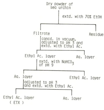
Figure 1 shows the
fractionation procedures of the etanolic extracts.
The last ethyl acetate layer was
evaporated to dryness and was used for the test. For some tests, the crude extracts were
dialyzed against water for overnight. Both dialyzate and diffusate were concentrated to
the original volume of the crude extracts by the evaporation in vacuum and served for
the use. Gel filtrations on Sephadex G-10, G-25 and Bio Gel P-4 of the crude extractswere carried out for some tests.
Hemolytic activity
The hemolytic activity was tested by the method of Fuijita and Nishimoto (1952)
with a slight modification. Tubes contained 0.5 ml of 2% rabbit erythrocytes and the
same volume of the test solution. The erythrocytes were washed in 0.9% saline untilall traces of hemoglobin were removed. The test solution was prepared from the ten
Kagoshima Univ. Res. Center S. Pac. Occasional Papers, No. 5. 1985
Dry powder of
sea urchin
extd, with 70% EtOH
Filtrate
n
Residueconed, in vacuum, adjusted to pH 3 and extd, with Ethyl Ac.
Ethyl flc, layer Aq. layer
extd. with NaHCO,
Aq, layer
Ethyl Ac, layer ( ETX )
of pH 9 J3
Ethyl Ac. layer adjusted to pH 3
and extd, with Ethyl Ac.
Aq, layer
21 I
Fig. I. Fractionation of the extracts from the sea urchin.
were dissolved in 10 ml water. The development of hemolysis at room temperature
(18-23°C) was observed every hour. Pure saponin (Merk) was used as the standard.
Toxicity to killifish
Ten individuals of adult killifish (Oryzias latipes) were put in a beaker containing 150 ml test solution. The test solution was prepared from the differently diluted the crude extracts. When the fish no longer responed to the nip with a forceps, it was
assumed to be dead.
Activity on heart
The effect of the extracts on a heart was investigated with the isolated perfused
heart from the bull frog (Rana catesbeiana). Both the vena cava inferior and the aorta
were connected to the separate perfusing cannulae and the tip of ventricle was con
nected to the lever of a tension transducer with which the cardiac contractions could
be recorded.
Effect on nerve and muscle
The extensor digitorum longus muscle with the nerve attached was dissected from
a bull frog and was set in a chamber in which the test solution was applied. The
proximal end of the muscle was fixed to the chamber and the distal end was connected
to an isometric transducer to record the contractions. The nerve was placed on a pair of electrodes and the muscle was placed in between another pair of electrodes. The
212 Kihara et al: Extract of Sea Urchin Toxins
combined stimulations to the nerve and muscle with the interval of 4 sec were applied
to respective electrodes. Stimulations were supramaximal for the respective nerve and
muscle, and were delivered in every 12 sec. The sciatic nerve was desheathed 2 cm in
length at the middle and immersed in a small chamber in which the test solution was
applied. The effect of the extracts on the nerve was examined by measuring the amplitude of action potentials.
Effect on miniature endplate potentials
The sartorius muscle with nerve was dissected from the frog (Rana nigromaculata) and mounted in a small acrylite chamber in which the last ethyl acetate layer in Fig. 1, the neurotoxic fraction, was applied. Miniature endplate potentials (MEPPs) were recorded from the surface muscle fibers by impaling with a microelctrode filled with 3 M KCI. The microelectrode was kept in a muscle fiber throughout the exchange of the surrounding solution. The effect of the extracts on MEPPs frequency was investigated.
Bath solution
Bath solution contained 115mM NaCl, 2.5 mM KCI, 1.5 mM CaCl2 and 5 mM Tris maleat buffer at pH 7.2.
Results
Hemolytic activity
The 100 fold diluted extracts brought complete hemolysis within 2 hr but the 200
dilution did not. The 100 and 50 fold dilutions of the dialyzate induced complete
hemolysis within 4 hr and 2 hr, respectively.
The diffusate induced no hemolysis.
These results suggested that the active substance for hemolysis is in the dialyzate and
not in the diffusate. As the 10 fold dilution of 0.125% saponin made complete
hemolysis within 4 hr, the hemolytic activity of the crude extracts was estimated to
correspond to 1.25% saponin.
Toxicity to killifish
The 200 and 100 fold dilutions of the crude extracts brought 2 and 9 dead fish,
respectively. Less than 400 fold dilution of the crude extracts brought no dead fish.
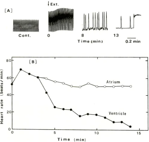
Activity on a heartThe amplitude of contractions of the frog heart, keeping a steady cardiac cycle in
the normal solution, gave rise to increase within a few second after the application ofthe crude extracts. Soon, both the systole and diastole became irregular, and finally the
systolic arrest took place (Fig. 2A). The atrial rhythmes were held nearly unchanged through, but the ventricular contractions became hard to follow and brought the irregular cardiac cycle. In Fig. 2B the less changed rhythms of the atrium and the
[A]
Kagoshima Univ. Res. Center S. Pac.. Occasional Papers, No. 5, 1985
| Ext.
Cont. 8 T i me (min ) 13 0.2 min 8 0 [B] i 1 c E 60 _ ,-, Atrium RI m * 40\
(0 *. 20n^^*—_^^^
Ventricle
-10 a) I^^"•n.
0 — _i 1 t 10 15 Time (min) 213Fig. 2. Effect of the crude extracts on the isolated bull frog heart.
(A) The test solution contained 70 fold dilution of the crude extracts for the use and
was exchanged with the normal solution at time 0. At around 13 min the systolic arrest
took place bui the irregular artial contraction remained. (B) The rates of atrium and
ventricle were presented successively after the application of the test solution containing
70 fold dilution of the crude extracts for the use. The heart rate was shown as beats per min.
significant decrease of the ventricular rhythms are shown. After the arrest with the crude extracts, the heart was able to contract again rhythmically after repeated washing with the normal solution. The dialyzate of the crude extracts brought the same effect
on the heart but the diffusate did not. The fraction numbers of gel filtrations for the
heart activity were coincident with those for the hemolytic activity (Table 1).
Effect on nerve and muscle
In the normal solution the muscular contractions by the stimulation to both the nerve and muscle were nearly the same and were steady. By the application of neurotoxic fraction, the tension by the stimulation to the muscle gave rise to decrease
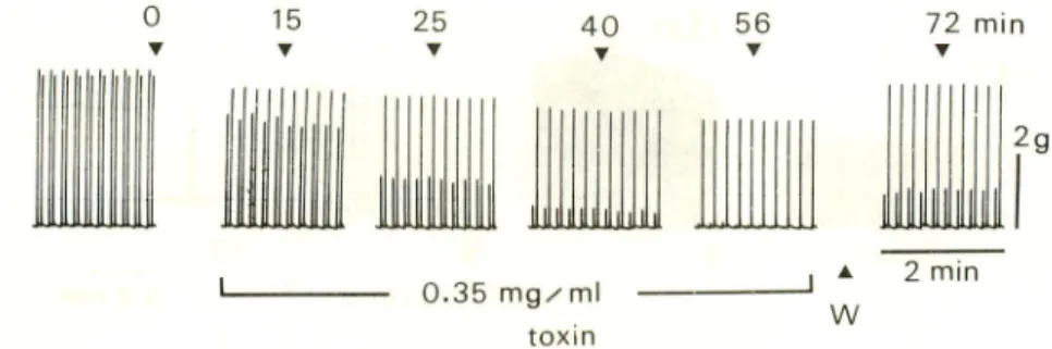
214 Kihara el al. : Extract of Sea Urchin Toxins 25 • 40 T liiilijljjlii 0.35 mg/ml toxin 56 Y w 72 min • 2 min
Fig. 3. Effect of the toxin on contraction of the nerve-muscle preparation.
Paired contractions by combined stimulation of the nerve and muscle in 4 sec interval
were induced in every 12 sec. The earlier and later contractions in the pair were induced
by the stimulation to the nerve and muscle, respectively. The earlier contraction decreased rapidly finaly disappearing and recovered partly, but the latter one decreased slightly and
recovered almost completely.
0 2
m
n i
0 4 0 6
toxin ( mg/ ml)
Fig. 4. Effect of the toxin on MEPP frequency.
The increase of MEPP frequency in the toxin containing solution and the recovery by washing with the normal solution are presented. Note the dose dependent increase of .MEPP.
the tension proceeded faster and significantly (Fig. 3). As the nervous conduction was
confirmed to change inappreciably, this decreased contraction by the nerve stimulation suggested the difficulty in neuromuscular transmission. By the washing with the
toxin-free solution, the contractions induced by the muscular stimulation recovered
almostly, but the neuromuscular transmission did partially.
Effect on MEPP frequencv
The neurotoxic fraction from the sea urchin increased the MEPP frequency
immediately and significantly after the application into the bath. The increasing effect
of this fraction on the MEPP frequency was presented in Fig. 4 in which the dose
Kagoshima Univ. Res. Center S. Pac. Occasional Papers. No. 5. 1985 100 100 1.5 Ca 0 Ca ID 0.15 0.3 0.45 0.6 0.15 0.3 0.45 0.6 Extract concentration tmg/mll
Fig. 5. The relation between MEPP frequency and the concentration of the toxin. The left part shows the MEPP frequency obtained with a normal Ca-* concentration
(1.5 mM) in the test solution, the right pan without Ca2'.
Table I. Effects of the crude extracts and their fractions.
Results of gel filtrations were shown in the tube numbers. ( f-) and ( ) represent effective and ineffective, respectively.
Amberlite IR 120B Dialysis . Dialyzate I Diffusate Gel filtration Sephadex G 10 Sephadex G-25 Bio Gel P 4
Hemolysis MEPP Heart
t-+
20 30 28 38 20 30
26 34 46 56 26 34
22 32 46 54 22 32
normal solution were observed. Neither the membrane potential of the impaled muscle
nor the amplitude and the time course of individual MEPP changed. When the logarithm of the MEPP frequency was plotted against the toxin concentration, a linear relation was observed (Fig. 5). The increasing effect of the neurotoxic fraction could
be seen without Ca2+ in the surrounding solution. An example in which Ca2+ was
replaced by Mg2+ was presented in Fig. 5. In Mg2+ solution the toxin increased the MEPP frequency dose dependently. The diffusate of the crude extracts was as active as the crude extracts on the increase of MEPP frequency but the dialyzate was ineffec
216 KiHARA et al.: Extract of Sea Urchin Toxins
those for the heart activity or hemolysis (Table 1).
Discussion
The various biologically active substances were reported to be widely distributed in sea animals (Halstead, 1965 ; Hashimoto, 1978). The present crude extracts from the
sea urchin may contain various substances.
The saponin is a common compound in sea stars and sea cucumbers, and has been reported to be present in some kinds of sea urchin (Ruggieri et al., 1970). The toxicity to killifish and the hemolytic activity on rabbit erythrocytes coincide with the saponin action. As the saponin has been known to be hardly dialyzable, the hemolytic activity of the dialyzate attributes to the action of saponin (Hashimoto and Yasumoto, 1960). Rio et al. (1965) have reported that the saponin of the sea star (Pycnopodia
helianthoides) made arrest of toad heart in systole. They ascribed this arrest to the altered permeability of the cell membrane. The effect of the crude extracts on a heart could be explained partly by the effect of saponin, because the saponin could change the permeability of the membrane by its detergent action. However, because the cardiac cycle is maintained by the coworking of several kinds of cardiac muscle, the further discussion on the effect of the crude extracts will be inadequate until further investiga
tion.
Friess et al. (1968) have reported that asterosaponin of the starfish (Asterias
amurensis) cause a conduction block and a powerful muscle contracture in the rat
phrenic nereye-diaphragm preparation. In our preliminary experiment, the crude
extracts also caused a conduction block and a muscle contracture in the frog nerve-muscle preparation. These conduction block and the muscle contracture could be
explained by the saponin action, presumably. As the saponin was isolated from the sea urchin in the latter investigation (unpublished data), several actions of the extracts can be explained by the saponin action. However, the suppressed neuromuscular transmis sion by the neuro toxic fraction (Fig. 3) cannot be ascribed to the saponin because it was excluded by the partial purification procedure. Therefore the toxin is some substance which acts on the neuromuscular junction.
The increasing action of the toxin on MEPP frequency is clear from Fig. 4. The
MEPP frequency has been known to be increased by depolarizing of the nerve terminal. And this increase in MEPP frequency requires Ca2+ in the external solution (Castillo
and Katz, 1954). The capability of the toxin to induce MEPP without Ca2+ is obvious
from Fig. 5. The depolarization of the nerve terminal by this toxin seems to be unlikely, although the proof is difficult, because no change of the membrane potential of the muscle was observed. Also MEPP frequency could be increased by the increased osmolarity of the surrounding solution (Blioch et tf/.,1968). As the osmotic pressure of the normal solution and the test solution were 228 and 232 mosm, respectively, the osmotic effect for the increased MEPP frequency was doubtful. As the detailed
Kagoshima Univ. Res. Center S. Pac. Occasional Papers. No. 5. 1985 217
investigation of the effect of the toxin on MEPP frequency will be descrived in other paper (Anraku et al., in cotributing), further discussion will not be presented here. The black widow spider venom is well known lo increase MEPP frequency (Lon-genecker et al., 1970). However, the spider venom has been reported to be protein, to require a divalent cation for its action and to be irreversible action. Hence, the present
neurotoxin is different from the spider venom.
Recently, Sevcik and Barboza (1983) reported that the crude extracts from the sponge, Tedania ignis, increase MEPP frequency without effects on the amplitude and shape. The compounds were estimated for the molecular weight to be 900 from the results of gel filtrations. The molecular weight and some of the biological activies were
similar to our neurutoxin. However, as both Tedania toxin and the present toxin are unpurified, the further discussion is difficult.
The chemical description of the present sea urchin toxin is premature, but it is
clearly low molecular substance from the results of gel filtrations. As the effect of the toxin on MEPP frequency is unigue, the designation of the toxin will be reasonable.
Acknowledgements-V^ c express our sincere thanks to Dr. U. Raj, University of the South Pacific, for collecting the sea urchin specimens. This work was supported by the Special Research Grant of the Ministry of Education, Science and Culture, Japan (The Scientific Survey of the South Pacific, organized by the Kagoshima University
Research Center for the South Pacific).
References
Alender. CB. (1967) A biologically active substances from the spines of two diadematia sea urchins. XxvAnimal Toxins, p. 145 (Russell, F.E. and Saunders, P. R., Ed.). O.xfordiPergamon Press.
Alender. C.B.. Feigen. G,A. and Tomita, J.T. (1965) Isolated and characterization of
sea urchin toxin. Toxicon 3.9.
Blioch, Z. V., Glagoleva, I.M., Liberman, E.A. and Nenashev, V.A. (1968) Astudy of the mechanism of guantal transmitter release at a chemical synapse, J. Physiol.
199. 1 1
Del castillo. J. and Katz. B. (1954) Changes in endplate activity produced by pre synaptic polarization. J. Rhysiol. 124, 586
Feigen, G.A., Sanz, E., Tomita. J.T. and Alender, CB. (1968) Studies on the mode of action of sea urchin toxin. II. Enzymatic and immunological behavior, Toxicon
6, 17
Fleming, W.J. and Howder, M.E.H. (1974) Partial purification and characterization of
a lethal protein from Tripneustes gratilla. Toxicon 12, 447
Friess, S.L., Standaert, F.G.,Whitcomb, E.R., Nigrelli, R.F., Chanley, J.D. and Sobot-ka, H. (1959) Some pharmacological properties of holothulin, an active
neurutox-218 KiHARA et al: Extract of Sea Urchin Toxins
in from the sea cucumber. Jour. Pharmac. and Exp. Ther. 126, 323
Friess, S.L., Durant, R.C. and Chanley, J.D. (1968) Further studies on biological
actions of steroidal saponins produced by poisonous echinoderms. Toxicon 6, 81 Fujita, M. and Nishimoto, K. (1952) On the biological assay of Japanese senega. J.
Pharm. Soc. Japan 72, 1645
Halstead, B.H. (1965) Poisonous and Venomous Marine Animals. Vol. 1, p. 537,
Washington, D.C.:U.S. Goverment Printing Office.
Hashimoto, Y. (1978) Marine Toxins, p. 275, Tokyo:Japan Scientific Societies Press. Hashimoto, Y. and Yasumoto, T. (1960) Confimation of saponin as a toxic principle
of starfish. Bull. Jap. Soc. of Sci. Pisheries 26, 1132
Kimura, A., Hayashi, H. and Kuramoto, M. (1975) Separation, purification and pharmacological action of toxic substances. Jap. J. Pharmacol. 25, 109
Longenecker, H.E.Jr., Hurlbut, W.P., Mauro, A. and Clark, A.W. (1970) Effects of black widow spider venom on the frog neuromuscular junction. Nature 225, 701 Misler, S. and Hurlbut, W.P. (1975) Action of black widow spider venom on guantized release of acetylcholine at the frog neuromuscular junction;Dependence upon external Mg2+. Proc. Natl. Acad. Sci. USA. 76, 991
Nigrelli, R.F. and Jakowska, S. (1960) Effects of holothulin, a steroid saponin from the Bahamian sea cucumber (Actinopyga agassizi) on biological systems. Ann. N.Y.
Acad. Sci. 90, 884
Rio, G.J., Stempien, M.F.Jr., Nigelli, R.F. and Ruggieri, G.D. (1965) Echinoderm toxins. I. Some biochemical and physiological properties of toxins from several species of Asteroidea. Toxicon 3, 147
Ruggieri, G.D., Nigrelli, R.F. and Stempien, M.F.Jr. (1970) some biochemical and physiological properties of extracts from several echinoderms. Toxicon 8, 149 Thron, CD., Durant, R.C. and Friess, S.L. (1964) Neurumuscular and cytotoxic effects
of holothurin A and related saponins at low concentration levels. Toxicol. Appl.
Pharm. 6,182
Yasumoto, T., Watanabe, T. and Hashimoto, Y. (1964) Physiological activities of starfish saponin. Bull. Jap. Soc. of Sci. Fisheries 30, 357