Availability of Isolated Cells of the Eel
Liver for Investigating Biologically Active
Substances Produced by Marine Invertebrates
著者
HAYASHI Seiichi, UEDA Yoshinori, ITAKURA
Takao, OOSHIRO Zentaro
journal or
publication title
鹿児島大学水産学部紀要=Memoirs of Faculty of
Fisheries Kagoshima University
volume
30
page range
349-356
別言語のタイトル
海産無脊椎動物の生産する生理活性物質検索におけ
るウナギ肝浮遊細胞の利用
Mem. Fac. Fish., Kagoshima Univ.
Vol.30 pp. 349-356 (1981)
Availability of Isolated Cells of the Eel Liver for
Investigating Biologically Active Substances
Produced by Marine Invertebrates
Seiichi Hayashi*, Yoshinori Ueda*, Takao Itakura* and Zentaro Ooshiro*
Abstract
To investigate biologically active sustances produced by marine invertebrates isolated cells of the eel liver were used. Effect of extracts of tissues on glucose release from the liver cells of the eel and on gluconeogenesis in the liver cells wereexamined. Extracts of eye-stalk, mit-gut gland, and abdominal ganglion of prawn had stimuratory effect on glucose release. Extracts of tissues of sea cucumber had inhibitory effect on glucose release and gluconeogenesis. Gluco neogenesis in the eel liver cells wasinhibited almost completely by the extract of digestive organ
of starfish.
The effect of extracts of tissues of marine invertebrates on glucose release from the liver cells of the eel (Anguilla japonica) and on gluconeogenesis in the eel liver was
investigated. It is known that Gastropoda and Bivalvia of Mollusca have neuro
endocrine system1) and Cephalopoda has some endocrine gland2). However, there is
little report on the metabolism of carbohydrate and amino acid in these Mollusca.
So no physiological effect of neuroendocrine and endocrinesystem on these metabolism
has known. Crustacea has endocrine and neuroendocrine system and physiological
function of these system has been well studied. For example, it is known that there
is hormones regulating the metabolism of carbohydrate and protein3) and
hyperglycemic hormone4) secreted from eyestalk.
To use isolated cells of the eel liver is not necessarilly good method to inverstigate
the presence or absence of hormones or biologically active substance in tissues of
invertebrates. However, since we can prepare rather easily cell-suspension of the eel liver and determine gluconeogenesis and glycogenosis in the cells very exactly,
we examined the effect of extracts of each tissues of marine invertebrate. Prawn, sea hares, sea cucumber, ligia, sea urchin, starfish, and sea squirts were used. Ex
tracts of kidney, pancreas, gill, brain, and pituitary of the eel were also investigated.
Experimental Procedures
Isolation of liver ceUs of the eel Japanese cultured eels weighing about 150 g
350 Mem. Fac. Fish., Kagoshima Univ. Vol. 30 (1981)
were purchased from the fish market. They were kept in freshwater aquaria and
fasted for 3 to 14 days before use.
Livers were excised from anethesized eels and perfused with modified Krebs-Ringer bicarbonate buffer (pH 7.4) containing collagenase (15 mg per 50 m/). The pro
cedures of the perfusion of liver and the isolation of liver cells have been described
previously5*6).
Measurement of the incorporation of 14C-labeUed substrates into glucose and determination of glucose Eel liver contains glycogen even after fasting for several months as reported by Dave et aP\ Similarly, glucose was released to the
incubation mixture from liver cells prepared from fasted eels6). Therefore, labelled
substrates were used for examining gluconeogenesis in liver cells. L-[U-14C]-Alanine
(50/iCi), [2-14C]-pyruvate (50 jiCi) and DL-[2-14C]-lactate were dissolved in 4 ml
of 0.5 M unlabelled substrate. L-[U-14C]-Alanine was taken to dryness by storage in vacuo over P205 for overnight to remove the ethanol before use.
The reaction mixture for gluconeogenesis contained, in a final volume of 2 ml, one ml of cell suspension, 20 fil of 14C-labelled substrate, 0.1 ml of extract and Ringer solution gassed with 95% 02 and 5% COz. The mixture was incubated at 30°C for
30 to 60 min. The reaction was initiated by the addition of cell suspension
prein-cubated at 30°C for 15 min and stopped by the addition of 0.2 ml of 60% HC104. After centrifugation at 3,000 rpm for 10min, the supernatant was neutralized with
0.5 ml of 2 M K2C03 and cooled. One ml of neutralized supernatant was applied
to a Dowex 1X8 column (0.6x5 cm, HCOO" form, 100-200 mesh) and Dowex
50X8 column (0.6x2 cm, H+ form, 200-400 mesh). 0.5 ml of 14C-glucose fraction
was added to 5 ml of toluene containing 4 g of 2, 5-diphenyloxazole (DPO), 0.1 g of
1, 4-bis (2-(5-phenyloxazoyl))benzene (POPOP) and 500 m/ of Triton X-100 per
litre. The radioactivity was measured using a Beckman LS-230 spectrophotometer. The reaction mixture for glucose release from eel liver-cells contained, in a final volume of 2 ml, one ml of cell suspension, 0.1 ml of extract and Ringer solution gassed
with 95% 02 and 5% C02. The neutralized supernatant obtained by the procedures
described above was used for determination of glucose by colorimetirc8) and enzymatic
methods9).
Preparation of extract
Each tissue was extracted by 4 vol of water or 4 vol of
6% HC104. After tissue was homogenized with ice water by Ultra Tarrax (Type ION), homogenate was centrifuged at 20,000 rpm for 1 h at 5°C. Supernatant was used for experiments. Homogenate of tissue by 6% HC104 was centrifuged at 3,000 rpm for 10 min and the supernatant was neutralized with 2 M K2C03. Neu
tralized supernatant was used for experiments.
When a small tissue such as abdominal ganglion of prawn or pituitary of eel was
homogenized, 10 to 20 vol of water or 6% HC104 was used. Tissue extract was
Hayashi-Ueda-Itakura-Ooshiro : Biologically active substancesproduced by marine invertebrates 351
Chemicals and enzymes L-[U-14C]-Alanine, [2-14C]-pyruvate, and DL-[2-14C]-lactate was purchased from RCC Amersham. ATP and NADP were obtained
from Oriental Yeast Co. CoUagenase from CI. histolyticum was obtained from P-L Biochemicals and glucose-6-phosphate dehydrogenase (from yeast) and hexokinase
(from yeast) were from Boehringer Mannheim. Other chemicals were purchased
from Wako Chemicals.
Results and discussion
Effects of extracts on glucose release from eel liver-cells As shown in
Table 1. and Fig. 1, extracts of eyestalk, mit-gut gland, and abdomianal ganglion of
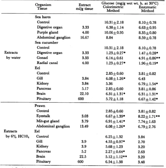
Table 1. Effect of extracts on glucose release from liver cells of the eel.
Organism Tissue Extract m//g tissue Glucose (mg/g wet wt. h. at 30°C) Colorimetric Enzymatic Method Method Sea hares Control 10.31±2.18 8.10±0.78 Digestive organ 3.33 6.38±1.14 6.63±0.95 Purple gland 4.00 10.06±0.95 8.35±0.80 Abdominal ganglion 16.67 8.84 8.59±0.78 Sea cucumber Control 10.31zh2.18 8.10±0.78
Extracts Digestive organ 3.33 1.23±0.27* 1.47±0.28*
by water Gonad 3.33 6.14±0.61 4.91 ±0.86** Radial canal 4.00 1.23±0.27* 1.96±0.19* Eel Control 2.85±0.60 3.81 ±0.82 GUI 3.84 6.08±1.26* 6.43 Kidney 3.84 5.36 6.79±1.54* Pancreas 5.17 2.85±0.60 3.81 ±0.86 Brain 22.10 6.31±1.31* 6.31±1.31* Pituitary 600 5.72±1.18 6.67±1.42* Prawn Control 2.85±0.60 3.81 ±0.82 Eyestalk 3.03 6.67 ±1.39* 8.22±1.71** Mit-gut gland 3.79 6.91±1.41* 7.74±1.63 Abdominal ganglion 13.49 6.08±1.26* 6.79±2.76 Extracts Eel by6%HC104 Control 6.25±1.32 3.84 Gill 3.9 4.55±0.97* 2.70 Kidney 3.9 5.68±1.23 3.20 Pancreas 5.2 2.27±0.64* 2.63 Brain 22.5 5.12±1.12** 3.20 Pituitary 600 6.54±1.38 3.48 * P<0.01 ** P<0.05
352 Control Eyestalk M1t-gut gland Abdominal ganglion S £ Digestive organ Digestive organ Radial canal Gonad Pancreas Kidney Pituitary
Mem. Fac. Fish., Kagoshima Univ. VoL 30 (1981)
TOO =* * P<0.01 t X ) Enzymatic determination Colorlmetric determination 200 300 1 f • —•— 1 ' . -** P<0.05
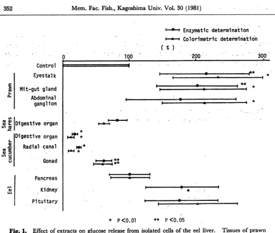
Fig. 1. Effect of extracts on glucose release from isolated cells of the eel liver. Tissues of prawn
were extraxted by 6% HClo4. Others were extracted by water.
prawn by 6% HC104 had an effect of increasing glucose release from liver cells.
These extracts stimulates glucose release by 2 times. Inhibitory effect of water-extract
of digestive organ and radial canal of sea cucumber was observed. They inhibited glucose release by 80 to 90%. Water-extracts of gill and brain of the eel had stim-uratory effect, but extracts of gill, brain and pancreas of the eel by 6% HC104 had
almost no effect. Since extracts of gill, brain and pancreas by 6% HC104 had almost
no effect on glucose release and extracts by water had stimuratory effect, these ef
fectors in extracts seemed to be something like protein.
As Kleinholz et aW reported that hyperglycemic hormones from eyestalk were
peptides, there are some possibility that the extract of eyestalk having stimulating
effect on glucose release contained some peptides like hyperglycemic hormones.
Simiraly there were some substances to stimulate glucose release from eel liver-cells in extracts of mit-gut gland and abdominal ganglion. It is unknown what kind of
substances they are.
It was found that the extracts of sea cucumber had inhibitory effect on glucose re
lease. This inhibitory effect may be due to some peptides like insulin, because there
Hayashi-UedaItakura'Ooshiro : Biologically activesubstances producedby marine invertebrates 353 invertebrate10). However, there is another possibility that this inhibitory effect is
due to saponin11).
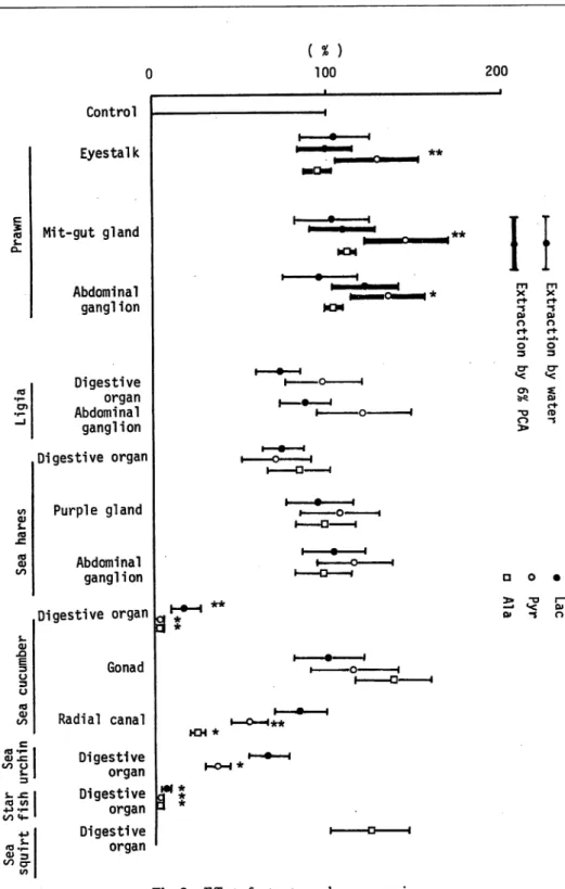
Effect of extracts on gluconeogenesis Extracts of eyestalk, mit-gut gland, and abdominal ganglion of prawn by 6% HC104 have stimulatoryeffect on gluconeogenesis
from 14C-pyruvate in the eel liver-cells as shown in Fig. 2. Water extracts ofdigestive
organ and radial canal of sea cucumber, and of digestive organ of sea urchin have inhibitory effect on gluconeogenesis from 14C-lactate, 14C-pyruvate, and 14C-alanine.
Extract of digestive organ of starfishes inhibited gluconeogenesis almost completely
(Table 2).
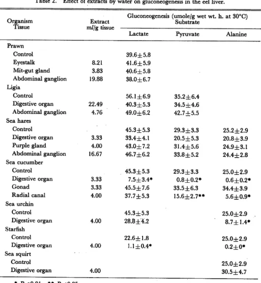
Table 2. Effect of extracts by water on gluconeogenesis in the eel liver.
Gluconeogenesis (umole/g wet wt. h. at 30°C)
Organism Extract Substrate
Tissue m//g tissue
Lactate Pyruvate Alanine
Prawn Control 39.6±5.8 Eyestalk 8.21 41.6±5.9 Mit-gut gland 3.83 40.6±5.8 Abdominal ganglion 19.88 38.0±6.7 Ligia Control 56.1 ±6.9 35.2±6.4 Digestive organ 22.49 40.3±5.3 34.5 ±4.6 Abdominal ganglion 4.76 49.0±6.2 42.7±5.5 Sea hares Control 45.3±5.3 29.3±3.3 25.2±2.9 Digestive organ 3.33 33.4±4.1 20.5 ±5.3 20.8±3.9 Purple gland 4.00 43.0±7.2 31.4±5.6 24.9±3.1 Abdominal ganglion 16.67 46.7±6.2 33.8±5.2 24.4±2.8 Sea cucumber Control 45.3 ±5.3 29.3±3.3 25.0±2.9 Digestive organ 3.33 7.5±3.4* 0.8±0.2* 0.6±0.2* Gonad 3.33 45.5±7.6 33.5±6.3 34.4±3.9 Radial canal 4.00 37.7±5.3 15.6±2.7** 5.6±0.9* Sea urchin Control 45.3±5.3 25.0±2.9 Digestive organ 4.00 28.8±4.2 8.7±1.4* Starfish Control 22.6±1.8 25.0±2.9 Digestive organ 4.00 1.1 ±0.4* 0.2±0* Sea squirt Control 25.0±2.9 Digestive organ 4.00 30.5±4.7 * P<0.01 ** P<0.05
354 c S- s-0) -Q E 3 U 3 a 10 <D t o c t o o rd to 4-> «r-</) a-en
Mem. Fac. Fish., Kagoshima Univ. Vol. 30 (1981)
Control Eyestalk Mit-gut gland Abdominal gang!ion Digestive organ Abdominal ganglion Digestive organ Purple gland Abdominal ganglion Digestive organ Gonad Radial canal Digestive organ Digestive organ Digestive organ 3 ( % ) 100 i • » MX Km t • i i — • -i — • — -i i o I I D 1 KM * I D 1 I • I I O I I rj 1 t •
Fig. 2. Effect of extracts on gluconeogenesis.
200 ——j
ii
-s -s Q» Q> n n d- c+ _ i . • j . o o 3 3 c r CX *< *< o> z ^ O) c+ "O n> o -s 3> D O • 3> -a r— —• *< o* o» -s r>Hayashi-Ueda-Itakura-Ooshiro : Biologically active substances produced by marine invertebrates 355
It is interesting that the extracts of eyestalk, mit-gut gland, and abdominal gland
had stimuratory effect on gluconeogenesis, since there was no reports that whether
hyperglycemic hormones in crustaceas stimulated gluconeogenesis or did not10).
These extracts stimulated glucose release from liver cells of the eel as well as glu
coneogenesis in the eel liver.
Yasumoto et al12> reported the presence of saponin in starfishes. Saponin has
hemolytic toxin11). Saponin seems to react with cell membrane and to inhibit metabolism in cell. The extract into which 6% of HC104 was added still had in
hibitory effect on gluconeogenesis by 49%. Similary the extract treated at 60°C
for 30 min still had inhibitory effect by 66%. After the gel-filtration by Sephadex G-75, fractions monitored at 244 nm had inhibitory effect. It is known that saponin
absorbs the light at 244 nm. Though the substance in the extract of starfish, which inhibited gluconeogenesis almost completely, was not identified as saponin, it is thought this inhibitory effect was due to saponin. Simiraly inhibitory effect of the extracts from sea cucumber seems due to saponin.
Acknowledgement
We wish to thank Dr. H. Hirata, S. Kadowaki, T. Nakazono, and T. Kasedo, the
Fisheries Research Laboratory, Kagoshima University, for their help to take samples.
References
1) Gabe, M. (1966): "Neurosecretion" (Pergamon, London, England).
2) Wells, M. J., and J. Wells (1959): Hormonal control of sexual maturity in Octopus. /. exp.
Biol., 36, 1-33.
3) Ramaurthi, R., Mumberch, M. W., and B. T. Scheer (1968): Endocrine control of glycogen
synthesis in crabs. Comp. Biochem. Physiol., 26, 311-319.
4) Kleinholz, L. H., Kinball, F.} and M. McGarvey (1967): Initial characterization and separation of hyperglycemic (diabetogenic) hormones from the crustacean eyestalk. Gen. comp.
Endocrinol., 8, 75-81.
5) Hayashi, S., and Z. Ooshiro (1975): Gluconeogenesis and glycolysis in isolated, perfused liver
of the eel. Bull. Japan. Soc. Sci. Fish., 41, 201-208.
6) Hayashi, S., and Z. Ooshiro (1978): Preparation of isolated cells of eel liver. Bull. Japan. Soc. Sci. Fish., 44, 499-503.
7) Dave, G., Jahansson-Sjoback, M.-L., Larsson, A., Lewander, K., and U. Lidman (1975): Metabolic and hematological effect of starvation in the European eel, Anguilla anguila, L. Comp. Biochem. Physiol., 52A, 423-430.
8) Sasaki, T. (1969): "Micro Determination for Clinical Chemistry", 160-167 (Kinpo Do, Kyoto,
Japan), (in Japanese)
9) Bergmeyer, H. U., Bernt, E., Schmidt, F., and H. Stork (1974): "Methods of Enzymatic Analysis", 1196-1201 (Academic Press, New York and London).
10) Nomura, T. (1978): "Biologically Active Substances Produced by Marine Organisms", 44-46
(Nan Kodo, Tokyo, Japan), (in Japanese)
107-356 Mem. Fac. Fish., Kagoshima Univ. Vol. 30 (1981)
114 (Nan Kodo, Tokyo, Japan), (in Japanese)
12) Yasumoto, T., Tanaka, M., and Y. Hashimoto (1966): Distribution of saponin in Echinoderms. Bull. Japan. Soc. Sci. Fish., 32, 673-676.