Studies on the Characteristics of the
Bacteriophages of Vibrio alginolyticus strain
B-1 Isolated from Kinko Bay
著者
NAKAMURA Kunihiko, KAKIMOTO Daiichi, SWAFFORD
James, JOHNSON Roy
journal or
publication title
鹿児島大学水産学部紀要=Memoirs of Faculty of
Fisheries Kagoshima University
volume
27
number
1
page range
59-64
別言語のタイトル
錦江湾で分離されたVibrio alginolyticus B-1バク
テリオファージの特性に関する研究
URL
http://hdl.handle.net/10232/13112
Vol. 27, No. 1, pp. 59-64 (1978)
Studies on the Characteristics of the Bacteriophages
of Vibrio alginolyticus strain B-l
Isolated from Kinko Bay
Kunihiko Nakamura,*1 Daiichi Kakimoto,*2
James Swafford,*8 and Roy Johnson*8
Abstract
Eight variant bacteriophages infectious for Vibrio alginolyticus strain B-l, isolated from Kinko Bay, were isolated and purified. The virion consisted of an icosahedral head of 700 A, tail length of 1400 A and large plate. These bacteriophages were distinguishable reciprocally on plaque morphology, burst size, heat sensitivity, and the quality of resistance against host bacteria. These bacteriophages seemed to be naturally occurring variants produced in a variable environment of the seashore.
Hosaka1* isolated and showed the morphology of Vibrio parahaemolyticus bacteri ophages. Hori and co-workers2) isolated the bacteriophages of V. parahaemoly
ticus from sea shores, stools of patients, and marine fish. Baross et al.3) demon-stated that shellfish which were shown invariably to harbor high titers of spe cific V. parahaemolyticus bacteriophages had the ability to transduce the agar hy-drolyzing characteristic to V. parahemolyticus under simulated in condition in aqu
arium oyster.
Kakimoto and Nagatomi4) isolated Vibrio strain B-l bacteriophage from Kinko bay. Nakamura et alP identified this Vibrio strain as Vibrio alginolyticus strain B-l. The phage makes various type of plaques on an agar plate in spite of re peated single plaque isolations. The authors tried to purify these phages and examine the characteristics of these V. alginolyticus strain B-l bacteriophages.
Materials and Methods
Bacterial strains: The host strain was isolated by Kakimoto and Nagatomi4) from Kinko Bay in 1970. Kakimoto et al.5) calculated the similarity index bet ween this strain and V. parahaemolyticus STO-5 for about 100 traits, including morphological, physiological, and biochemical observations and reported that this strain was a biotype of V. parahaemolyticus. Nakamura et al.B* identified this
*x Department of Bacteriology, Faculty of Medicine, Kagoshima University, Kagoshima 890. *2 Laboratory of Microbiology, Faculty of Fisheries, Kagoshima University, 4-50-20
Shimoa-rata Kagoshima.
*3 Department of Botany and Microbiology, Arizona State University. Tempe, Arizona
60 Mem. Fac. Fish., Kagoshima Univ. Vol. 27, No. 1 (1978)
strain as Vibrio alginolyticus. Authentic strains of V. alginolyticus 370, 5556, and
138-2 were obtained from Dr. Shimizu at the Unniversity of Tokyo.
V. parahae
molyticus STO-5 was obtained from Dr. Sakai at Hokkaido University.
V. cholerae
ATCC 145035 was obtained from American Type Culture Collection.Medium: The medium routinely used was made by dissolving the following
components in one liter of distilled water; NaCl 30g; KCl 0.7g; MgCl2 6H20 10. 8g; MgS04 7H20 5.4g; CaCl2 2H20 l.Og; yeast extract l.Og; polypeptone 5.0g
(ZoBell 2216 E Medium).
Purification of Bacteriophages: The phage lysates of V. alginolyticus strain B-l
had been purified from the phage lysate which was preparated by Kakimoto and
Nagatomi4), by the double layer method and were maintained at 15 C. Accord
ingly, the phages isolated from the centers of plaques with an inoculating nee
dle were incubated with host bacteria at 37 C for 5 hrs and chloroform was added in the proportion of 1 to 10.
Then 0. 5 % of soft agar containing both supernatant of the chloroform-treat
ed lysate and host bacteria were poured onto plates.
Plaques of this phage
tend to deformed on moist agar plate and distinct plaques were difficult to ob tain. Therefore, plates were dried at 37 C overnight before use. These techn iques were repeated many times for additional phage types and subsequent pu rification depended on plaque morphology.
Isolated bacteriophages were examined using 10 min exposures to room tem peratures, 60 C, 70 C, and 80 C. Heat sensitivity at 60 C was carried out by sampling at 10 min intervals for 60 min. Sensitivity of the phages to saturated chloroform, 1 % phenol and 0. 25 % formaldehyde was also examined.
Plaques were stained by the method of Jackson7> and photographed for co mparative morphology. Bacteriophage samples for electron microscopic obser vation were prepared by centrifuging for 3 hrs at 35000 r.p.m. on Beckman Model E ultracentrifuge. The bacteriophage pellet was washed twice in 1^ ammonium acetate, pH 7.0. Lysates were placed on 300 mesh copper grids
supported with carbon-stabilized formvar and negatively stained with 1 % sodium phosphotungstate, pH 7. 0.
Results and Discussion
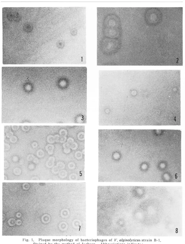
Eight bacteriophages infectious for V. alginolyticus strain B-l were isolated and
purified from a lysate of V. alginolyticus strain B-l. Fig. 1 shows the diverse
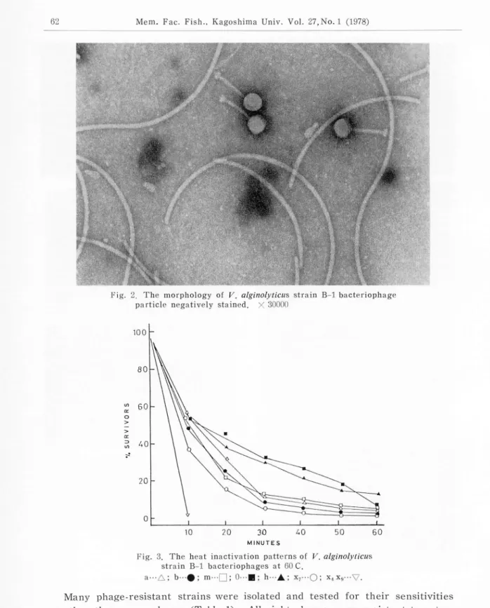
plaque morphology of these phages. No morphological difference was noted among these bacteiophages. The typical shape of these bacteriophages is shown in Fig. 2. The virion consists of an icosahedral head of 700 A, a tail length of 1400 A and a large tail plate. The shape and size of this virion were identical with those infectious for V. parahaemolyticus strains reported by Sklarow
et al.s). All these were uniformly inactive on V. parahaemolyticus STO-5, V. algino
Fig. 1. Plaque morphology of bacteriophages of V. alginolyticus strain B-l.
Stained by the method of Jackson. Abbreviations indicate:
l---a phage; 2-b phage; 3---0 phage; 4--m phage; 5-h phage; 6--x4 phage; 7--x7 phage; 8--x9 phage.
62 Mem. Fac. Fish., Kagoshima Univ. Vol. 27, No. 1 (1978)
Fig. 2, The morphology of V. alginolyticus strain B-l bacteriophage
particle negatively stained. X 30000
100
2 40
Fig. 3. The heat inactivation patterns of V. alginolyticus
strain B-l bacteriophages at 60 C.
a---A; b-#; m—|~J; 0---B; h--A; x7---0; x,x,---V.
Many phage-resistant strains were isolated and tested for their sensitivities
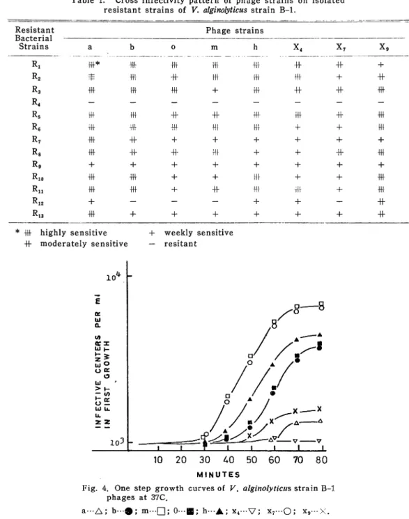
satur-Table 1. Cross infectivity pattern of phage strains on isolated resistant strains of V. alginolyticus strain B-l.
Resistant Phage strains
Strains a b 0 m h x4 X7 X9 Ri Iff* fff fff fff fff -ff Hf + R2 $ +H- -ff ttf fff fff + Hf R3 R4 R5 fff !ff 4H- + •Hf +f -ff fff Hf fH- 4f -ff -Hf Iff Hf -Hf R6 +H- fff -fff Hf Hf + 4- fff R7 -fff -ff + + + + H- + R8 fff -ff Hf Hf + -f Hf •Hf R9 + -f + + + + + + Rio •Hf fff + + Hf + -f •Hf Rn fff fff + Hf •Hf Hf + fff R12 + - - - + + - Hf R13 fff + H- + + -f + -ff * fff highly sensitive Hf moderately sensitive + weekly sensitive — resitant
10*.
I D-—8 Ml0
A
/•
UI H 0 a: / / / 0 / / / LJ / / / > 1- / / •INFECTI
INFIRS
O/
/ 8 /x xA—*
- H ^L^m ^ ^ AV X7 XT t 1 r 1 1 1 1 1 10 20 30 40 50 60 70 80 MINUTESFig. 4. One step growth curves of V. alginolyticus strain B-l
phages at 37C.
a---A; b---0; m.-n; 0—•; h—A; x4--V; x7---0; x9---X.
ated chloroform and sensitive to both \% phenol and 0.25 96 formaldehyde. All of the bacteriophages used for heat sensitivity measurment were inactivated at 70 C for 10 min. Fig. 3 shows the heat inactivation time of these
bacteriopha-64 Mem. Fac. Fish., Kagoshima Univ. Vol. 27, No. 1 (1978)
ges at 60 C. Phages X4 and X9 were inactivated significantly more rapidly than
the other phages. The burst sizes of the phages are shown in Fig. 4. From these observations, phages isolated were presumed to be dissimilar because of their plaque morphology, heat sensitivity and burst size.
In spite of isolating from the center of the plaque with an inoculating needle, almost these bacteriophage variants except for original phage occurred natural
ly during purification. For this reason, the enrichment process of the phages had to be done in a short time incubation. The numbers of the morphologically
same plaques were increased depending upon short time enrichment process and many times purification. However we couldn'f get really purified bacterio
phages of strain B-l.
The variability of these phages may depend on both short generation time of
the host bacteria and lysogenecity of these bacteriophages. Acknowledgements
K. Nakamura in master course studies of Faculty of Fisheries has performed at Arizona State University under the patronage of the student exchange program of the Japanese education administration. We are thankful to Mr. Uehara, for his skilled and conscientious aid in electron microscopic observation at Kagoshima
University.
References
1) Hosaka, Y. (1966) : The morphology of V in phage 14. Electron micrographs biology-2 Hitachi Electron Microscope.
2) Hori, M., K. Maeshima, S. Nakanishi, Y. Iida and T. Teramoto. (1967) : Studies on bacteriophages of Vibrio parahaemolyticus. uVibrio parahaemoly}icusn 2, 147-165.
3) Baross, J. A., J. Liston and R. Y. Morita. (1974): Some implications of genetic exchange among marine vibrios, including Vibrio parahaemolyticus, naturally occurring in the Pacific oyster. "International symposium on Vibrio parahaemolyticus19 129-137.
4) Kakimoto, D., and H. Nadatomi. (1971) : Study of bacteriophages in the Kinko Bay.
Bull Jap. Soc. Fish. 38, 271-278.
5) Kakimoto, D., K. Nakamura, and Y. Tanaka. (1972): Studies on the vibrios and pseudomonads living in the Kinko Bay. Mem. Fac. Fish. Kagoshima. Unive. 21,109-112. 6) Nakamura, K., D. Kakimoto, J. Swafford and R. Johnson. (1979): in press. 7) Jackson, R. H. (1967) : Differential staining of bacteriophage plaques for photograph.
Can. J. Microbiol, 13, 117-118.
8) Sklarow, S. S., R. R. Colwell, G. B. Chapman and S. Zane (1973): Characteristics of
a Vibrio parahaemolyticus bacteriophage isolated from Atlantic coast sediment. Can. J.