Title
Analysis of matrix metalloproteinase (MMP-8 and MMP-2) activity in gingival crevicular fluid from children with Down's syndrome
Author(s) Alternative
Yamazaki-Kubota, T; Miyamoto, M; Sano, Y; Kusumoto, M; Yonezu, T; Sugita, K; Okuda, K; Yakushiji, M; Ishihara, K
Journal Journal of periodontal research, 45(2): 170-176 URL http://hdl.handle.net/10130/1476
Right
This is the pre-peer reviewed version of the following article: J Periodontal Res. 2010
Apr;45(2):170-6, which has been published in final form at
Analysis of Matrix Metalloproteinase (MMP -8 and MMP-2)
Activity in Gingival Crevicular Fluid from Children with Down’s
Syndrome
Yamazaki-Kubota, T1, 2, Miyamoto, M3, Sano, Y1, Kusumoto, M.4, Yonezu, T1, Sugita, K5, Okuda, K3, Yakusiji, M1, Ishihara, K2, 3*
1
Department of Pediatric Dentistry, Tokyo Dental College, 1-2-2 Masago, Mihama-ku, Chiba 261-8502, Japan
2
Oral Health Science Center, Tokyo Dental College, 1-2-2 Masago, Mihama-ku, Chiba 261-8502, Japan
3
Department of Microbiology, Tokyo Dental College, 1-2-2 Masago, Mihama-ku, Chiba 261-8502, Japan
4
Tsuruga Institute of Biotechnology, Toyobo Co., Ltd., Tsuruga, Japan
5
Division of Child Health, Faculty of Education, Chiba University
Running title: MMP production in Down's syndrome patients
Key word: Down’s syndrome; periodontal disease; matrix metalloproteinase; subgingival bacterial flora
*
Corresponding author Department of Microbiology
Tokyo Dental College, 1-2-2 Masago Mihama-ku, Chiba
261-8502, Japan
TEL: +81-43-270-3741 FAX: +81-43-270-3742 e-mail: [email protected]
Abstract
Background and Objectives: High levels of colonization by periodontopathic bacteria and a high prevalence of chronic inflammatory periodontal disease have been reported in children with Down's syndrome. Matrix metalloproteinases (MMPs) are mediators of extracellular matrix degradation and remodeling, and are deeply involved in the course of periodontal disease. To clarify the relationship between Down's syndrome and periodontitis, we investigated levels of MMP-2 and MMP-8 in gingival crevicular fluid (GCF) and detection of periodontopathic bacteria from subgingival plaque.
Materials and Methods: GCF and plaque samples were isolated from central incisors. Levels of MMPs were evaluated by ELISA and periodontopathic bacteria were detected by polymerase chain reaction.
Results: Levels of MMP-2 and MMP-8 in Down's syndrome patients were higher than those in healthy controls. In the Down’s syndrome group, increases in these MMPs were observed in GCF from patients with an Oral Hygiene Index score of less than 2 and in GCF from bleeding on probing- negative sites. Detection rate of periodontopathic bacteria in Down's syndrome patients was higher than that in the controls. MMP-2 levels in sites harboring Porphyromonas gingivalis or Aggregatibacter (Actinobacillus) actinomycetemcomitans were lower than those without these microorganisms.
Conclusion: These results suggest an increase in MMP-2 and MMP-8 in Down's syndrome patients, regardless of whether inflammation of periodontal tissue is present or not.
INTRODUCTION
Down’s syndrome (DS) is an autosomal chromosomal anomaly resulting from trisomy of chromosome 21 (1). Several reports have described a high prevalence of chronic inflammatory periodontal disease in children with DS (2-5). In DS, the frequency of periodontitis lesions with alveolar bone loss is higher around the mandibular incisors than around the first maxillary molars (4). Several systemic deficiencies, including defective neutrophil chemotaxis and a reduction in IgM production have been reported in DS (2, 6).
Matrix metalloproteinases (MMPs) are a group of endogenous proteinases that contribute to the degradation of the extracellular matrix and basement membrane components (7). MMPs are believed to act as mediators of extracellular matrix degradation and remodeling and be deeply involved in the course of periodontal disease (8). Human periodontal tissue cells such as fibroblasts, keratinocytes, macrophages, polymorphonuclear leukocytes (PMNs) and endothelial cells are all capable of expressing and releasing MMPs, and a number of studies have suggested that MMP expression and levels are good indicators for a clinical diagnosis of periodontal disease (9, 10). MMP-2 is a gelatinase which mainly cleaves type IV collagen and degrades native fibrillar interstitial collagens (11-13). Gelatinases are believed to play an important role in tissue destruction in periodontitis (14-18). MMP-8 is a PMN-type collagenase involved in periodontal tissue degradation in periodontal disease (10). PMN-type collagenase is stored in specific granules within PMNs, and is released rapidly when PMNs are triggered. Sorsa et al. (19) demonstrated that the major collagenase involved in periodontitis was MMP-8.
Recent studies have reported significantly higher levels of MMP-8 in saliva in DS children than in controls (20). MMP-2 levels in cultured gingival fibroblasts from Down’s
syndrome patients were significantly higher than in controls (21). In addition,
Porphyromonas gingivalis, Tannerella forsythia, Treponema denticola, Prevotella nigrescens, Campylobacter rectus, Aggregatibacter (Actinobacillus) actinomycetemcomitans and Capnocytophaga were detected with significantly greater frequency in Down’s syndrome
patients (22, 23). Periodontal microorganisms have been reported to induce release of MMP-8 and MMP-9 in the gingival crevice via activation of host immune response (24, 25). However, the reason for this increase in level of MMPs in DS patients remains to be clarified. The aim of this study was to investigate MMP-2 and MMP-8 expression in gingival crevicular fluid (GCF) in DS patients without periodontitis to clarify which factors were involved in increased levels of MMPs in DS patients.
MATERIALS AND METHODS
Patients. Fourteen children with DS (9 boys and 5 girls; mean age 12.7 years, range 6 to 18 years) and 14 healthy children (8 boys and 6 girls; mean age 12.2 years, range 7 to 17 years) were enrolled in this study. The DS children were selected at random from the Department of Pediatrics, Dental Faculty, Tokyo Dental College and a school for physically handicapped children at Chiba University. None of the DS patients or healthy children were on any medication over the 3- month period prior to sampling, and were otherwise healthy. Informed consent was obtained from the parents of the children, and approval for the study from the Tokyo Dental College Ethical Committee.
Clinical Examination of Periodontal Status . All clinical examinations were performed after collection of GCF. Clinical examination consisted of evaluation according to the Oral
Hygiene Index (OHI, (26)), probing pocket depth (PD), and bleeding on probing (BOP). After GCF collection, PD and BOP in all teeth were recorded and the presence or absence of gingivitis determined. PD was defined as the distance from the free gingival margin to the base of the periodontal pocket for each tooth. BOP was scored as presence or absence of bleeding within 30 sec of probing. Any patients manifesting disease at the time of examination were excluded from the study.
Analysis of metalloproteinase production Maxillary central incisors were chosen for sampling of GCF as this reduced the risk of contamination of saliva. GCF was collected by means of durapore filter membranes (pore size: 0.22 µm; Millipore Corp., Bedford, MA, USA) without touching the marginal gingival. GCF samples were collected first to avoid any tendency the site might have for bleeding on/after plaque sampling and probing. The site was isolated with cotton rolls, and the surface gently dried to avoid contamination by saliva. A durapore membrane was inserted 1 mm into the sulcus and left in place for 5 minutes. The membrane was then placed in a microcentrifuge tube containing 500 µl phosphate buffered saline (pH 7.4) and stored at -80°C until use in determining MMP-2 and MMP-8 levels. Where there was visible contamination with blood, the strips were discarded. In the case of visible contamination, GCF samples were collected on another day.
The samples were thawed at room temperature before determination of MMP levels. Levels of MMP-2 and MMP-8 were determined using the Human Biotrack ELISA system (GE Health Care, Tokyo, Japan) according to the manufacturer’s instructions, and the results were expressed as ng/membrane. The detection limits for MMP-2 and MMP-8 with the kits used were given by the manufacturer as 1.5-24 ng and 0.25-4 ng, respectively.
Detection of bacteria from subgingival plaque . Subgingival plaque was collected from the maxillary central incisors. After careful removal of supragingival plaque deposits, sampling sites were isolated with cotton rolls, gently air-dried and subgingival plaque was collected with 2 sterile paper points. Samples were isolated from the maxillary central incisors, and 3-4 samples were collected from each person. Paper points from all 3 experimental sites were pooled in 200 µl boiling buffer containing 20mM Tris-HCl buffer (pH8.5), 2mM EDTA and 1% triton X-100. The suspension was treated at 100oC for 10 min and supernatant was obtained by centrifugation. Genomic DNA from the supernatant was isolated by phenol extraction. Polymerase chain reaction (PCR) was used to detect microorganisms. P. gingivalis and A. actinomycetemcomitans were detected according to
the method of Ashimoto et al (27). The primers used for Treponema denticola were
5'-GCGAACAGATATTTGACATAACTAGGGAAG-3' and
5'-CTATTCTTTCGCTTGACCATATTATTGTCC-3'; amplicon length was 155 bp. The
primers used for Campylobacter rectus were
5'-AACTTCTCTATCCGATTACCGCTTAAG-3' and 5'-TACTAGCCAAGGCATCCACCACTTAC-3'; amplicon length was 203 bp. Primers were designed based on the 16S and 23S rRNA sequences of T. denticola and C. rectus, respectively, in GenBank at the National Center of Biotechnology Information (Bethesda, MD, USA). The specificity of these primers was confirmed against 39 oral bacterial species. PCR for these microorganisms was performed as previously reported for Fusobacterium
nucleatum/periodonticum (28). Obtained products were analyzed using 2% agarose gel
electrophoresis.
healthy control group were determined using the Student t-test. Differences in MMPs were determined by a one-way ANOVA followed by the Student-Newman-Keuls test to make multiple comparisons among groups in DS patients separated by oral condition and in controls.
RESULTS
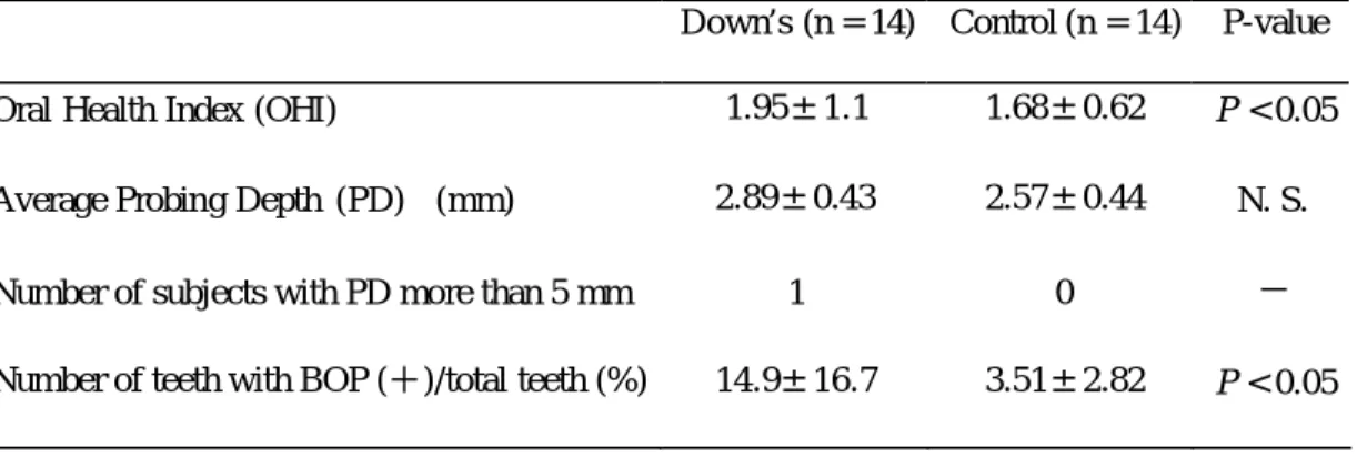
Clinical findings A summary of the OHI, PD and BOP in both the DS and control groups is given in Table 1. In the DS group, one patient had 5- mm deep gingival pockets. No significant difference was found in PD between the DS patients and the controls. OHI in DS patients was significantly higher (p < 0.05) than that in the controls. The level of BOP-positive sites was significantly higher (p < 0.05) in the DS patients than in the controls. The mean and SD of probing depth of sampling sites in DS patients and controls were 2.9 + 0.58 and 2.9 + 0.56, respectively. , These values were close to average of full mouse as shown in Table 1.
Production of MMP-2 and MMP-8 In the GCF samples, levels of MMP-2 and MMP- 8 were significantly higher in the DS patients (p < 0.01) than in the controls (Fig 1). It is possible that this difference in MMP levels between the two groups was influenced by OHI and BOP values. Therefore, MMP levels were compared after dividing the DS patients based on levels of OHI and BOP. Fig. 2 shows levels of MMP-2 revealed after dividing the DS patients into high and low OHI groups. When the DS group was separated into two group s, one with an OHI score of 2 or less and the other with a score of more than 2, levels of MMP-2 and MMP-8 in both groups were significantly higher than those in control patients.
No statistically significant difference was observed in MMP level between high and low OHI in DS patients (p < 0.01). Levels of MMP2 and MMP-8 at BOP-positive and - negative sites in DS patients were higher than those in the control patients. No statistically significant difference, however, was found in MMP2 and MMP-8 levels between BOP-positive and -negative sites in DS patie nts (Fig. 3, p<0.001).
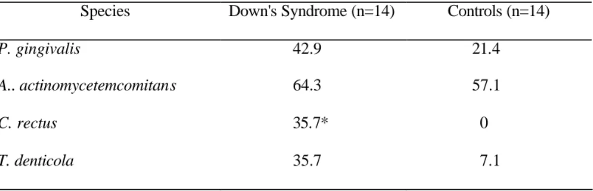
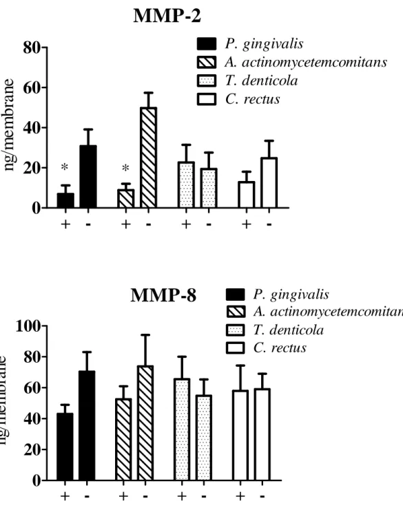
Bacterial Prevalence in DS and Controls We compared prevalence of subgingival bacterial species between the DS group and the controls (Table 3). The detection rate for all periodontopathic bacteria tested was higher in DS patients than in the control group, although this was only statistically significant for C. rectus. MMP levels in DS patient s with or without each periodontopathic bacterium are shown in Fig. 4. Levels of MMP-2 were significantly lower in DS patients harboring
P. gingivalis or A. actinomycetemcomitans than in those who were not (P<0.001).
DISCUSSION
A high susceptibility to and prevalence of periodontitis in DS patients has been reported (29). However, the reason for this high susceptibility and prevalence remains to be clarified. In the present study, we showed an increase in levels of MMP-2 and MMP-8 in DS patients compared with in healthy controls. This result agrees with that of a previous report in DS patients (20, 21). MMP-2 and MMP-8 were reported to show an increase in chronic periodontitis and aggressive periodontitis patients (18, 19, 30). It is possible that an increase in MMPs in gingival tissue induces periodontal destruction. In the present study, we evaluated levels of MMP-2 and -8 using a membrane filter and expressed as ng/membrane. An increase in the vascular permeability of blood vessels following gingival stimulation was reported (31). This suggests that MMP levels in GCF are influenced by
stimulation during sampling. Therefore, MMP levels in unstimulated GCF may better reflect MMPs levels in gingival tissue. Although collection of GCF using filter paper is frequently used (30, 32, 33), further study employing measurement of real concentrations of MMPs in unstimulated GCF is required.
In the present study, the ratio of BOP-positive sites in DS patients was somewhat higher than that in healthy controls, indicating gingival inflammation in DS patients; although average PD in DS patients was the same as that in the controls. MMP-8 is a neutrophil-derived collagenase. It is possib le that the higher levels of MMP-2 and MMP-8 seen in DS patients in this study were a result of inflammation caused by poorer oral hygiene than that in the healthy controls. To eliminate the effects of level of oral hygiene or inflammation on increase in MMP production in DS patients, the DS patients were divided by OHI or BOP score at the sampling site. DS patients with an OHI score of less than 2 also showed significantly higher levels of MMP-2 and MMP-8 than healthy subjects. Levels of these MMPs showed no statistically significant difference between patients with an OHI score of less than 2 and those with a score of 2 or more in DS patients. MMP-2 and MMP-8 levels in DS patients at BOP-negative sites were significantly higher than those in the controls. No difference was found in these levels between BOP-positive and -negative sites in DS patients. These results suggest a difference in production of MMP-2 and MMP-8 in DS patients, regardless of inflammatory response. MMP-2 was reported to be constitutively expressed at low levels in the periodontium (34) The increase in MMP-2 observed in this study agrees with that seen in a previous report by Komatsu et al. (21). They reported an increase in MMP-2 by increase in MMP-2 mRNA expression in fibroblasts from DS patients
in vitro. It is possible that there is a difference in MMP-2 production between DS patients
neutrophil-derived proteinase (8). The present results showed no significant difference in MMP-8 levels between DS patients with an OHI score of less than 2 and an OHI score of 2 or more, or between BOP-positive and BOP-negative sites in DS patients. This suggests that a factor other than infiltration by neutrophils during inflammation is involved in inducing an increase in levels of MMP-8. These observations suggest that level of oral hygiene and inflammation are not the only cause of increase in MMPs in DS patients.
Detection rates of periodontopathic bacteria in DS patients were higher than those in the control patients. The profile was similar to that in a report by Amano et al. (22). Interestingly, if the DS patients were separated by detection of microorganisms, levels of MMP-2 and MMP-8 were lower in DS patients harboring A. actinomycetemcomitans and P.
gingivalis. Differences in MMP-2 were statistically significant (P < 0.001). Lipopoly
saccharide of A. actinomycetemcomitans was reported to increase production of MMP-2 in fibroblasts (35). P. gingivalis significantly up-regulated MMP-2 and MMP-9 mRNA expression by oral epithelial cells (36). The present result s disagree with those of these earlier reports. However, Garlet et al. (37) reported a correlation between an increase in MMP-1, -2, and -9 and interleukin 1β, tumor necrosis factor α and interferon γ in A.
actinomycetemcomitans-infected mice. The expression of MMP-2 in their study showed a
progressive increase from 24 hr until 15 days after infection, with a decrease at 30 and 60 days. P. gingivalis and A. actinomycetemcomitans have factors which induce an immunoresponse, including lipopolysaccharide and immunosuppressive factors (38-40). Immune dysfunction was also reported in Down’s syndrome (41). It is possible that immunomodula tion by these microorganism or immune dysfunction affected production of MMP-2. However, further analysis with a larger number of samples is required to confirm this reduction. Levels of MMP-8 in subjects infected by periodontopathic bacteria were
slightly lower or the same as those in subjects free of infection. Most MMP-8 in periodontal lesions is released from neutrophils, and MMP-8 has been reported to be associated with severity of chronic periodontitis (10). Defect in and dysfunction of bactericidal capacities have been described (6, 42). These types of dysfunction may affect levels of MMP-8 in DS patients. In addition, colonization by periodontopathic bacteria showed no effect on MMP levels. This also suggests that inflammation caused by periodontopathic bacterial colonization does not involve an increase in MMP-2 and MMP-8 in DS patients. Colonization by P. gingivalis and A. actinomycetemcomitans have been reported to induce immune response in many studies (43). This discrepancy also suggests immune dysfunction. To clarify the dys function of the immune response in DS patients, further analysis is required comparing condition of periodontal tissue and immune response in DS patients.
The present result s showed an increase in MMP-2 and MMP-8 in DS patients. Recently, Bildt reported that pro-MMP-2, active MMP-2 and the MMP-2 complex were higher in patients with chronic periodontitis than in healthy subjects, suggesting the involvement of these metalloproteinases in periodontitis (44). Involvement of MMP-8 production in periodontitis has also been reported (8). However, the present results indicate an increase in MMPs, regardless of whether inflammation of gingival tissue is present or not. Taken together with the results of earlier studies, the results of the present study suggest that an increase in MMP-2 and MMP-8 levels is involved in higher susceptibility to and prevalence of periodontitis in DS patients, although further analysis with a larger population will be required for clarification.
Acknowledgment: The authors would like to thank Associate Professor Jeremy Williams, Tokyo Dental College, for his assistance with the English of this manuscript. This research
was supported by an Oral Health Science Center Grant from Tokyo Dental College, and a
“High-Tech Research Center” Project for Private Universit ies matching fund subsidy from
the Ministry of Education, Culture, Sports, Science and Technology of Japan, 2006-2010
Reference
1. Desai SS. Down syndrome: a review of the literature. Oral Surg Oral Med Oral Pathol
Oral Radiol Endod 1997; 84: 279-285.
2. Cohen MM, Winer RA, Schwartz S, Shklar G. Oral aspects of mongolism. I. Periodontal disease in mongolism. Oral Surg Oral Med Oral Pathol 1961; 14: 92-107.
3. Barnett ML, Press KP, Friedman D, Sonnenberg EM. The prevalence of periodontitis and dental caries in a Down's syndrome population. J Periodontol 1986; 57: 288-293.
4. Modeer T, Barr M, Dahllof G. Periodontal disease in children with Down's syndrome.
Scand J Dent Res 1990; 98: 228-234.
5. Meyle J, Gonzales JR. Influences of systemic diseases on periodontitis in children and adolescents. Periodontol 2000 2001; 26: 92-112.
6. Izumi Y, Sugiyama S, Shinozuka O, Yamazaki T, Ohyama T, Ishikawa I. Defective neutrophil chemotaxis in Down's syndrome patients and its relationship to periodontal destruction. J Periodontol 1989; 60: 238-242.
7. Nagase H, Visse R, Murphy G. Structure and function of matrix metalloproteinases and TIMPs. Cardiovasc Res 2006; 69: 562-573.
8. Sorsa T, Tjaderhane L, Konttinen YT, et al. Matrix metalloproteinases: contribution to pathogenesis, diagnosis and treatment of periodontal inflammation. Ann Med 2006; 38: 306-321.
9. Kiili M, Cox SW, Chen HY, et al. Collagenase-2 (MMP-8) and collagenase-3 (MMP-13) in adult periodontitis: molecular forms and levels in gingival crevicular fluid and immunolocalisation in gingival tissue. J Clin Periodontol 2002; 29: 224-232.
10. Sorsa T, Uitto VJ, Suomalainen K, Vauhkonen M, Lindy S. Comparison of interstitial collagenases from human gingiva, sulcular fluid and polymorphonuclear leukocytes. J
Periodontal Res 1988; 23: 386-393.
11. Imai K, Ohta S, Matsumoto T, et al. Expression of membrane-type 1 matrix metalloproteinase and activation of progelatinase A in human osteoarthritic cartilage. Am
J Pathol 1997; 151: 245-256.
12. Aimes RT, Quigley JP. Matrix metalloproteinase-2 is an interstitial collagenase. Inhibitor- free enzyme catalyzes the cleavage of collagen fibrils and soluble native type I collagen generating the specific 3/4- and 1/4- length fragments. J Biol Chem 1995; 270: 5872-5876.
13. Konttinen YT, Ceponis A, Takagi M, et al. New collagenolytic enzymes/cascade identified at the pannus- hard tissue junction in rheumatoid arthritis: destruction from above. Matrix Biol 1998; 17: 585-601.
14. Birkedal-Hansen H, Moore WG, Bodden MK, et al. Matrix metalloproteinases: a review.
Crit Rev Oral Biol Med 1993; 4: 197-250.
15. Ingman T, Sorsa T, Lindy O, Koski H, Konttinen YT. Multiple forms of gelatinases/type IV collagenases in saliva and gingival crevicular fluid of periodontitis patients. J Clin
Periodontol 1994; 21: 26-31.
16. Makela M, Salo T, Uitto VJ, Larjava H. Matrix metalloproteinases (MMP-2 and MMP-9) of the oral cavity: cellular origin and relationship to periodontal status. J Dent Res 1994; 73: 1397-1406.
17. Korostoff JM, Wang JF, Sarment DP, Stewart JC, Feldman RS, Billings PC. Analysis of in situ protease activity in chronic adult periodontitis patients: expression of activated MMP-2 and a 40 kDa serine protease. J Periodontol 2000; 71: 353-360.
18. Ejeil AL, Igondjo-Tchen S, Ghomrasseni S, Pellat B, Godeau G, Gogly B. Expression of matrix metalloproteinases (MMPs) and tissue inhibitors of metalloproteinases (TIMPs) in healthy and diseased human gingiva. J Periodontol 2003; 74: 188-195.
19. Sorsa T, Ding YL, Ingman T, et al. Cellular source, activation and inhibition of dental plaque collagenase. J Clin Periodontol 1995; 22: 709-717.
20. Halinen S, Sorsa T, Ding Y, et al. Characterization of matrix metalloproteinase (MMP-8 and -9) activities in the saliva and in gingival crevicular fluid of children with Down's syndrome. J Periodontol 1996; 67: 748-754.
21. Komatsu T, Kubota E, Sakai N. Enhancement of matrix metalloproteinase (MMP)-2 activity in gingival tissue and cultured fibroblasts from Down's syndrome patients. Oral
Dis 2001; 7: 47-55.
22. Amano A, Kishima T, Kimura S, et al. Periodontopathic bacteria in children with Down syndrome. J Periodontol 2000; 71: 249-255.
23. Barr-Agholme M, Dahllof G, Linder L, Modeer T. Actinobacillus actinomycetemcomitans, Capnocytophaga and Porphyromonas gingivalis in subgingival
plaque of adolescents with Down's syndrome. Oral Microbiol Immunol 1992; 7: 244-248. 24. Soder B, Airila Mansson S, Soder PO, Kari K, Meurman J. Levels of matrix
metalloproteinases-8 and -9 with simultaneous presence of periodontal pathogens in gingival crevicular fluid as well as matrix metalloproteinase-9 and cholesterol in blood. J
Periodontal Res 2006; 41: 411-417.
25. Yamazaki T, Miyamoto M, Yamada S, Okuda K, Ishihara K. Surface protease of
Treponema denticola hydrolyzes C3 and influences function of polymorphonuclear
leukocytes. Microbes Infect 2006; 8: 1758-1763.
status. J Am Dent Assoc 1960; 61: 172-179.
27. Ashimoto A, Chen C, Bakker I, Slots J. Polymerase chain reaction detection of 8 putative periodontal pathogens in subgingival plaque of gingivitis and advanced periodontitis lesions. Oral Microbiol Immunol 1996; 11: 266-273.
28. Kobayashi N, Ishihara K, Sugihara N, Kusumoto M, Yakushiji M, Okuda K. Colonization pattern of periodontal bacteria in Japanese children and their mothers. J
Periodontal Res 2008; 43: 156-161.
29. Reuland-Bosma W, van Dijk J. Periodontal disease in Down's syndrome: a review. J Clin
Periodontol 1986; 13: 64-73.
30. Alfant B, Shaddox LM, Tobler J, Magnusson I, Aukhil I, Walker C. Matrix metalloproteinase levels in children with aggressive periodontitis. J Periodontol 2008; 79: 819-826.
31. Egelberg J. Permeability of the dento-gingival blood vessels. IV. Effect of histamine on vessels in clinically healthy and chronically inflamed gingivae. J Periodontal Res 1966; 1: 297-302.
32. Pirhan D, Atilla G, Emingil G, Sorsa T, Tervahartiala T, Berdeli A. Effect of MMP-1 promoter polymorphisms on GCF MMP-1 levels and outcome of periodontal therapy in patients with severe chronic periodontitis. J Clin Periodontol 2008; 35: 862-870.
33. Grayson R, Douglas CW, Heath J, Rawlinson A, Evans GS. Activation of human matrix metalloproteinase 2 by gingival crevicular fluid and Porphyromonas gingivalis. J Clin
Periodontol 2003; 30: 542-550.
34. Visse R, Nagase H. Matrix metalloproteinases and tissue inhibitors of metalloproteinases: structure, function, and biochemistry. Circ Res 2003; 92: 827-839.
lipopolysaccharide regulates matrix metalloproteinase, tissue inhibitors of matrix metalloproteinase, and plasminogen activator production by human gingival fibroblasts: a potential role in connective tissue destruction. J Cell Physiol 2007; 212: 189-194.
36. Andrian E, Mostefaoui Y, Rouabhia M, Grenier D. Regulation of matrix metalloproteinases and tissue inhibitors of matrix metalloproteinases by Porphyromonas
gingivalis in an engineered human oral mucosa model. J Cell Physiol 2007; 211: 56-62.
37. Garlet GP, Cardoso CR, Silva TA, et al. Cytokine pattern determines the progression of experimental periodontal disease induced by Actinobacillus actinomycetemcomitans through the modulation of MMPs, RANKL, and their physiological inhibitors. Oral
Microbiol Immunol 2006; 21: 12-20.
38. Page RC. The role of inflammatory mediators in the pathogenesis of periodontal disease.
J Periodontal Res 1991; 26: 230-242.
39. Shenker BJ, Slots J. Immunomodulatory effects of Bacteroides products on in vitro human lymphocyte functions. Oral microbiol Immunol 1989; 4: 24-29.
40. Shenker BJ, McKay T, Datar S, Miller M, Chowhan R, Demuth D. Actinobacillus
actinomycetemcomitans immunosuppressive protein is a member of the family of
cytolethal distending toxins capable of causing a G2 arrest in human T cells. J Immunol 1999; 162: 4773-4780.
41. Cuadrado E, Barrena MJ. Immune dysfunction in Down's syndrome: primary immune deficiency or early senescence of the immune system? Clin Immunol Immunopathol 1996; 78: 209-214.
42. Gregory L, Williams R, Thompson E. Leucocyte function in Down's syndrome and acute leukaemia. Lancet 1972; 1: 1359-1361.
periodontopathic bacteria and its role in the pathogenesis of periodontitis. Periodontol
2000 1997; 14: 79-111.
44. Bildt MM, Bloemen M, Kuijpers-Jagtman AM, Von den Hoff JW. Collagenolytic fragments and active gelatinase complexes in periodontitis. J Periodontol 2008; 79: 1704-1711.
Table 1. Clinical data (mean ± SD) from DS patients and controls
Down’s (n = 14) Control (n = 14) P-value Oral Health Index (OHI) 1.95±1.1 1.68±0.62 P < 0.05
Average Probing Depth (PD) (mm) 2.89±0.43 2.57±0.44 N. S. Number of subjects with PD more than 5 mm 1 0 − Number of teeth with BOP (+)/total teeth (%) 14.9±16.7 3.51±2.82 P < 0.05
Table 2 Detection rate (%) of specific periodontal pathogens in subgingival plaque samples
Species Down's Syndrome (n=14) Controls (n=14)
P. gingivalis 42.9 21.4
A.. actinomycetemcomitans 64.3 57.1
C. rectus 35.7* 0
T. denticola 35.7 7.1
Figure legend
Fig. 1. MMP-2 and MMP-8 levels (ng/membrane ± SE) in GCF. A. MMP-2 levels in GCF from DS patients and controls
B. MMP-8 levels in GCF from DS patients and controls.
C. MMP-2 levels in GCF by BOP. (§p < 0.001 compared to control) D. MMP-8 levels in GCF by BOP. (§p < 0.001 compared to control)
E. MMP-2 levels (ng/membrane ± SD) in GCF by OHI. Patients were separated into two group by score of OHI 2 and less or more than 2. (§p < 0.001 compared to control)
F. MMP-8 levels (ng/membrane ± SD) in GCF by OHI. Patients were separated into two group by score of OHI 2 and less or more than 2. (§p < 0.001 compared to control)
A and B were analyzed by t-test (*p < 0.001)
C-E was analyzed by one-way repeated measures ANOVA for inter- group comparisons. Student-Newman-Keuls test was used for multiple comparisons (§p < 0.001).
Fig. 2. Comparison of MMP-2 and MMP-8 production (ng/membrane ± SE) based on detection of periodontopathic bacteria (*p < 0.001 by t-test compared with control).
Fig. 1
MMP-2
BOP (+) BOP (-) Control 0 10 20 30 40 ng /m e m br a n e MMP-8
BOP (+) BOP (-) Control 0 20 40 60 80 100 ng/m e m b rane
§
§
§
§
MMP-8OHI > 2 OHI < 2 Control 0 20 40 60 80 100 ng /m e m br a n e MMP-2
OHI > 2 OHI < 2 Control 0 10 20 30 40 50 ng /m e m br a n e
§
§
A
B
C
D
E
F
MMP-8 Down's Control 0 20 40 60 80 ng/m em b ra n e MMP-2 Down's Control 0 10 20 30 n g /m e m b ran e*
*
n=5 n=9 n=14 n=5 n=9 n=14 n=5 n=9 n=14 n=5 n=9 n=14Fig. 2