福島県立医科大学 学術機関リポジトリ
This document is downloaded at: 2021-11-08T00:02:22Z
Title Lectin-dependent inhibition of antigen-antibody reaction:
Application for measuring α2,6-sialylated glycoform of transferrin( 本文 )
Author(s) 星, 京香
Citation
Issue Date 2014-03-25
URL http://ir.fmu.ac.jp/dspace/handle/123456789/611
Rights This thesis/dissertation is modified from "J Biochem. 2013 Sep;154(3):229-32. doi: 10.1093/jb/mvt065. © The Authors 2013. Published by Oxford University Press".
DOI
Text Version ETD
Lectin-dependent inhibition of antigen-antibody reaction: Application for measuring α2,6-sialylated glycoform of transferrin
SSAレクチンによる抗原-抗体反応の阻害:
髄液中α2,6-シアル酸化トランスフェリン測定への応用
福島県立医科大学医学部 生化学講座 星 京香
Contents
1. Abstract... 1
2. Introduction... 2
3. Results 3.1 Inhibition of anti-transferrin antibody binding to transferrin by lectins …….... 4
3.2 Reactivity of SSA lectin and antibodies against glycoforms of transferrin ……. 5
3.3 Reactivity of SSA lectin and antibodies against glycoforms of α2-macroglobulin……….. 5
3.4 Reactivity of SSA lectin and antibodies against crude CSF……….... 6
3.5 SSA-ALI as a high throughput method for diagnosing iNPH ………... 6
4. Discussion ... 9
5. Materials and Methods 5.1 Patients ………. 11
5.2 Lectins ………... 12
5.3 Transferrin and α2-Macloglobulin isoforms ………12
5.4 Western blotting and lectin-probed ELISA ……….... 12
5.5 Lectin inhibition assay by ELISA ……….. 13
5.6 Fractionation of Tf isoforms in human CSF………... 14
5.7 SSA inhibition assay by automated latex-agglutination immunoassay ……… 15
6. Acknowledgments ……… 16
7. Abbreviations ……… 17
8. References ………. 18 9. Figures and Figure legends ………. 21
1. Abstract
We previously found two isoforms of transferrin, i.e., non-sialylated Tf and α2,6-sialylated Tf, in cerebrospinal fluid (CSF). The ratio of isoforms
(α2,6-sialylated/non-sialylated Tf) was a marker for idiopathic normal pressure hydrocephalus (iNPH), a dementia-inducing disorder. I have recently found that binding of anti-transferrin antibody (Tf-ab) to α2,6-sialylated Tf is inhibited by Sambucus sieboldiana Agglutinin (SSA) lectin, which recognizes the α2,6-sialylated epitope. This finding lends itself to automated latex-agglutination immunoassay (ALI), using SSA lectin as an inhibitor specific for α2,6-sialylated Tf. Namely, concentration of α2,6-sialylated Tf is estimated by ALI signal difference in the presence and absence of
SSA lectin (SSA-ALI). The concentration of α2,6-sialylated Tf measured by SSA-ALI (n = 47) is correlated with those by ELISA (r2 = 0.8554, p < 0.01), which was previously developed for measuring α2,6-sialylated Tf. The ratio of α2,6-sialylated to non-sialylated, calculated on SSA-ALI signals, shows significant difference between iNPH (n = 28) and non-iNPH (n = 19) (Mann-Whitney U tests: p = 0.015), indicating that the SSA-ALI is a simple and rapid method for measuring the ratio of α2,6-sialylated/non-sialylated Tf.
2. Introduction
Idiopathic normal pressure hydrocephalus (iNPH) is a dementia-inducing disorder associated with gait disturbance and urinary incontinence (1, 2). The disease is caused by abnormal metabolism in the cerebrospinal fluid (CSF), which induces ventriculomegaly observable by computed tomography (CT) or magnetic resonance imaging (MRI).
We previously reported that cerebrospinal fluid (CSF) contains two transferrin isoforms: one had α2,6-sialylated N-glycans whereas the other had non-sialylated N-glycans. Glycan analyses revealed that the non-sialylated form had biantenary asialo- and agalacto-complex type N-glycans, which carried bisecting β1,4-Nacetylglucosamine and core α1,6-fucose (3, 4). Sialylated form had biantennary complex type N-glycans that carry sialyl α2,6-galactose residues.
Non-sialylated Tf was decreased in iNPH and the ratio of isoforms (α2,6-sialylated/non-sialylated Tf) differentiated iNPH from Alzheimer’s disease. For analysis of the isoform ratio, we measured the isoforms by Western blotting (4) or a lectin-probed ELISA (5), in which anti-transferrin antibody (Tf-ab) and lectins were used for detecting protein and glycan portions of Tf, respectively. These methods, however, are not clinically beneficial to use because they are time- and labour-consuming.
Automated Latex-agglutination Immunoassay (ALI) is a rapid and simple method for measuring protein concentration including serum and CSF proteins (6). For
measuring Tf, small latex particles carrying Tf-ab are mixed with Tf in the specimen and the resulting aggregates are photometrically quantified (7). The assay, however, does not distinguish α2,6-sialylated and non-sialylated Tf, because Tf-ab equally reacts with both isoforms.
In the present study I demonstrate that even polyclonal antibody is inhibited by lectin binding; i.e., SSA (Sambucus sieboldiana Agglutinin) lectin, which binds α2,6-sialylated epitope, inhibits the binding of polyclonal Tf-ab to α2,6-sialylated Tf.
The finding is applied for measuring α2,6-sialylated Tf by ALI, resulting in confirmation of our previous conclusion that the alteration of Tf isoforms in CSF is a diagnostic marker for iNPH.
3. Results
3.1 Inhibition of anti-transferrin antibody binding to transferrin by lectins
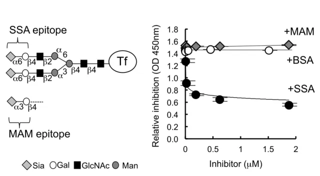
Human serum transferrin (Tf) has biantennary complex type N-glycans that carry Siaα2,6Galβ1,4GlcNAc residues, i.e., epitopes for SSA lectin (Fig. 1A). I tested whether or not SSA inhibited binding of anti-transferrin antibody (Tf-ab) to human serum transferrin. ELISA was used for estimating the antigen-antibody reaction. A capture antibody, polyclonal Tf-ab (Cappel), was pre-treated with 1 mM sodium periodate (NaIO4) to oxidize the exocyclic glycerol side chain of sialic acid, abolishing SSA epitopes on the antibody (8). The periodate-treated antibody was coated on a microtiter plate. Antigen (10 ng/mL of serum Tf) was mixed with a various concentration of SSA lectin, and then the mixture was applied to the plate. After extensive washes, Tf signal was detected with HRP-conjugated anti-human Tf antibody (Bethyl laboratories). As shown in figure 1B, SSA inhibited Tf signal in a dose-dependent manner. The signal (O.D. 1.5) decreased almost linearly in a range of 2
~ 20 nM of SSA and then saturated in a range of 0.2 ~ 1.86 µM (ca. O.D. 0.6). Based on the observation, 65 nM of SSA was used in the following inhibition assay (SSA-ELISA). Neither Maackia amurensis (MAM) lectin nor BSA inhibited the Tf signal. This result is consistent with the observation that human Tf carries Siaα2,6Galβ1,4GlcNAc, an SSA epitope, but not Siaα2,3Galβ1,4GlcNAc, a MAM epitope. Human transferrin has two N-glycans, which could be partially α2,6-sialylated.
Precise estimation of SSA inhibition signal of these isoforms will need to be examined
with a set of the polyclonal antibodies.
By ELISA, I tested three kinds of antibodies in an SSA inhibition assay; i.e., goat polyclonal Tf-ab from Cappel, rabbit polyclonal Tf-ab from DAKO Co., Ltd. and goat polyclonal Tf-ab from Bethyl laboratories. Their signals associated with serum Tf were inhibited by SSA lectin; ca. 50-60 % inhibition for Cappel antibody (Fig. 1B) and ca. 30% inhibition for DAKO and Bethyl antibodies. The three polyclonal antibodies were more or less inhibited regardless of the species from which each was derived.
3.2 Reactivity of SSA lectin and antibodies against glycoforms of transferrin
To examine glycan specificities in SSA inhibition, I prepared a series of glycan isoforms (glycoforms) of Tf. To abolish SSA epitopes, human serum Tf was treated with 1 mM sodium periodate (NaIO4) to oxidize the exocyclic glycerol side chain of sialic acid. Tf was also treated with sialidase, yielding asialo-Tf. The asialo-Tf was further treated with galactosidase to prepare agalacto-Tf. As shown in figure 2A, SSA does not inhibit Tf-ab binding to NaIO4-treated, asialo- or agalacto-Tf, but does inhibit serum Tf by 62%, suggesting that the terminal α2,6-sialic acid epitopes on Tf are essential for the inhibition by SSA.
3.3 Reactivity of SSA lectin and antibodies against glycoforms of α2-macroglobulin I tested whether SSA inhibition was observed for another α2,6-sialylated glycoprotein, α2-macroglobulin (α2M). SSA inhibition was examined with ELISA,
using anti-α2M antibody (α2M-ab) and various glycoforms of α2M. SSA inhibited α2M-ab binding to α2M by 65%, but did not inhibit NaIO4-treated, asialo- or agalacto-α2M (Fig. 2B), suggesting that, regardless of antigen proteins, SSA inhibits antigen-antibody reactions by binding to α2,6-sialylated-glycans on antigens.
3.4 Reactivity of SSA lectin and antibodies against crude CSF
I next applied SSA inhibition assay for human crude CSF. As shown in figure 3A, human CSF contains both α2,6-sialylated and non-sialylated Tf (an upper and lower band, respectively, on an immunoblot). The crude human CSF was applied to an SSA-agarose column to fractionate α2,6-sialylated Tf and non-sialylated Tf, which were recovered in lactose-eluted and flow-thorough fraction, respectively (Fig. 3A).
SSA inhibition is detected with α2,6-sialylated Tf in the eluted fraction, but not detected with non-sialylated Tf in flow-thorough fraction (Fig. 3B). These results suggest that SSA inhibition is applicable for measuring α2,6-sialylated Tf in fractionated CSF. In addition, the ELISA signal in human crude CSF was partially inhibited by adding SSA, indicating that SSA inhibition is applicable even for crude CSF.
3.5 SSA-ALI as a high throughput method for diagnosing iNPH
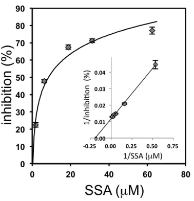
Next I examined whether SSA also inhibits Tf-ab binding to α2,6-sialylated Tf in an automated latex-agglutination immunoassay (ALI), which is a rapid and simple method
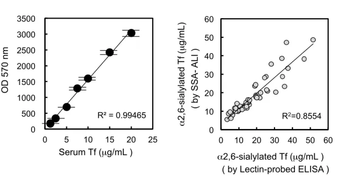
for measuring protein concentration (6). For measuring Tf, small latex particles carrying Tf-ab are mixed with Tf in the specimen and the resulting aggregates are photometrically quantified. In the assay, specimen was mixed with SSA in a flow cell, and then Tf-ab-coated latex beads were added. For SSA inhibition in ALI, the polyclonal Tf-ab from DAKO was used due to its highest sensitivity. ALI signals with serum Tf were inhibited by SSA lectin in a saturable manner (Fig. 4). Double reciprocal plot analysis revealed that the maximal inhibition is 77% and half maximal inhibition is observed at 8.4 µM of SSA lectin. Based on this observation, 62.5 µM of SSA lectin was used in the following inhibition assay (SSA-ALI). A calibration line for quantifying α2,6-sialylated Tf was linear at least in a range of 1.25 ~ 20 µg/ML (Fig. 5A). I then
applied SSA-ALI to crude human CSF. Dilution linearity was observed in a range of 0.125 ~ 0.5 µL of crude CSF. When 0.25 and 0.5 µg of α2,6-sialylated Tf were added to 0.1 µL of CSF, signal recoveries were 95.7 + 11.6 % and 97.5 + 10.5 % of the expected value, respectively, suggesting that the interfering substances in CSF, if any, are negligible. Day-to-day variation of the assay was less than 8%. By SSA-ALI, I measured concentration of α2,6-sialylated Tf in crude CSF from 47 individuals. The specimen was also measured by a lectin-probed ELISA (5), which was developed for estimating concentration of α2,6-sialylated Tf in our previous study. The results of SSA-ALI with 47 individuals were correlated with those of the lectin-probed ELISA (R2
= 0.8554, p < 0.01) (Fig. 5B). Thus the SSA-ALI could be a simple and rapid method for measuring α2,6-sialylated isoform of Tf in CSF. In addition, the SSA-ALI was successfully applied to diagnosis of a dementia-inducing disorder, idiopathic normal
pressure hydrocephalus, which shows abnormal CSF metabolism and increase of α2,6-sialylated /non-sialylated Tf ratio (Fig. 6).
4. Discussion
Fucosylated alpha-fetoprotein (AFP) is an early marker of hepatocellular carcinoma (9, 10). It is notable that non-fucosylated AFP is not the specific marker, indicating that glycoform-specific detection of AFP is essential for accurate diagnosis. The diagnostic isoform, fucosylated AFP, is recognized by Lens culinaris Agglutinin (LCA) lectin (10).
Suzuki et al. found that LCA lectin inhibited the binding of anti-AFP monoclonal antibodies to the fucosylated AFP (11). Only a few monoclonal antibodies were inhibited by LCA, but most were not. Indeed, only two out of thirty MoAbs were inhibited; one was inhibited by 36% and the other was 59% (12). They speculated that LCA binding to the core fucose sterically hindered neighboring antigen epitopes that were recognized by the limited number of monoclonal antibodies (11). It is notable that SSA lectin binds to the most distal glycan epitopes, terminal α2,6-sialic acids, whereas LCA binds proximally. Tf glycan complexed with SSA at the terminal α2,6-sialic acids may be flexible enough to hinder a large part of antigen epitopes, which are recognized by a polyclonal Tf-ab. Further analysis is required to clarify the molecular mechanisms of lectin inhibition of antibody binding.
A variety of methods have been reported to measure distinct types of glycoforms of Tf: Western blotting (4), lectin-probed ELISA (5), an anion exchange chromatography (13), isoelectric focusing (14), and capillary electrophoresis (15). The SSA-ALI we developed is more time- and labour-saving than these other methods.
SSA inhibition assay was applied to detect α2,6-sialylated α2M as well as α2,6-sialylated Tf at least in ELISA. The assay may be applicable for other
glycoprotein markers that have α2,6-sialylated glycan. For example, α2,6-sialylated carcinoembryonic antigen is elevated in serum of colon cancer patients with liver metastases (16). Haptoglobin with α2,6-sialylated glycans is significantly increased in serum of patients with prostate cancer versus those with benign prostate diseases (17).
Measurement of these α2,6-sialylated glycoproteins by SSA inhibition assay or SSA-ALI would be useful for diagnosing these diseases.
5. Materials and Methods 5.1 Patients
This study included 28 patients with iNPH, including 14 males and 14 females (median age 76 years; range 64 ~ 85 years). The patients were diagnosed using the Clinical Guidelines for iNPH issued by the Japanese Society of Normal Pressure Hydrocephalus (18). The non-iNPH group consisted of 19 patients, including 10 males and 9 females (median age 76 years; range 64 ~ 85 years). Briefly, the diagnosis of possible iNPH was based on the clinical symptoms and signs of gait disturbance, dementia, and urinary incontinence, as well as computed tomography (CT) and magnetic resonance imaging (MRI) findings of enlarged ventricles (Evans index of > 0.3). A bolus infusion test and tap test (removal of 30 mL of CSF) were performed routinely as necessary to determine probable iNPH in all patients. The tap test-negative patients were classified as non-iNPH. The probable iNPH patients were subjected to a CSF shunt operation. The diagnosis of definite iNPH was based on improvement of the symptoms at a month after the operation. The study identified 28 patients who fit the above-mentioned criteria.
Shunt non-responders were classified as non-iNPH patients.
This study, including the process of securing informed consent, was approved by the Ethics Committee of Fukushima Medical University (approval number 613), which is guided by local policy, national laws, and the World Medical Association Declaration of Helsinki.
5.2 Lectins
SSA and MAM were purchased from J-Oil Mills, Kanagawa, Japan.
5.3 Transferrin and α2-Macloglobulin isoforms
Serum Tf and plasma α2M purified from human blood were purchased from Sigma-Ardrich, St. Louis, MO, USA. Serum Tf or plasma α2M was treated with sialidase (Nacalai Tesque, Kyoto, Japan) to remove terminal sialic acids, generating asialo-Tf or asialo-α2M (19). Asialo-Tf or asialo-α2M was further treated with galactosidase (Seikagaku Corporation, Tokyo, Japan) to remove terminal galactoses, yielding agalacto-Tf or agalacto-α2M.
5.4 Western blotting and lectin-probed ELISA
For detecting Tf isoforms, Western blotting was optimized as described before (4) and a lectin-probed ELISA was previously developed (5). Briefly, Tf-ab was coated on a 96-well C8 Maxisorp immunomodule plate (Nunc, Roskidle, Denmark). CSF samples were appropriately diluted with TBST containing 0.5 mM EDTA and applied to the plate and incubated at 4°C overnight. After three washes with TBST, the plate was incubated sequentially with a biotin-SSA (Seikagaku Corporation, Tokyo, Japan) and a horseradish peroxidase-labeled streptavidin (Takara-Bio Inc., Kyoto, Japan). After three washes with TBST, the plate was incubated with the TMB substrate (Kirkegaard and Perry Laboratories, Gaithersburg, MD, USA) and the absorbance at 450 nm was measured in each well by a plate reader (Bio-Rad Laboratories, Hercules, CA, USA).
5.5 Lectin inhibition assay by ELISA
Tf-ab (Cappel; ICN Pharmaceuticals, Aurora, OH, USA) and α2M-ab (R&D Systems, Minneapolis, MN) were treated with 1 mM sodium periodate to oxidize the exocyclic glycerol side chain of sialic acid (8), thus abolishing SSA epitopes on the antibody. The
periodate-treated antibody was coated on a 96-well C8 Maxisorp immunomodule plate.
Samples were appropriately diluted with TBST, mixed with 65 nM of SSA lectin as an inhibitor of Tf-ab or α2M-ab binding (or BSA as a negative control), and incubated at room temperature for 1h. The mixture was applied to the plate and incubated at 4°C overnight. After five washes with TBST, the plate was incubated with HRP-conjugated anti-human Tf antibody (Bethyl laboratories, Montgomery, TX, USA) or α2M-ab (GeneTex, San Antonio, TX) for 1h. After washing 5 times with TBST, the plate was incubated with the TMB substrate, and the absorbance at 450 nm was measured. In similar assay, MAM lectin was examined for their inhibitory potencies in Tf-ab binding.
5.6 Fractionation of Tf isoforms in human CSF
The crude human CSF was applied to an SSA-agarose (J-Oil Mills) column to fractionate α2,6-sialylated Tf and non-sialylated Tf, which were recovered in lactose-eluted and flow-thorough fraction, respectively. After removal of lactose, both fractions were subjected to SSA-ELISA assay.
5.7 SSA inhibition assay by automated latex-agglutination immunoassay
The antibody-sensitized latex particles were prepared as follows. The 190 nm polystyrene latex particles suspended in Buffer A (25 mM HEPES pH 7.5, 1 mM EDTA 2Na, 0.05% NaN3) were mixed with the equal volume of Buffer A containing the polyclonal Tf-ab (Dako, Glostrup, Denmark) that was pretreated with 1 mM sodium periodate. The final concentrations of the antibody and the particles were 1.5 mg/mL and 0.25% (w/v), respectively. The mixture was stirred at room temperature for 1 h. The antibody-sensitized particles were centrifuged at 59,000 x g for 40 min. The precipitated particles were suspended by sonication in 0.5% Block Ace in Buffer B (25 mM HEPES pH 7.5, 150 mM NaCl, 1 mM EDTA-2NA, 0.05% NaN3), and then centrifuged at 59,000 x g for 40 min. The precipitated particles were resuspended with Buffer B at the final concentration of 0.05%.
The immuno-agglutination reaction was started by mixing 2 µL of sample solution with 100 µL of the particle reagents and 100 µL of SSA (or BSA) in Buffer B. The immuno-agglutination reaction was monitored at a wavelength of 570 nm with a Hitachi Autoanalyzer H7180 (Hitachi, Tokyo, Japan). Human serum Tf was used as a standard.
6. Acknowledgments
I would like to express my sincere gratitude to Professor Yasuhiro Hashimoto for providing me the opportunity to perform this study. I am deeply grateful to Drs. Keiro Shirotani, Yoshinobu Kariya, Kiyomitsu Nara and Hiromi Ito for critical advices. I thank Drs. Masamichi Nagae, (RIKEN), Kana Matsumoto, (RIKEN), Yoshiki Yamaguchi, (RIKEN), Atsushi Kuno, (AIST), Hisashi Narimatsu, (AIST) for their valuable cooperation in my experiments, Madoka Nakajima, (Juntendo University School of Medicine, JUSM), Masakazu Miyajima, (JUSM), Hajime Arai, (JUSM) for providing CSF samples. I also thank Drs. Kenta Noda and Tosihide Miura of Nittobo Medical Co., Ltd. for supporting the ALI analysis. I acknowledge Professor Kenneth
Nollet for editorial advice. This work was supported by Japanese governmental grants from NEDO [MG project]; MHLW [grant number 2006-Nanchi-Ippan-017]; JST [grant numbers AS221Z00232F, AS231Z01053, 241FT0255, and 149]; MEXT [a
Grant-in-Aid for Scientific Research on Innovative Areas, grant number 23110002 (Deciphering sugar chain-based signals regulating integrative neuronal functions) and a Grant-in-Aid for Scientific Research, grant number 23590367].
7. Abbreviations:
AFP, alpha-fetoprotein; ALI, automated latex-agglutination immunoassay; α2M-ab, anti-α2-macroglobulin antibody; CSF, cerebrospinal fluid; Gal, Galactose; iNPH;
idiopathic normal pressure hydrocephalus, LCA, Lens culinaris Agglutinin; MAM, Maackia amurensis; SSA, Sambucus sieboldiana Agglutinin; Tf-ab, anti-transferrin antibody
Conflict of interest None declared.
8. References
1. Gallia, G.L., D. Rigamonti, and M.A. Williams, The diagnosis and treatment of idiopathic normal pressure hydrocephalus. Nat Clin Pract Neurol, 2006. 2(7): p.
375-81.
2. Relkin, N., et al., Diagnosing idiopathic normal-pressure hydrocephalus.
Neurosurgery, 2005. 57(3 Suppl): p. S4-16; discussion ii-v.
3. Hoffmann, A., et al., 'Brain-type' N-glycosylation of asialo-transferrin from human cerebrospinal fluid. FEBS Lett, 1995. 359(2-3): p. 164-8.
4. Futakawa, S., et al., A unique N-glycan on human transferrin in CSF: a possible biomarker for iNPH. Neurobiol Aging, 2012. 33(8): p. 1807-15.
5. Shirotani, K., et al., High Throughput ELISAs to Measure a Unique Glycan on Transferrin in Cerebrospinal Fluid: A Possible Extension toward Alzheimer's Disease Biomarker Development. Int J Alzheimers Dis, 2011. 2011: p. 352787.
6. Komoriya, T., et al., Development of a high-sensitivity latex reagent for the detection of C-reactive protein. J Immunol Methods, 2011. 373(1-2): p. 63-6.
7. Bernard, A., K.S. Chia, and R. Lauwerys, Latex immunoassay of transferrin in urine. J Immunol Methods, 1991. 144(1): p. 49-55.
8. Yamaji, T., et al., Application of a multivalent glycoprobe: characterization of sugar-binding specificity of Siglec family proteins. Methods Enzymol, 2003.
363: p. 104-13.
9. Debruyne, E.N. and J.R. Delanghe, Diagnosing and monitoring hepatocellular carcinoma with alpha-fetoprotein: new aspects and applications. Clin Chim Acta, 2008. 395(1-2): p. 19-26.
10. Toyoda, H., T. Kumada, and T. Tada, Highly sensitive Lens culinaris agglutinin-reactive alpha-fetoprotein: a new tool for the management of hepatocellular carcinoma. Oncology, 2011. 81 Suppl 1: p. 61-5.
11. Suzuki, Y., et al., Close topographical relationship in alpha foetoprotein (AFP) between a lens culinaris binding glycan and the epitope recognized by AFP-reactive monoclonal antibody, 18H4. Br J Cancer, 1987. 55(2): p. 147-52.
12. Taketa, K., et al., Lectin-dependent modulation of interaction between human alpha-fetoprotein and its monoclonal antibodies. Epitope mapping. Tumour Biol, 1998. 19(4): p. 318-28.
13. Jeppsson, J.O., H. Kristensson, and C. Fimiani, Carbohydrate-deficient transferrin quantified by HPLC to determine heavy consumption of alcohol. Clin Chem, 1993. 39(10): p. 2115-20.
14. Hackler, R., et al., Investigation by isoelectric focusing of the initial carbohydrate-deficient transferrin (CDT) and non-CDT transferrin isoform fractionation step involved in determination of CDT by the ChronAlcoI.D. assay.
Clin Chem, 2000. 46(4): p. 483-92.
15. Lanz, C., et al., Improved capillary electrophoresis method for the determination of carbohydrate-deficient transferrin in patient sera. Electrophoresis, 2004.
25(14): p. 2309-18.
16. Yamashita, K., et al., Expression of Sia alpha 2-->6Gal beta 1-->4GlcNAc residues on sugar chains of glycoproteins including carcinoembryonic antigens in human colon adenocarcinoma: applications of Trichosanthes japonica agglutinin I for early diagnosis. Cancer Res, 1995. 55(8): p. 1675-9.
17. Kazuno, S., et al., Multi-sequential surface plasmon resonance analysis of haptoglobin-lectin complex in sera of patients with malignant and benign prostate diseases. Anal Biochem, 2011. 419(2): p. 241-9.
18. Ishikawa, M., Clinical guidelines for idiopathic normal pressure hydrocephalus.
Neurol Med Chir (Tokyo), 2004. 44(4): p. 222-3.
19. Harada, H., et al., Assay of glycosidase by lectin affinity high-performance liquid chromatography. J Chromatogr, 1987. 407: p. 299-304.
3 6
SSA epitope
β4 β4 β4
β4 β2 β2 α6
α6
α α
Tf
β4 α3
MAM epitope
GlcNAc Man Sia Gal
A
0.0 0.2 0.4 0.6 0.8 1.0 1.2 1.4 1.6 1.8
0 0.5 1 1.5 2
Inhibitor (µM)
Relative inhibition (OD 450nm)
B
+SSA +BSA +MAM
Fig. 1. Lectin epitopes on N-glycans of Tf and lectin-dependent inhibition of Tf-ab binding to Tf in ELISA.
Human serum Tf has biantennary complex type N-glycans that carry sialyl-α2,6-galactose β1,4GlcNAc residues, i.e., SSA epitopes (A) : Sia, sialic acid; Gal, galactose; GlcNAc, N- acetylglucosamine; and Man, mannose. An MAM epitope, a sialyl-α2,3-galactose β1,4GlcNAc residue, is also illustrated. Effect of lectin addition on Tf-ab binding to Tf was examined in ELISA (B). ELISA signals were plotted in the presence of SSA (closed circle), MAM (gray square) or BSA (open circle) (Mean + S.D.). Inhibition was examined in the range of 2 nM ~ 1.86 mM of lectins.
9. Figures and Figure legends
0 20 40 60 80 100 120
0 20 40 60 80 100 120
A B
Serum Tf
asialo- agalacto- NaIO4
treated-
+SSA +BSA
Serum Tf ELISA signal (%) (SSA-dependent inhibition)
Transferrin α 2-macroglobulin
ELISA signal (%) (SSA-dependent inhibition)
Plasma α2M
asialo- agalacto- NaIO4
treated-
+SSA +BSA
Plasma α2M !
Fig. 2. Glycan specificity of SSA-dependent inhibition of antigen-antibody reactions in ELISA.
SSA was tested for inhibition of antibody binding to glycan-isoforms of Tf (A) or α2M (B). The proteins were treated with sodium periodate to oxidize sialic acid (NaIO4 treated-). The proteins were also treated with sialidase to remove sialic acid (asialo-), and further treated with galactosidase to remove galactose (agalacto-). SSA Inhibition (%) is defined using BSA as a control (Mean + S.D.).
0 20 40 60 80 100 120
75 - (KDa)
+SSA +BSA
Crude CSF
Eluted fraction Flow-
through
ELISA signal (%) (SSA-dependent inhibition)
α2,6-Sialylated Tf Non-sialylated Tf
A
B
Crude CSF
Eluted fraction Flow-
through Crude
CSF
Fig. 3. Depletion of α2,6-sialylated Tf from human CSF and its effect on SSA- dependent inhibition.
Crude human CSF was applied to an SSA-agarose column and flow-through fraction was prepared. Bound proteins were specifically eluted with lactose. The crude CSF, flow- through fraction, and eluted fractions were subjected to immunoblotting, using Tf-ab (A).
The fractions were assayed for SSA-dependent inhibition in ELISA (B).
0 10 20 30 40 50 60 70 80 90
0 20 40 60 80
SSA ( µ M)
inhibition (%)
1/SSA (µM)
1/inhibi/on (%)
0.00 0.01 0.02 0.03 0.04
-‐0.25 0.00 0.25 0.50 0.75
Fig. 4. Dose dependency of SSA concentration on inhibition of Tf-ab binding to Tf in ALI.
Inhibition (%) is defined as difference in ALI signals between the presence and absence of SSA. A dose dependency was examined in the range of 1.9 mM - 62.5 mM of SSA and the results are plotted in a double reciprocal manner in an insert .
( by SSA- ALI )
A B
0 10 20 30 40 50 60
0 10 20 30 40 50 60 R2=0.8554 R² = 0.99465
0 500 1000 1500 2000 2500 3000 3500
0 5 10 15 20 25
α2,6-sialylated Tf (µg/mL)
α2,6-sialylated Tf (µg/mL ) ( by Lectin-probed ELISA ) Serum Tf (µg/mL )
OD 570 nm
Fig. 5. SSA-dependent inhibition of Tf-ab binding to Tf in ALI
Based on SSA-dependent inhibition of ALI signals, a calibration line is prepared in a range of 1.25 - 20 mg/mL, using serum Tf as a standard (A). α2,6-Sialylated Tf in crude CSF (n
= 47) was measured by SSA-ALI or lectin-probed ELISA, which was previously developed for measuring α2,6-sialylated Tf. ALI data are plotted against lectin-probed ELISA data (B).
non-iNPH iNPH 0
2 4 6 8
10 non-iNPH
iNPH
ratio of α2,6-sialylated / non-sialylated
n = 19 n = 28
Fig. 6. Ratio of α2,6-sialylated Tf/non-sialylated Tf in CSF from iNPH and non-iNPH.
The ratio of α2,6-sialylated Tf to non-sialylated Tf was plotted. Non-iNPH (n = 19) is represented with open circles and iNPH (n = 28) is represented with closed circles. Mean + S.D. is indicated with an error bar. The ratio of non-iNPH and iNPH are significantly different (p = 0.015, Mann-Whitney U tests).