Acta Med. Nagasaki 26 : 4-8
Hypomagnesemia due to Renal Magnesium
Wastage of Unknown Etiology
Michiko TAURA, Kenshi SATO, Isao MORIMOTO, Motomori IZUMI, Toshitaka USA, Renju MAEDA
First Department of Internal Medicine, Nagasaki University School of Medicine
Received for publication, April 7, 1981 This manuscript was presented at the 53th meeting of
Japan Endocrine Society in October 1980.
A twenty-two year old woman, investigated because of tetanic convulsion and ref- ractory hypocalcemia, was shown to have hypomagnesemia, hypokalemia, hypocalciuria and hypophosphaturia. The patient sometimes had loose stool but no steatorrhea. Bowel absorption test of D-Xylose, 131I-triolein and 47Ca were in the normal range. Relative hypermagnesuria was observed despite of hypomagnesemia. Renal function was normal.
Parathyroid hormone level was quite low but showed a rapid increase after the injection of magnesium supplement. Normal responsiveness to parathyroid hormone was observed in the kidney. Oral administration of magnesium supplement resulted in relief of sub- jective symptoms as well as normal serum levels of calcium and potassium. During this period, serum magnesium rose up but never reached the normal level. These findings suggested that renal magnesium wastage of unknown etiology was the significant factor contributing to the hypomagnesemia and that hypocalcemia was probable due to impaired serection of PTH. The mechanism of the hypokalemia observed with the magnesium depletion remains unknown.
INTRODUCTION
A decreased serum level of magnesium may accompany a considerable number of pathologic conditions. Hypomagnesemia of unknown etiology, however, is unusual2>s>i3).
The present communication deals with a 22 year old woman who was found to have pri- mary renal wastage of magnesium. Hypocalcemia and hypokalemia are well-recognized manifestation of magnesium deficiency')"). The pathophysiologic mechanism of these con-
ditions are discussed.
Adress: 7-1 Sakamoto-machi, Nagasaki, 852
田浦 紀 子,佐 藤 賢 士,森 本 勲夫,和 泉 元 衛,宇 佐 利 隆,前 田 蓮 十
CASE REPORT
A 22-year-old woman was admitted to the hospital because of tetany and generalized convulsion in September 1977. There was parental consanguinity ; they were first cousins.
She was born at a normal delivery. Her parents, two sisters and brother didn't have tetany or hypomagnesemia. Since weaning, she had had frequent loose stools. At age of 11 and 14, she had suffered generalized convulsions. At age of 20, hypocalcemia was found. Despite calcium administration, she had frequent tetany and refractory hypocal- cemia. She had stiff hands virtually all day.
On admission, she was 139 cm in height and 42 kg in weight. Chvostek's sign and Trousseau's sign were positive. The remainder of the physical examination was normal.
Proteinuria, hematuria and aminoaciduria were not observed. The Fishberg urine con- centration test and phenolsulfonphthalein test were within the normal range. Steatorrhea was not observed. Serum calcium, phosphorus, and potassium were 7.9 mg/dl, 3.2 mg/d and 3.5 mEq/L respectively. BUN was 6.0 mg/dl. Creatinine was 0.4 mg/dl. Creatinine clearance was 114 ml/min. Urinary excretion of calcium and phosphorus on an ordinary diet were 20-60 mg/day and 30-400 mg/day respectively. Potassium clearance was 11.4 ml/min. No abnormalities were found on bone X-ray films, G-I series, Ba enema, and C. T. scanning of the pancreas. Bowel absorption tests were normal on 131I-triolein, D-
Xylose, and 47Ca. Pancreozymin-Secretin test was normal. No abnormalities were found in Arginine infusion test, TRH test, LH-RH test and rapid ACTH test.
Calcium gluconate was administered intravenously to improve tetany and hypocal- cemia. Clinical symptoms were relieved temporarily but hypocalcemia didn't improve with
it. On the other hand, hypomagnesemia with inappropriately high excretion of magnesium in urine and low PTH level despite hypocalcemia were observed. Magnesium sulfate (10 mEq) was drip-infused intravenously. The serum PTH level was 0.2 ng/ml before magnesium administration (normal range :0.5-1.0 ng/ml). At 15 minutes after the initi- ation of magnesium infusion, PTH rose up to 0.7 ng/ml. Serum calcium level during this test showed slight decrease (Fig. 1). The Ellsworth-Howard test was performed.
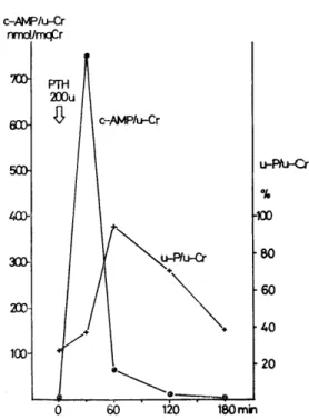
Bovine PTH (200 units) was injected intramuscularly. Cyclic AMP/creatinine and phos- phorus/creatinine in urine responded with a 130 times increase and a 4.6 times increase respectively (Fig. 2).
Oral therapy was started with 25 mEq of magnesium sulfate and the dose was gradually increased up to 90 mEq a day. Serum magnesium rose only to subnormal values.
Serum calcium and potassium also returned to normal with this magnesium supplement.
Magnesium excretion in the urine reached as much as 300 mg a day although the serum magnesium remained subnormal. However, her clinical condition improved and she only had stiff hands occasionally (Fig. 3).
Fig. 1 Magnesium infusion induced a tem- porary increase in serum PTH level
which was initially low. Serum calcium
showed a slight decrease at that time.
Fig. 2 Excretion of c-AMP and phosphorus in urine increased normally after the admi-
nistration of bovine PTH 200 unit.
Fig. 3 Serum magnesium level rose only to subnormal values despite supplemental administration of magnesium while urinary excretion of magnesium increased
during it. Serum calcium level was normalized by magnesium supplement
alone.
DISCUSSION
Hypomagnesemia has been described in association with many disease state, for example : malabsorption syndrome, hyperaldosteronism, primary hyperparathyroidisrn, chronic alcoholism, hyperthyroidism, and various malignancies. In our case, ordinary renal function tests were normal and renal loss of potassium, sodium, calcium and phos- phorus were not observed. Urinary excretion of magnesium was, however, inappropriately high despite hypomagnesemia. Large dose of magnesium supplement didn't normalize serum magnesium completely and high excretion of magnesium in the urine continued.
The mechanism of renal magnesium wastage remained unexplained. Though evidence of malabsorption, steatorrhea and intestinal protein loosing were not demonstrated, she had loose stool frequently. This situation might worsen the hypomagnesemia and induce tetany.
We considered that the main cause of hypomagnesemia in this patient was primary renal magnesium wastage though an accurate balance study of magnesium was not performed.
Hypocalcemia is well known in magnesium deficiency. Many investigators have suggested mechanism involving parathyroid hormone in the hypocalcemia of magnesium depletion. Some investigators suggested end-organ unresponsiveness to PTH10314) and some an impaired production and/or secretion of PTH1)3'4>11>ls). Others considered both mech- anisms') . In our case, responsiveness to PTH in the kidney was normal ;excretion of c-AMP and phosphorus in the urine in the Ellsworth-Howard test was normal. Serum PTH was quite low despite of hypocalcemia but responded to acute magnesium adminis- tration. These findings suggested.that secretion of PTH was reduced but the capacity to produce it was maintained. Magnesium therapy normalized serum calcium and potassium levels without any other supplements. Thus magnesium ion was found necessary for normal calcium and potassium homeostasis. Temporary hypocalcemia after magnesium infusion was also observed in our case. It has been well documented that the parenteral adminis- tration of magnesium reduces serum calcium, partly through a calciuric effect and possibly by causing a shift of calcium into cell"". ANASTI) considered that the early fall in serum calcium was due to a shift of extracellular calcium into cells, produced by an increase in either parat.1iyroid hormone or magnesium or both ; the decline in serum calcium observed after infusion of magnesium was too rapid to be accounted for by an increase in urinary calcium.
Hypokalemia is a well-recognized manifestation of magnesium deficiency. Our case also showed hypokalemia which was normalized by magnesium administration. We cannot currently explain the mechanism of this hypokalemia.
ACKNOWLEDGMENT
The authors greatly appreciate Dr. K. H. Clifton, R. E. R. F. Chief of Research, for his critical review of the manuscript.
REFERENCES
1) ANAST C. S. et al (1976) : Impaired Release of Parathyroid hormone in magnesium deficiency. J. Clin. Endocrinol. Metab. 42, 707-717
2) CAPT. RICHARD M., et al (1966): Hypomagnesemia of unknown etiology. Am. J.
Med. 41, 645-656
3) CHASE L. R. et al (1974): Secretion and metabolic efficacy of parathyroid hormone in patients with severe hypomagnesemia. J. Clin. Endocrinol. Metab. 38, 363-371 4) HABENER, J. E. et al (1976): Relative effectiveness of magnesium and calcium on
the secretion and biosynthesis of parathyroid hormone in vitro. Endocrinology 98, 197-
202
5) JONES, K. H. et al (1966) : Effect of infusions of magnesium and of calcium in parathyroid insufficiency. Clin. Sci. 30, 139-150
6) LEVI, J. et al (1974): Hypocalcemia in magnesium depleted dogs: Evidence for re- duced responsiveness to parathyroid gland function. Metabolism 23, 323-334
7) MENDEL, L. B. et al (1909) : The paths of excretion for inorganic compound IV.
The excretion of magnesium. Am. J. Physiol. 25: 1-22
8) PAUNIER L. et al (1968) : Primary hypomagnesemia with secondary hypocalcemia in an infant. Pediatrics 41, 385-402
9) RANDALL, R. E. Jr. et al (1959) : Magnesium depletion in man. Ann. Int. Med.
50, 257-287
10) REDDY, C. R. et al (1973): Studies on mechanism of hypocalcemia of magnesium depletion. J. Clin. Invest. 52, 3000-3010
11) RUDE, R. K. et al (1978): Parathyroid hormone secretion in magnesium deficiency.
J. Clin. Endocrinol. Metab. 47, 800-806
12) SHILS, M. E. (1969): Experimental human magnesium depletion. Medicine 48, 61- 85
13) SKYBERG, D. et al (1968) : Neonatal hypomagnesemia with selective malabsorption of magnesium -- a clinical entity. Scan. J. Clin. Lab. Invest. 21, 355-363
14) STEP, H. et al (1969): Hypocalcemia due to hypomagnesemia and reversible para- thyroid hormone unresponsiveness. J. Clin. Endocrinol. 29, 842-848
15) SUH, S. M. et al (1973) : Pathogenesis of hypocalcemia in primary hypomagnesemia:
Normal end-organ responsiveness to parathyroid hormone, impaired parathyroid gland function. J. Clin. Invest. 52, 153-160