Development of single chain Fv antibody (scFv)
which recognizes O-glycns on Adult T-cell
Leukemia (ATL) cell surface for targeted
therapy
著者
TODAKA Taro, MUCHIMA Kaname, WAKAO Masahiro,
MATSUMOTO Hikaru, ITO Yuji, SUDA Yasuo
journal or
publication title
The Research Reports of the Faculty of
Engineering, Kagoshima University
volume
58
page range
65-65
year
2017-03
NDSU-KU Joint Symposium on Biotechnology, Nanomaterials and Polymers, North Dakota, 15-16 Oct, 2015
Development of single chain Fv antibody (scFv) which recognizes O-glycns on
Adult T-cell Leukemia (ATL) cell surface for targeted therapy
Taro Todaka*, Kaname Muchima*, Masahiro Wakao**, Hikaru Matsumoto*, Yuji Ito***, Yasuo
Suda****
Abstract
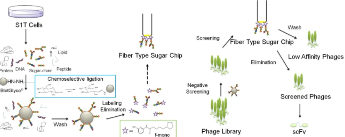
Adult T-cell leukemia (ATL) is a blood cancer caused by the infection of retrovirus human T-cell lymphotropic virus type-1 (HTLV-1). For an individual infected with HTLV-1, the risk of developing ATL is estimated about 5%. Approximately 10 to 20 million individuals are estimated to be infected with HTLV-1 worldwide. In Japan, about 1 million individuals are infected with HTLV-1, and ca. 1,000 patients of ATL are died each year. Mechanism of carcinogenesis has not yet been revealed, and antibodies for therapeutic use of ATL have not been established. In this study, we developed single chain Fv antibodies (scFv) which recognize O-glycans (O-linked sugar-chains, O-SCs) on ATL cell surface for an effective marker or its novel drug for targeted therapy of ATL using our nano-biotechonology. O-SCs were extracted from cell surface of S1T, which is HTLV-1 infected T-cell line established from an ATL patient. To prepare a fiber type Sugar Chip, obtained O-SCs were conjugated with our original fluorescent linker molecule (designated as f-mono) [1]. O-SC ligand conjugates were then immobilized on the terminal of optical fiber (diameter: 50 mm) via gold nano-particles. Human B-cell derived phage library was firstly negatively screened using peripheral blood mononuclear cells (PBMC) of the healthy volunteer to eliminate phages which bound to PMBC. The phages were then applied to the Sugar Chip to screen the phages specifically bound to O-SCs on S1T cell by the localized plasmon resonance (LPR) method. Obtained O-SC-binding phages were transfected to Escherichia coli, TG1 to prepare monoclonal phages according to the standard protocol. The monoclonal phages were transfected to E.coli, HB2151 to express scFv, and eight kinds of scFvs were isolated and purified. The all purified scFvs showed single band (about 27 kDa) in the SDS-PAGE stained with CBB. By flow cytometry analysis, one scFv, named TS1T-1, bound to S1T and other ATL cell lines, but did not to CEM or other leukemia cell lines, suggesting specific binding to ATL cells. The precise structure of the TS1T-1 binding sugar-chain is currently evaluated with our array-type Sugar Chip and surface plasmon resonance (SPR) imaging.
Fig. Schematic image of preparing O-SC binding scFv. Reference
[1] Sato M, Ito Y, Arima N, Baba M, Sobel M, Wakao M, Suda, Y., J. Biochem. 2009, 146, 33-41.