Activity and Left Ventricular Remodeling in Patients With Chronic Heart Failure
A Propensity Score-Matched Analysis
Hirokazu Sano, MD, Shu Kasama, MD, Shinichiro Fujimoto, MD, Takuji Toyama, MD, Noriaki Takama, MD, Norimichi Koitabashi, MD, Shuichi Ichikawa, MD, Yasuyuki Suzuki, MD,
Naoya Matsumoto, MD, Yuichi Sato, MD, and Masahiko Kurabayashi, MD
Abstract:Statin therapy reduces enhanced cardiac sympathetic nerve activity (CSNA) in patients with heart disease, and prevents left ventricular (LV) remodeling in chronic heart failure (CHF) patients.
We sought to evaluate the effects of statin therapy on CSNA, as evaluated by 123I-metaiodobenzylguanidine (MIBG) scintigraphy, and LV remodeling in CHF patients.
This study was sub-analysis of our previous report of the result that the serial123I-MIBG studies were the most useful prognostic indicator in CHF patients. Patients with CHF (n¼208; left ventricular ejection fraction
<45%) but no cardiac events for at least 5 months before the study, were identified according to their history of decompensated acute heart failure requiring hospitalization. The patients underwent123I-MIBG scintigraphy and echocardiography immediately before hospital discharge and after 6 months. The delayed % denervation, delayed heart/mediastinum count (H/M) ratio, and washout rate (WR) were determined by123I-MIBG scintigraphy. The LV end-diastolic volume (EDV) and end-systolic volume (ESV) were also determined by echocardiography. We selected 164 patients and used propensity score matching to compare patients who received oral statin (n¼82), and those who did not (n¼82).
The changes in123I-MIBG scintigraphic parameters improved, and in echocardiographic LVEDV and LVESV reduced in the statin group com- pared with those in the non-statin group. Moreover, there were significant correlations between changes in the123I-MIBG scintigraphic findings and those in the LVEDV (% denervation,r¼0.534,P<0.001; H/M ratio, r¼ 0.516,P<0.001; and WR,r¼0.558,P<0.001); or the LVESV (%
denervation,r¼0.479,P<0.001; H/M ratio,r¼ 0.450,P<0.001; and WR,r¼0.520,P<0.001) in the statin group. In contrast, there was no relationship between these parameters in the non-statin group.
Statin therapy not only improved CSNA, but also reduced LV volume, in other wards, prevented LV remodeling in CHF patients.
(Medicine93(27):e214)
Abbreviations: ACE = angiotensin-converting enzyme, ARB = angiotensin receptor blocker, CHF = chronic heart failure, CSNA = cardiac sympathetic nerve activity, EDV = end-diastolic volume, EF
= ejection fraction, ESV = end-systolic volume, H/M = heart/
mediastinum count, LDL-C = low-density lipoprotein cholesterol, LV = left ventricular, MIBG = meta-iodobenzylguanidine, SPECT = single photon emission computed tomography, TDS = total defect score, WR = washout rate.
INTRODUCTION
T he 3-hydroxyl-3-methylglutaryl-coenzyme A reductase inhibitors (statins) reduce mortality and morbidity in var- ious patients, including those with dyslipidemia, ischemic heart disease, and cerebrovascular disease.1 – 3 Statins effectively lower low-density lipoprotein cholesterol (LDL-C) level; in addition, statins have other potentially favorable ‘‘pleotropic’’
effects in patients with chronic heart failure (CHF).
4,5Activation of the cardiac sympathetic nerve activity (CSNA) is a cardinal pathophysiological abnormality associ- ated with human heart failure.
6Therefore, plasma norepi- nephrine concentrations affect the prognosis of CHF patients.
7Myocardial imaging with
123I-metaiodobenzylguani- dine (MIBG), an analogue of norepinephrine, is useful for detecting abnormalities in the myocardial adrenergic nervous system in CHF patients.
8,9Many studies have suggested that treatment of heart failure can improve CSNA, as evaluated by cardiac
123I-MIBG scintigraphy.
10 – 23On the other hand, statin therapy reduces enhanced CSNA in patients with CHF.
5Moreover, this agent is reported to prevent left ventricular (LV) remodeling in these patients.
24Although favor- able effects of statin therapy have been established, little is known about the effects of treatment with statin on cardiac
123I-MIBG scintigraphic changes and LV parameters in patients with CHF.
Accordingly, this study was performed, using our pre- viously reported data,
25to determine whether statin therapy improves CSNA as evaluated by
123I-MIBG scintigraphy, and whether this agent prevents LV remodeling in CHF patients.
MATERIALS AND METHODS Study Patients and Protocol
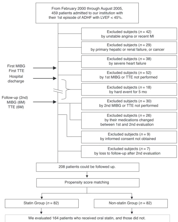
From February 2000 through August 2005, 459 patients were admitted to our institution with their first episode of
Editor: Undurti Narashima Das.Received: August 12, 2014; revised: October 3, 2014; accepted: October 6, 2014.
From the Department of Medicine and Biological Science (Cardiovascular Medicine), Gunma University Graduate School of Medicine, Maebashi, Japan (HS, SK, TT, NT, NK, MK); Department of Cardiovascular Medicine, Cardiovascular Hospital of Central Japan (Kitakanto Cardiovas- cular Hospital), Gunma, Japan (SK, SI); Department of Cardiology, Juntendo University Graduate School of Medicine, Tokyo, Japan (SF), Department of Cardiology, Nihon University School of Medicine, Tokyo, Japan (NM, YS).
Correspondence: Shu Kasama, Department of Medicine and Biological Science (Cardiovascular Medicine), Gunma University Graduate School of Medicine, 3-39-22 Showa-machi, Maebashi, Gunma, 371- 8511, Japan (e-mail: [email protected]).
The authors have no funding and conflicts of interest to disclose.
Copyright#2014 Wolters Kluwer Health | Lippincott Williams & Wilkins.
This is an open access article distributed under the Creative Commons Attribution-NoDerivatives License 4.0, which allows for redistribution, commercial and non-commercial, as long as it is passed along unchanged and in whole, with credit to the author.
ISSN: 0025-7974
DOI: 10.1097/MD.0000000000000214
decompensated acute heart failure with a LV ejection fraction (EF) of less than 45%, according to the inclusion criteria described in our previous study.
25This study was sub-analysis using our previous database.
25Chest radiography, standard elec- trocardiography, echocardiography were performed in all of the patients. In the acute phase, all patients were treated with standard heart failure treatment including intravenous diuretics, vasodila- tors (carperitide, nicorandil, nitroglycerin, and so on), and if necessary, dopamine or dobutamine was added to maintain the blood pressure. Patients were excluded from the study if they had unstable angina or recent acute myocardial infarction, and had performed any coronary revascularization procedures within 3 months (42 patients were excluded), and had primary hepatic failure, renal failure, or active cancer (29 patients). Moreover, patients with severe heart failure requiring mechanical support (intraaortic balloon pumping, left ventricular assist device, or cardiac resynchronization therapy) or patients requiring heart transplantation were also excluded (38 patients) (Figure 1).
During the stable period, the patients were treated with standard oral medications for heart failure, including angioten- sin-converting enzyme (ACE) inhibitors, angiotensin receptor blockers (ARBs), beta-adrenergic blocking agents, and diuretics.
None of the patients was treated with tricyclic antidepressants or other serotonin reuptake inhibitors. We performed
123I-MIBG scintigraphy and echocardiography just before hospital dis- charge. However, 52 patients were excluded from this study because scintigraphy or echocardiography had not been per- formed during the hospitalization. The medical management of the patients was directed by an internist or cardiologist from our institution, and
123I-MIBG scintigraphic and echocardio- graphic parameters were available to them. In this study, 18 patients were excluded because there were hard events (n
¼10; cardiac events, n
¼5; cerebral events, and n
¼3; other events) for 5 months after enrollment.
The
123I-MIBG scintigraphy and echocardiography were repeated about 6 months after hospital discharge (mean:
6.4 months). Patients were excluded from the study if the second evaluation had not been performed (30 patients), or if their medication changed between the first and the second evaluation (26 patients). The study was approved by the ethics review board of our institution, and informed written consent was obtained from all patients. Nine patients were excluded because informed consent was not obtained. Moreover, 7 patients were excluded because we lost to follow-up after second evaluation.
We followed up 208 patients who had highly reliable information.
The 208 study patients consisted of 130 men and 78 women with a mean age of 68.6 years (range 35–87 years).
To evaluate whether the statin treatment affected the CSNA and LV remodeling in our patients with CHF, we stratified our patients into statin (n
¼82), and non-statin groups (n
¼82), using propensity score matching (Figure 1). The statin agents included in this study were rosuvastatin (n
¼22), atorvastatin (n
¼20), fluvastatin (n
¼16), pravastatin (n
¼10), simvastatin (n
¼9), and pitavastatin (n
¼5). In addition, we did not select the statin treatment according to the clinical features in our CHF patients. For our study protocol,
25statin was started during hospitalization, and this drug was continued follow-up period.
Therefore, in other words, in the statin group, oral adminis- tration of statin was continued during the study period.
123
I-MIBG Scintigraphy
The
123I-MIBG imaging method used has already been previously described.
25,26In brief, the
123I-MIBG was obtained
from a commercial source (FUJIFILM RI Pharma Co. Ltd, Tokyo, Japan). At 15 minutes and 4 hours after injection, anterior planar and single photon emission computed tomographic (SPECT) images were obtained with a single-head gamma camera (Mil- lennium MPR, GE Medical Systems, Waukesha, WI).
The heart/mediastinum count (H/M) ratio was determined from the anterior planar delayed
123I-MIBG image using the standard method. The washout rate (WR) was calculated from early and delayed planar images. Regional tracer uptake was assessed semiquantitatively using a 5-point scoring system (0, normal to 4, no uptake) in 17 segments on the delayed SPECT image as recommended by the American Heart Association.
27The total defect score (TDS) was calculated as the sum of all defect scores. The TDS was converted to the percentage of the total denervated myocardium (% denervation). The % denerva- tion was calculated using the following formula: TDS/68 (maximum score
¼4 17) 100. At our laboratory, the refer- ence range of the %denervation values is from 6 to 18; the delayed H/M ratio range from 2.18 to 2.70; and the normal WR range from 20% to 30%, as previously reported.
25,26Echocardiography
Echocardiography was performed using standard methods.
Two experienced independent echocardiography technicians who were blinded to the study methods performed all of the measurements. The LV end-diastolic volume (EDV), LV end- systolic volume (ESV), and LVEF were calculated using the 2D-biplane method, as previously reported.
22Serial Changes Between the First and Second Scintigraphic and Echocardiographic Parameters
Changes between the first and second
123I-MIBG scinti- graphic (% denervation, H/M ratio, and WR) and echocardio- graphic parameters (EDV, ESV, and LVEF) were calculated using the following formula: delta (X)
¼[(X) value after 6 months] [baseline value of (X)], where (X)
¼123I-MIBG scintigraphic or echocardiographic parameters.
Statistical Analysis
The analyses were performed using SPSS 16.0 (SPSS Inc, Chicago, IL), or SAS version 9.1 (SAS Institute Inc, Cary, NC).
Numerical results were expressed as the mean SD. In all the analyses,
P<0.05 was considered statistically significant. A propensity-matched analysis was conducted to minimize the selection bias for statin administration.
28To obtain the propen- sity score for the probability that statin would be administered, multivariate logistic regression analyses were conducted. The propensity score was based on the following variables: age, sex, ischemic etiology, smoking, New York Heart Association (NYHA) functional classes, acute phase treatments,
123I-MIBG scintigraphic and echocardiographic parameters, and presence of diabetes and hypertension. The patients in the statin and non- statin groups were matched 1:1 to 2 digits.
Categorical data were compared between the 2 groups using
2-sided chi-square tests, and differences between continuous
variables were evaluated using the unpaired
ttest. NYHA func-
tional classes were compared using the Wilcoxon matched pairs
signed rank test. In patients who underwent a second assessment,
changes from the baseline were evaluated within each treatment
group using a paired
ttest and between the 2 groups using 2-way
ANOVA. Linear regression analysis was performed to determine
the relationship between continuous variables.
To evaluate the contribution of the degree of change in WR (ie, delta-WR), univariate and stepwise multivariate analyses were used to examine the variable of interest (Table 3). More- over, in order to evaluate the effects of addition of statin to the beta-blocker on CSNA, each patients group treated with beta- blocker (n
¼77), and treated without beta-blocker (n
¼87) were evaluated by the same analysis (Tables 4 and 5, respectively).
RESULTS Clinical Characteristics
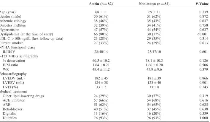
No significant differences in clinical characteristics (except dyslipidemia) or cardiac medications were found
between the 2 groups. At baseline, the % denervation, H/M ratio, WR, LVEDV, LVESV, LVEF, NYHA functional class, and the frequency rates of follow-up LDL-C levels
>100 mg/dLwere similar between the 2 groups (Table 1).
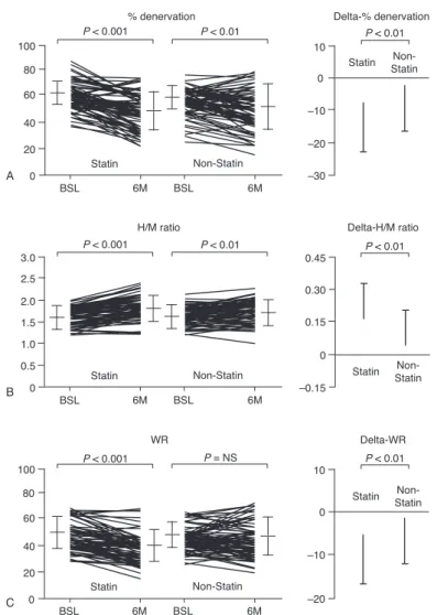
Comparison of Cardiac
123I-MIBG Scintigraphic Findings Before and 6 Months After Treatment Figure 2 and Table 2 provide a summary of the % denervation, H/M ratios, and WR values. In both groups, % denervation was significantly decreased after 6 months relative to the baseline values. However, the delta-% denervation in the statin group was significantly lower than that in the non-statin
From February 2000 through August 2005,459 patients admitted to our institution with their 1st episode of ADHF with LVEF < 45%.
First MIBG First TTE
Follow-up (2nd) MIBG (6M)
TTE (6M) Hospital discharge
208 patients could be followed up.
We evaluated 164 patients who received oral statin, and those did not.
Propensity score matching
Statin Group (n = 82) Non-statin Group (n = 82)
Excluded subjects (n = 42) by unstable angina or recent MI
Excluded subjects (n = 29)
by primary hepatic or renal failure, or cancer
Excluded subjects (n = 52) by 1st MIBG or TTE not performed
Excluded subjects (n = 38) by severe heart failure
Excluded subjects (n = 18) by hard event for 5 mo Excluded subjects (n = 30) by 2nd MIBG or TTE not performed
Excluded subjects (n = 26) by their medications changed between 1st and 2nd evaluation
Excluded subjects (n = 9) by informed consent not obtained
Excluded subjects (n = 7) by loss to follow-up after 2nd evaluation
FIGURE 1. Flow diagram of participants in current study. ADHF¼acute decompensated heart failure, LVEF¼left ventricular ejection fraction, MI¼myocardial infarction, MIBG¼metaiodobenzylguanidine scintigraphy, TTE¼transthoracic echocardiography, 6 M¼after 6 months of hospital discharge.
group. In both groups, the H/M ratios were significantly increased after 6 months compared with the baseline values.
However, the delta-H/M ratios were significantly higher in the statin group than those in the non-statin group. Finally, the WR in the statin group was significantly decreased after 6 months relative to the baseline values. In contrast, in the non-statin group, no significant differences were observed between the baseline and 6 months posttreatment values. Moreover, delta- WR was significantly lower in the statin group than in the non- statin group.
Comparison of Echocardiographic Findings Before and 6 Months After Treatment
Table 2 also provides a summary of the LVEDV, LVESV, and LVEF. In both groups, LVEDV and LVESV were signifi- cantly decreased and LVEF was significantly increased after 6 months relative to the baseline values. The changes in LVEDV and LVESV were significantly greater in the statin group than those in the non-statin group. The change in LVEF in the statin group tended to be more favorable than that in the non- statin group, but these changes were not statistically significant.
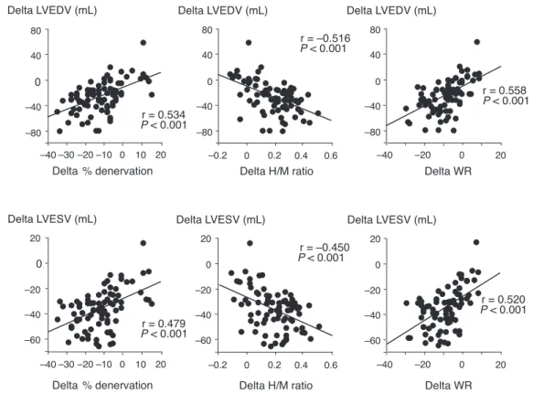
Relationship Between LV Volume and
123I-MIBG Scintigraphic Findings Before and After
Treatment
There were significant correlations between changes in the
123
I-MIBG scintigraphic findings and those in the LVEDV (%
denervation,
r¼0.534,
P<0.001; H/M ratio,
r¼ 0.516, P<0.001; and WR,
r¼0.558,
P<0.001); or the LVESV (%
denervation,
r¼0.479,
P<0.001; H/M ratio,
r¼ 0.450, P<0.001; and WR,
r¼0.520,
P<0.001) in the statin group (Figure 3). In contrast, there was no relationship between these parameters in the non-statin group.
Evaluation of Factors Predicting Decreased Delta-WR
Table 3 shows the results of the univariate and multivariate analyses to assess factors predicting an increase in delta-WR. In the univariate analysis, age, non-beta-blocker treatment, and non-statin treatment were predictive factors. The stepwise multivariate analysis also identified age, non-beta-blocker treat- ment, and non-statin treatment as significant independent pre- dictors of increasing delta-WR in the CHF patients.
In the patients treated with beta-blocker, in the univariate analysis, age and non-statin treatment were predictive factors.
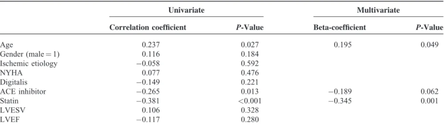
The stepwise multivariate analysis also identified age and non- statin treatment as significant independent predictors of increas- ing delta-WR (Table 4). In the patients treated without beta- blocker, in the univariate analysis, age, non-ACE inhibitor treatment, and non-statin treatment were predictive factors.
The stepwise multivariate analysis identified age and non-statin treatment as significant independent predictors of increasing delta-WR (Table 5).
TABLE 1. Clinical Characteristics of the Patients
Statin (n¼82) Non-statin (n¼82) P-Value
Age (year) 6811 6911 0.739
Gender (male) 50 (61%) 51 (62%) 0.872
Ischemic etiology 38 (46%) 35 (43%) 0.637
Diabetes mellitus 32 (39%) 34 (41%) 0.750
Hypertension 47 (57%) 44 (54%) 0.637
Dyslipidemia (at the time of entry) 66 (80%) 30 (37%) <0.001
LDL-C>100 mg/dL (last follow-up data) 23 (28%) 29 (35%) 0.314
Current smoker 27 (33%) 24 (29%) 0.613
NYHA functional class
II/III/IV 28/40/14 25/47/10 0.601
I-123 MIBG scintigraphy
% denervation 60.510.2 58.110.3 0.126
H/M ratio 1.640.21 1.660.20 0.506
WR 49.411.2 47.99.6 0.379
Echocardiography
LVEDV (mL) 18245 18139 0.866
LVESV (mL) 12438 12340 0.901
LVEF(%) 337 338 0.743
Medical treatment
Other lipid-lowering drugs 24 (29%) 30 (37%) 0.319
ACE inhibitor 57 (66%) 54 (68%) 0.616
ARB 51 (62%) 54 (65%) 0.625
Beta-blocker 40 (51%) 37 (45%) 0.638
Digitalis 13 (16%) 16 (20%) 0.539
Diuretics 76 (93%) 76 (93%) 1.000
Values are meanSD or number (%).
ACE¼angiotensin-converting enzyme, ARB¼angiotensin-receptor blocker, H/M¼heart/mediastinum count, LDL-C¼low-density lipoprotein cholesterol, LVEDV¼left ventricular end-diastolic volume, LVEF¼left ventricular ejection fraction, LVSDV¼left ventricular end-systolic volume, MIBG¼meta-iodobenzylguanidine, NYHA¼New York Heart Association, WR¼washout rate.
TABLE 2. Changes in % Denervation, Heart/Mediastinum Count Ratio, Washout Rate, Left Ventricular Volume, and Left Ventricular Ejection Fraction in Both Groups
Statin Non-statin
Baseline 6 months Delta Baseline 6 months Delta
I-123 MIBG scintigraphy
% denervation 60.510.2 48.413.6 12.111.4 58.110.3 52.015.3y 6.112.0z H/M ratio 1.640.21 1.810.26 0.170.15 1.660.20 1.740.24y 0.080.16z
WR 49.411.2 40.011.6 9.48.9 47.99.6 45.713.6 2.210.8z
Echocardiography
LVEDV (mL) 18245 15949 2331 18139 16740y 1428§
LVESV (mL) 12438 9744 2629 12340 10738y 1623§
LVEF(%) 337 4010 88 338 389y 55
Values are meansSD.
H/M¼heart/mediastinum count, LVEDV¼left ventricular end-diastolic volume, LVEF¼left ventricular ejection fraction, LVSDV¼left ventricular end-systolic volume, MIBG¼meta-iodobenzylguanidine, WR¼washout rate.
P<0.001 vs baseline.
yP<0.01 vs baseline.
zP<0.01 vs statin group.
§P<0.05 vs statin group.
0
–20 –10
–30 10
Delta-% denervation
Statin Non- Statin
WR
0
–10
–20 10
Delta-WR H/M ratio
0.30
0 0.15
–0.15 0.45
Delta-H/M ratio
1.0 2.0 3.0
0.5 1.5 2.5
0
% denervation P < 0.001
100
BSL 6M
BSL 6M
Statin Non-Statin 0
20 40 60 80
A
B
C
BSL 6M
BSL 6M
Statin Non-Statin
BSL 6M
BSL 6M
Statin Non-Statin
Statin Non- Statin
Statin Non- Statin 100
0 20 40 60 80
P < 0.01 P < 0.01
P = NS
P < 0.001 P < 0.01 P < 0.01
P < 0.001 P < 0.01
FIGURE 2. Comparison of cardiac123I-metaiodobenzylguanidine scintigraphic findings for % denervation (A), H/M ratio (B), and WR (C) in the 2 groups. BSL¼baseline. H/M¼heart/mediastinum count. WR¼washout rate. 6 M¼after 6 months of therapy.
DISCUSSION
The patients were stratified into the statin and non-statin groups using propensity score matching. The
123I-MIBG scinti- graphic and echocardiographic parameters showed improve- ment in both groups, with more favorable changes in the statin group. There were significant correlations between changes in the
123I-MIBG scintigraphic findings and LV volumes in the
statin group. Moreover, stepwise multivariate analyses showed that non-statin treatment had an independent and significant negative relationship with delta-WR in CHF patients.
Inflammatory cytokines play an important role in the development and progression of CHF. They have been impli- cated in the development of LV remodeling, endothelial dys- function, and increased cardiac myocyte apoptosis.
29As statins
TABLE 3. Univariate and Multivariate Linear Model of Delta-WR
Univariate Multivariate
Correlation coefficient P-Value Beta-coefficient P-Value
Age 0.234 0.003 0.206 0.003
Gender (male¼1) 0.131 0.096
Ischemic etiology 0.030 0.702
NYHA 0.023 0.769
Digitalis 0.102 0.183
ACE inhibitor 0.115 0.141
Beta blocker 0.404 <0.001 0.372 <0.001
Statin 0.293 <0.001 0.236 0.001
LVESV 0.025 0.751
LVEF 0.029 0.710
ACE¼angiotensin-converting enzyme, LVEF¼left ventricular ejection fraction, LVSDV¼left ventricular end-systolic volume, NYHA¼New York Heart Association.
r = 0.534 P < 0.001
Delta % denervation Delta LVEDV (mL)
Delta H/M ratio r = –0.516 P < 0.001 Delta LVEDV (mL)
0
–60 –40 –20 20
r = 0.479 P < 0.001 Delta LVESV (mL)
Delta H/M ratio r = –0.450 P < 0.001 0
–60 –40 –20 20
Delta LVESV (mL)
Delta WR
r = 0.520 P < 0.001 0
–60 –40 –20 20
Delta LVESV (mL) Delta WR
r = 0.558 P < 0.001 Delta LVEDV (mL)
–20 0
–40 20
80 40
–80 –40 0
80 40
–80 –40 0 80
40
–80 –40 0
–40 –30 –10 –20 0 10 20 –0.2 0 0.4 0.2 0.6
–20 0
–40 20
–40 –30 –10 –20 0 10 20 –0.2 0 0.4 0.2 0.6 Delta % denervation
FIGURE 3. Correlations between the changes of123I-MIBG scintigraphic findings and left ventricular end-diastolic volume (LVEDV) (Top), or left ventricular end-systolic volume (LVESV) (Bottom) after statin therapy in patients with chronic heart failure. Delta LVEDV¼the value of LVEDV after treatmentpretreatment value of LVEDV, Delta % denervation¼the value of % denervation after treatmentpretreat- pretreatment value of % denervation, Delta H/M ratio¼the value of H/M ratio after treatmentpretreatment value of H/M ratio, Delta WR¼the value of WR after treatmentpretreatment value of WR, Delta LVESV¼the value of LVESV after treatmentpretreatment value of LVESV. H/M ratio¼heart/mediastinum count ratio, WR¼washout rate.
are well known to have anti-inflammatory effects and down- regulate inflammatory cytokines in failing heart,
30it may attenuate LV global remodeling. In general, increasing of LV volume (ie, progression of LV remodeling) has been shown to be associated with the poor prognosis in patients with CHF.
31Therefore, increasing effort has been directed toward pharma- cological attenuation of LV volume for failing human hearts.
Node et al
24reported a significant reduction in LV volumes in patients with CHF after statin therapy compared with placebo.
Similarly, in this study, LVEDV and LVESV were significantly decreased after the 6 months treatment in the statin group compared with the non-statin group. Therefore, our findings suggest that addition of statin to standard therapy can prevent LV remodeling in patients with CHF.
123
I-MIBG is an analogue of the adrenergic neuron-block- ing agent guanethidine, which is thought to utilize the same myocardial uptake and release mechanisms as norepi- nephrine.
32Therefore, cardiac
123I-MIBG imaging is a useful tool for detecting abnormalities of the myocardial adrenergic nervous system in CHF patients.
8,9Furthermore, many reports have suggested that the treatment of CHF with ACE inhibi- tors,
10–12ARBs,
16– 19beta-blockers,
12–15or spironolactone
19– 22can improve CSNA, based on cardiac
123I-MIBG scintigraphic findings. However, little is known about the effects of
statin therapy on CSNA in CHF patients. In this study, we examined whether statin therapy improved the
123I-MIBG scintigraphic parameters in our CHF patients. We found that the statin group showed improvement compared with the non- statin group. Moreover, the stepwise multivariate analyses revealed that the non-statin treatment had an independent and significant relationship with increasing delta-WR in the CHF patients. Given our previously reported observation that delta- WR is the best currently available prognostic indicator for CHF,
25our findings demonstrated for the first time that statin may be the available agent for improving CSNA and for preventing cardiac events of patients with CHF. Furthermore, both groups treated with and without beta-blocker, multivariate analyses revealed that the non-statin treatment had an independent and significant relationship with increasing delta-WR. Therefore, the statin treatment may reduce enhanced CSNA even if beta-blocker is not administrated.
It has been reported that the release of norepinephrine is enhanced, and uptake of norepinephrine is also prevented, in the failing heart.
33Kang et al
34demonstrated that the release and uptake of norepinephrine are modulated by activation of ATP- sensitive potassium channels in experimental rat models. As statin is reported to activate ATP-sensitive potassium channels and have also cardioprotective properties,
35it may attenuate
TABLE 5. Univariate and Multivariate Linear Model of Delta-WR in the Patients Treated Without Beta-Blocker
Univariate Multivariate
Correlation coefficient P-Value Beta-coefficient P-Value
Age 0.237 0.027 0.195 0.049
Gender (male¼1) 0.116 0.184
Ischemic etiology 0.058 0.592
NYHA 0.077 0.476
Digitalis 0.149 0.221
ACE inhibitor 0.265 0.013 0.189 0.062
Statin 0.381 <0.001 0.345 0.001
LVESV 0.106 0.328
LVEF 0.117 0.280
ACE¼angiotensin-converting enzyme, LVEF¼left ventricular ejection fraction, LVSDV¼left ventricular end-systolic volume, NYHA¼New York Heart Association.
TABLE 4. Univariate and Multivariate Linear Model of Delta-WR in the Patients Treated With Beta-Blocker
Univariate Multivariate
Correlation coefficient P-Value Beta-coefficient P-Value
Age 0.239 0.032 0.211 0.045
Gender (male¼1) 0.120 0.297
Ischemic etiology 0.111 0.335
NYHA 0.098 0.396
Digitalis 0.160 0.166
ACE inhibitor 0.158 0.182
Statin 0.402 <0.001 0.404 <0.001
LVESV 0.168 0.145
LVEF 0.180 0.117
ACE¼angiotensin-converting enzyme, LVEF¼left ventricular ejection fraction, LVSDV¼left ventricular end-systolic volume, NYHA¼New York Heart Association.
CSNA. Therefore, we hypothesize that statin therapy can improve CSNA in patients with CHF. However, further study will be required to confirm this hypothesis.
In this study, there were significant correlations between changes in the LV volume and the
123I-MIBG scintigraphic parameters after treatment with statin in patients with CHF.
However, no significant correlations were found in the non- statin group. With respect to the influence of statin, it is still unclear whether attenuation of LV volume, ie, due to the anti- remodeling effect of statin,
29increases myocardial uptake of norepinephrine or whether increased myocardial uptake of norepinephrine leads to attenuation of LV volume. Therefore, further studies are necessary to clarify the relationship between the attenuation of LV volume and the increased myocardial uptake of norepinephrine.
Statins are classified either as hydrophilic (eg, rosuvastatin and pravastatin) and lipophilic (eg, atorvastatin, fluvastatin, simvastatin, and pitavastatin) according to the difference in their aqueous solubility. Differences in the pharmacologic properties of hydrophilic and lipophilic statins were identified in experimental
36and clinical studies.
37However, in the present study, no significant differences were found between the hydro- philic and lipophilic statin therapies in terms of changes in the
123
I-MIBG scintigraphic parameters. However, in the future, studies with a larger numbers of population should be conducted to examine the effects of statin on CSNA and to compare the effects of hydrophilic and lipophilic statins in CHF patients.
Currently, many independent reports from different cen- ters around the world support the idea that
123I-MIBG myo- cardial scintigraphy provides useful information for assessing patients with heart disease. The imaging modality appears valuable in predicting prognoses and estimating the efficacy of a therapy. However, quantitative
123I-MIBG parameters differ between institutions and between instruments, and the tracer is not widely available. For these reasons, cardiac
123I- MIBG has yet to achieve broad clinical acceptance; thus, few multicenter trials using the imaging modality have been con- ducted.
38 – 40Therefore, the evidence supporting the clinical value of this imaging technique remains inadequate, requiring worldwide multicenter clinical trials involving larger numbers of patients to establish the efficacy of this imaging modality.
CONCLUSIONS
The patients with CHF were divided into the statin group and the non-statin group by using propensity score matching.
The
123I-MIBG scintigraphic and echocardiographic parameters were improved in both groups but showed more favorable changes in the statin group. There were significant correlations between changes in the
123I-MIBG scintigraphic findings and LV volumes in the statin group. These findings indicate that statin therapy can improve cardiac sympathetic nerve activity and prevent LV remodeling in patients with CHF.
REFERENCES
1. Sacks FM, Pfeffer MA, Moye LA, et al. The effect of pravastatin on coronary events after myocardial infarction in patients with average cholesterol levels. Cholesterol and Recurrent Events Trial investiga- tors.N Engl J Med.1996;335:1001–1009.
2. Prevention of cardiovascular events and death with pravastatin in patients with coronary heart disease and a broad range of initial cholesterol levels. The Long-Term Intervention with Pravastatin in Ischaemic Disease (LIPID) Study Group.N Engl J Med.
1998;339:1349–1357.
3. Shepherd J, Cobbe SM, Ford I, et al. Prevention of coronary heart disease with pravastatin in men with hypercholesterolemia. West of Scotland Coronary Prevention Study Group.N Engl J Med.
1995;333:1301–1307.
4. Pliquett RU, Cornish KG, Peuler JD, et al. Simvastatin normalizes autonomic neural control in experimental heart failure.Circulation.
2003;107:2493–2498.
5. Gao L, Wang W, Li YL, et al. Simvastatin therapy normalizes sympathetic neural control in experimental heart failure: roles of angiotensin II type 1 receptors and NAD(P)H oxidase.Circulation.
2005;112:1763–1770.
6. Thomas JA, Marks BH. Plasma norepinephrine in congestive heart failure.Am J Cardiol.1978;41:233–243.
7. Cohn JN, Levine TB, Olivari MT, et al. Plasma norepinephrine as a guide to prognosis in patients with chronic congestive heart failure.
N Engl J Med.1984;311:819–823.
8. Henderson EB, Kahn JK, Corbett JR, et al. Abnormal I-123 metaiodobenzylguanidine myocardial washout and distribution may reflect myocardial adrenergic derangement in patients with conges- tive cardiomyopathy.Circulation.1988;78:1192–1199.
9. Merlet P, Valette H, Dubois-Rande JL, et al. Prognostic value of cardiac metaiodobenzylguanidine imaging in patients with heart failure.J Nucl Med.1992;33:471–477.
10. Takeishi Y, Atsumi H, Fujiwara S, et al. ACE inhibition reduces cardiac iodine-123-MIBG release in heart failure.J Nucl Med.
1997;38:1085–1089.
11. Kasama S, Toyama T, Kumakura H, et al. Effects of perindopril on cardiac sympathetic nerve activity in patients with congestive heart failure: comparison with enalapril.Eur J Nucl Med Mol Imaging.
2005;32:964–971.
12. Toyama T, Aihara Y, Iwasaki T, et al. Cardiac sympathetic activity estimated by 123I-MIBG myocardial imaging in patients with dilated cardiomyopathy after beta-blocker or angiotensin-converting enzyme inhibitor therapy.J Nucl Med.1999;40:217–223.
13. Toyama T, Hoshizaki H, Seki R, et al. Efficacy of carvedilol treatment on cardiac function and cardiac sympathetic nerve activity in patients with dilated cardiomyopathy: comparison with metoprolol therapy.J Nucl Med.2003;44:1604–1611.
14. Yamazaki J, Muto H, Kabano T, et al. Evaluation of beta-blocker therapy in patients with dilated cardiomyopathy—clinical meaning of iodine 123-metaiodobenzylguanidine myocardial single-photon emission computed tomography.Am Heart J.2001;141:645–652.
15. Kasama S, Toyama T, Hatori T, et al. Evaluation of cardiac sympathetic nerve activity and left ventricular remodelling in patients with dilated cardiomyopathy on the treatment containing carvedilol.Eur Heart J.2007;28:989–995.
16. Kasama S, Toyama T, Kumakura H, et al. Addition of valsartan to an angiotensin-converting enzyme inhibitor improves cardiac sympa- thetic nerve activity and left ventricular function in patients with congestive heart failure.J Nucl Med.2003;44:884–890.
17. Kasama S, Toyama T, Kumakura H, et al. Effects of candesartan on cardiac sympathetic nerve activity in patients with congestive heart failure and preserved left ventricular ejection fraction.J Am Coll Cardiol.2005;45:661–667.
18. Kasama S, Toyama T, Hatori T, et al. Comparative effects of valsartan with enalapril on cardiac sympathetic nerve activity and plasma brain natriuretic peptide in patients with congestive heart failure.Heart.2006;92:625–630.
19. Kasama S, Toyama T, Sumino H, et al. Additive effects of spironolactone and candesartan on cardiac sympathetic nerve activity and left ventricular remodeling in patients with congestive heart failure.J Nucl Med.2007;48:1993–2000.
20. Kasama S, Toyama T, Sumino H, et al. Effects of mineralocorticoid receptor antagonist spironolactone on cardiac sympathetic nerve activity and prognosis in patients with chronic heart failure.Int J Cardiol.2013;167:244–249.
21. Kasama S, Toyama T, Kumakura H, et al. Spironolactone improves cardiac sympathetic nerve activity and symptoms in patients with congestive heart failure.J Nucl Med.2002;43:1279–1285.
22. Kasama S, Toyama T, Kumakura H, et al. Effect of spironolactone on cardiac sympathetic nerve activity and left ventricular remodeling in patients with dilated cardiomyopathy.J Am Coll Cardiol.
2003;41:574–581.
23. Kasama S, Toyama T, Hatori T, et al. Effects of torasemide on cardiac sympathetic nerve activity and left ventricular remodelling in patients with congestive heart failure.Heart.2006;92:1434–1440.
24. Node K, Fujita M, Kitakaze M, et al. Short-term statin therapy improves cardiac function and symptoms in patients with idiopathic dilated cardiomyopathy.Circulation.2003;108:839–843.
25. Kasama S, Toyama T, Sumino H, et al. Prognostic value of serial cardiac 123I-MIBG imaging in patients with stabilized chronic heart failure and reduced left ventricular ejection fraction.J Nucl Med.
2008;49:907–914.
26. Kasama S, Toyama T, Sumino H, et al. Serial cardiac 123I- metaiodobenzylguanidine scintigraphic studies are more useful for predicting cardiac death than one-time scan in patients with chronic heart failure: sub-analysis of our previous report.Nucl Med Commun.2010;31:807–813.
27. Cerqueira MD, Weissman NJ, Dilsizian V, et al. Standardized myocardial segmentation and nomenclature for tomographic imaging of the heart: a statement for healthcare professionals from the Cardiac Imaging Committee of the Council on Clinical Cardiology of the American Heart Association.Circulation.2002;105:539–542.
28. Luellen JK, Shadish WR, Clark MH. Propensity scores: An introduction and experimental test.Eval Rev.2005;29:530–558.
29. Mann DL. Inflammatory mediators and the failing heart: past, present, and the foreseeable future.Circ Res.2002;91:988–998.
30. Lefer DJ. Statins as potent antiinflammatory drugs. Circulation.
2002;106:2041–2042.
31. Neglia D, Michelassi C, Trivieri MG, et al. Prognostic role of myocardial blood flow impairment in idiopathic left ventricular dysfunction.Circulation.2002;105:186–193.
32. Wieland DM, Wu J, Brown LE, et al. Radiolabeled adrenergic neuron-blocking agents: adrenomedullary imaging with [131I]iodo- benzylguanidine.J Nucl Med.1980;21:349–353.
33. Burgdorf C, Dendorfer A, Kurz T, et al. Role of neuronal KATP channels and extraneuronal monoamine transporter on norepinephr- ine overflow in a model of myocardial low flow ischemia.J Pharmacol Exp Ther.2004;309:42–48.
34. Kang CS, Chen CC, Lin CC, et al. Effect of ATP-sensitive potassium channel agonists on sympathetic hyperinnervation in postinfarcted rat hearts.Am J Physiol Heart Circ Physiol.
2009;296:H1949–H1959.
35. Lee TM, Lin MS, Chang NC. Effect of pravastatin on sympathetic reinnervation in postinfarcted rats.Am J Physiol Heart Circ Physiol.
2007;293:H3617–H3626.
36. Sakamoto K, Mikami H, Kimura J. Involvement of organic anion transporting polypeptides in the toxicity of hydrophilic pravastatin and lipophilic fluvastatin in rat skeletal myofibres.Br J Pharmacol.
2008;154:1482–1490.
37. Sakamoto T, Kojima S, Ogawa H, et al. Usefulness of hydrophilic vs lipophilic statins after acute myocardial infarction: subanalysis of MUSASHI-AMI.Circ J.2007;71:1348–1353.
38. Jacobson AF, Senior R, Cerqueira MD, et al. Myocardial iodine-123 meta-iodobenzylguanidine imaging and cardiac events in heart failure. Results of the prospective ADMIRE-HF (AdreView Myocar- dial Imaging for Risk Evaluation in Heart Failure) study.J Am Coll Cardiol.2010;55:2212–2221.
39. Boogers MJ, Borleffs CJ, Henneman MM, et al. Cardiac sympathetic denervation assessed with 123-iodine metaiodobenzylguanidine ima- ging predicts ventricular arrhythmias in implantable cardioverter- defibrillator patients.J Am Coll Cardiol.2010;55:2769–2777.
40. Nakata T, Nakajima K, Yamashina S, et al. A pooled analysis of multicenter cohort studies of 123I-mIBG imaging of sympathetic innervation for assessment of long-term prognosis in heart failure.
JACC Cardiovasc Imaging.2013;6:772–784.