IJOMS Vol 19-4-38 IJOMS Vol 19-4-38
288 Int J Oral-Med Sci 19(4):288–294, 2021
Original Article
Effects of Orthodontic Appliances on Sleep Bruxism Episodes
Saori Iwamoto,
1and Takashi Iida
21Nihon University Graduate School of Dentistry at Matsudo, Orthodontics, Matsudo, Chiba 271–8587, Japan
2Division of Oral Function and Rehabilitation, Department of Oral Health Science, Nihon University School of Dentistry
at Matsudo, Matsudo, Chiba 271–8587, Japan
Introduction
Definition of Sleep bruxism is that masticatory muscle activity occurs during sleep, and is characterized as rhythmic or non-rhythmic(1). To identify the expression mechanisms of sleep bruxism, our previous human study investigated the effect of sleep bruxism for the central and peripheral nervous system(2, 3). These studies sug-gested that sleep bruxism is associated with significant changes not only in the central nervous system as neuro-plastic change, but also in the peripheral nervous system as motor learning(2, 3). However, to identify the
expres-sion mechanism of sleep bruxism in human, it is essential to investigate the peripheral nervous system(e.g. orofa-cial area)related to sleep bruxism using several method-ological experimental protocol. Harada et al. investigated the effects of wearing a stabilization splint and a palatal splint without occlusal contact for sleep bruxism by a portable electromyographic(EMG)device, and they sug-gested that although both splints reduced the masticatory EMG activities associated with sleep bruxism, the effect of both splints for sleep bruxism was transient(4). Other studies also demonstrated that some kinds of splint re-duce the masticatory EMG activities associated with sleep bruxism(5 –7). These results suggest that ortho-dontic devices may also affect the masticatory EMG ac-tivities associated with sleep bruxism. Although Castro-Correspondence to:
Takashi Iida
E-mail : iida.takashi96@nihon-u.ac.jp doi:10.5466/ijoms.19.288
Article History Abstract
Received 27 November 2020 Accepted 25 December 2020
Purpose: The main aim of this study was to investigate the effect of wearing an ortho-dontic appliance(OA)on masticatory electromyogram(EMG)activities associated with sleep bruxism by a portable EMG device.
Materials and Methods: Eight individuals needing orthodontic treatment using a multi-bracket OA participated in this study. All participants used a portable EMG de-vice during sleep for 7 consecutive nights before orthodontic treatment and 7 consecu-tive nights immediately after wearing OA. The portable EMG device monitored EMG activity to detect temporalis muscle activities during clenching or grinding. Seven indi-viduals compared the occlusal contact condition during tooth clenching between with and without OA.
Results: Sleep bruxism episodes on Day 2, Day 3, and Day 4 immediately after wearing OA were significantly lower than before wearing OA(P < 0.05). There were no signifi-cant differences in the coefficients of variation of sleep bruxism episodes between be-fore and immediately after wearing OA. Negative correlation were found between the number of sleep bruxism events and NRS scores(R2 = 0.42). There were no significant
differences in the occlusal contact condition during tooth clenching between with and without OA.
Conclusions: The present findings suggests that pain caused by multi-bracket OA af-fects to reduce masticatory EMG activities associated with sleep bruxism only short term.
Keywords: sleep bruxism, orthodontic appliance,
portable electromyogram device, occlusal contact area,
florio et al. showed that clear aligners affected EMG signals during sleep(8), no studies have investigated the effect of multi-bracket orthodontic appliances. If wearing an multi -bracket orthodontic appliance also affects to sleep bruxism, the attachment of all oral appliances(e.g. stabilization splints, palatal splints without occlusal con-tact, clear aligners, and orthodontic appliances, etc.)re-gardless occlusal contact can be potentially affected the sleep bruxism episodes.
The purpose of the study 1 was to investigate the ef-fect of wearing an orthodontic appliance on the occlusal contact condition and the masticatory EMG activities during tooth clenching. The purpose of the study 2 was to investigate the effect of wearing an orthodontic appli-ance on the masticatory EMG activities associated with sleep bruxism using a portable EMG device.
Materials and Methods Study 1
Seven participants(2 men; 5 women; mean age 27.5±2.1 years)with no history of medical or psychological disor-ders and without abnormality in stomatognathic function participated to evaluate the effect of wearing an ortho-dontic appliance on the occlusal condition. The Ethics Committee of the Faculty of Dentistry, Nihon University Matsudo(EC19-031), based on the guidelines set forth in the Declaration of Helsinki had given prior approval for the study.
To prepare the sham orthodontic appliance, a dentition model of each participant was created. The sham ortho-dontic appliance according to multi-bracket orthoortho-dontic appliance(bracket; Standard Edgewise Bracket, TOMY International Inc., Tokyo, Japan, wire; Stainless Straight wire. 018inches, TOMY International Inc., Tokyo, Japan)
was attached with temporary sealing material(PRG Pro-tect Seal, Shofu Co., Ltd., Kyoto, Japan)(Fig. 1). The wire was bending along the dentition without the orthodontic force from the wire.
Each muscle EMG during maximum voluntary tooth clenching(MVC)for 3 s was measured three times with and without the sham orthodontic appliance from all par-ticipants. Bilateral EMG activities from the masseter muscles and the temporalis muscles were measured from disposable surface EMG electrodes(NM31; Nihon Kohden, Tokyo, Japan). Bandpass-filter of EMG signals was set from 20 Hz to 1 kHz. Filtered EMG signals were record-ed by PowerLab for offline analysis(Power Lab, Bio Re-search Center, Nagoya, Japan). To quantify the muscle activities, the root mean square(RMS)EMG amplitude in each 3-s period were calculated from filtered EMG sig-nals on each channel.
The occlusal contact area and point was synchronously recorded during 3 s of MVC by a silicone bite registra-tion material(Blue Silicone, GC, Tokyo, Japan), according to our previous studies(9, 10). To proceed the occlusal analysis, the silicone bite registration material were trimmed according to our previous studies(9, 10). The occlusal contact area and contact points was calculated from trimmed silicone bite registration material by an oc-clusal analysis device(BITEEYE BE -I, GC, Tokyo, Ja-pan).
Study 2
Eight malocclusion patients(4 men; 4 women; mean age 23.6±1.5 years)needs orthodontic treatment with an or-thodontic appliance were recruited as participants in this study. Exclusion criteria of this experiment were defined as any medical or psychological disorders. The Ethics Fig. 1. Intraoral photograph with(A)and without the sham orthodontic
IJOMS Vol 19-4-38 IJOMS Vol 19-4-38
290 Int J Oral-Med Sci 19(4):288–294, 2021
Committee of the Faculty of Dentistry, Nihon University Matsudo(EC18-007), based on the guidelines set forth in the Declaration of Helsinki had given prior approval for the study. Eight participants used a portable EMG device to measure temporal muscle activities during sleep for 7 consecutive nights before orthodontic treatment and 7 consecutive nights immediately after wearing an ortho-dontic appliance. The time between before and immedi-ately after wearing an orthodontic appliance was set as more than a month. The orthodontic appliance was at-tached with adhesives(Trancebond XT, 3M ESPE, St. Paul, MN).
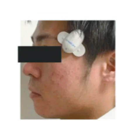
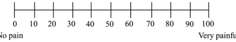
Temporalis EMG activities were recorded by a porta-ble EMG device(BUTLER GrindCare, Sunstar Suisse SA, Etoy, Switzerland)during sleep at home on 7 consecutive nights before and immediately after wearing the ortho-dontic appliance(Fig. 2A). The single -channel portable EMG device monitored EMG activity to detect temporalis muscle activities during clenching or grinding. The EMG signal was bandpass -filtered after sampling at 2 kHz. Baseline EMG amplitudes were calculated from the 80 fil-tered signal points immediately before a preset point. When the EMG amplitude was greater than 3 times the standard deviation calculated from the baseline the EMG amplitudes, the portable EMG device defined it as a sleep bruxism episode. Sleep bruxism episodes per hour in each night was counted by the portable EMG device and stored on application(Fig. 2B). To evaluate the variability of sleep bruxism episodes in each night, the coefficient of variation(CV)about sleep bruxism episodes in each night was calculated from the sleep bruxism episodes mea-sured by the portable EMG device from Day 1 to Day 7. In each day, participants scored the perceived pain inten-sity caused by the attachment of multi-bracket orthodon-tic appliance on numerical rating scale(NRS)(Fig. 3). Im-mediately after wearing the orthodontic appliance. The coefficient of determination between the number of sleep bruxism events and NRS scores was calculated in each day from all participants.
Statistical analysis
All experimental data are described as the median and interquartile range. To check normality and homoscedas-ticity, the Shapiro-Wilk test and Levene’s test applied, re-spectively. Since variables of the Shapiro-Wilk test and Levene’s test were non -normality(P = 0.04)and
hetero-scedasticity(P = 0.03), non-parametric analyses were ap-plied. RMS EMG amplitude, occlusal contact area, and oc-clusal contact point during MVC with and without sham orthodontic appliance were analyzed using Wilcoxon’s rank -sum test. Wilcoxon signed -rank test was used to compare sleep bruxism episodes and the CVs of sleep bruxism episodes in each night before and immediately after orthodontic appliance use. Pearson’s regression anal-ysis was used to analyze the correlation between pain scores and the number of sleep bruxism episodes. Signifi-cant defined as P values less than 0.05.
Results Study 1
Table 1 shows the comparison of EMG activities on bi-Fig. 2. Portable EMG device on temporalis muscle(A)and
lateral masseter and temporal muscles during MVC be-tween with and without the sham orthodontic appliance. There were no significant differences in RMS -EMG am-plitudes on each muscle during MVC between with and without the sham orthodontic appliance. Table 2 shows a comparison of the occlusal contact area and the occlusal contact point during MVC with and without the sham or-thodontic appliance. There were no significant differences in occlusal contact area and occlusal contact point during MVC between with and without the sham orthodontic appliance(P = 0.73, P = 0.98, respectively).
Study 2
Figure 4 shows the comparison of sleep bruxism epi-sodes per hours between before and immediately after wearing the orthodontic appliance on the same
partici-pant in each day. Sleep bruxism episodes on Day 2, Day 3, and Day 4 immediately after wearing the orthodontic ap-pliance were significantly lower than before wearing the orthodontic appliance(P < 0.05). Figure 5 shows the com-parison of the CVs of bruxism episodes between before and immediately after the orthodontic appliance for each day. There were no significant differences in the CVs of bruxism episodes between before and immediately after wearing the orthodontic appliance(P = 0.65). Figure 6 shows correlation between pain score and number of sleep bruxism episodes. Negative correlation were found between the number of sleep bruxism episodes and NRS scores(R2 = 0.42).
Discussion
The present study indicated for the first time that Fig. 3. Scoring the perceived pain caused by the installation of multi-bracket
orthodon-tics on the Numeric Rating Scale(NRS)
Table 1. Comparison of electromyogram(EMG)activities of bilateral masseter and temporal muscles during maximum voluntary tooth clenching between with and without the sham orthodontic appliance Abbreviations: RMS, root mean square.
Table 2. Comparison of occlusal contact area and occlusal contact point during maximum voluntary tooth clenching with and without the sham orthodontic appliance.
IJOMS Vol 19-4-38 IJOMS Vol 19-4-38
292 Int J Oral-Med Sci 19(4):288–294, 2021
wearing an orthodontic appliance does not reduce occlu-sal contact area and the occluocclu-sal contact point but reduc-es the masticatory EMG activitireduc-es associated with sleep bruxism episodes within 48 hours.
Study1 demonstrated that wearing an orthodontic ap-pliance does not affect masticatory EMG activities, occlu-sal contact area, and occluocclu-sal contact points during MVC. Although past studies investigated the effects of oral ap-pliances on sleep bruxism episodes(4, 5), the oral appli-ances in these studies changed the occlusal condition. However, according to the study by Harada et al., use of
a palatal splint without occlusal contact reduced the mas-ticatory EMG activities associated with sleep bruxism(4). The present results suggest that the main factor reduc-ing sleep bruxism episodes is not the changes in occlusal contact, but the use of an oral appliance.
Harada et al. suggested that use of stabilization splints and a palatal splint without occlusal contact reduced the masticatory EMG activities associated with sleep brux-ism(4). However, this study compared the sleep bruxism episodes for each week. The present results showed that sleep bruxism episodes on Day 2, Day 3, and Day 4 with Fig. 4. Comparison of sleep bruxism episodes between before
and immediately after wearing the orthodontic appli-ance in each day(median and interquartile range). The horizontal line in the box plot is the median. * P < 0.05
Fig. 5. Comparison of coefficient of variations of sleep brux-ism episodes between before and immediately after the orthodontic appliance in each day(median and in-terquartile range).
The horizontal line in the box plot is median.
an orthodontic appliance were significantly less than without an orthodontic appliance, and may suggest that all oral appliances(e.g. stabilization splints, palatal splints without occlusal contact, clear aligners, and orthodontic appliances, etc.)reduce the masticatory EMG activities associated with sleep bruxism within 48 hours. However, use of an orthodontic appliance device can induce pain (11–13). Our present study demonstrated that negative
correlation were found between pain score and number of sleep bruxism events. Our present study suggests that pain due to orthodontic appliances affect to reduce the masticatory EMG activities associated with sleep brux-ism. In addition, although there were no significant differ-ences in bruxism episodes between each day before wearing the orthodontic appliance, frequency of bruxism episodes on each day before wearing the orthodontic ap-pliance was not suitable due to a daily variation. Further studies are needed to estimate the validity of daily varia-tion about bruxism episodes using same portable EMG device in healthy participants.
A limitation of this study is that the participants may not have been pure sleep bruxers. Some studies have demonstrated that the threshold of sleep bruxism epi-sodes was 20 times / hour(14, 15). Although our present study did not make the criteria about the threshold of sleep bruxism episodes to recruit the participants, the mean rate of sleep bruxism episodes in the present study was fortunately more than that. Since participants need-ed to apply orthodontic appliances in portable EMG re-cording during night, the sample size was relatively small and subjects could not be grouped as bruxers and non-bruxers in this study. Further studies are needed to investigate the effect of a multi-bracket orthodontic ap-pliance for sleep bruxism in patients with patho-bruxism (16). Second, although portable EMG device have high
validity as the measurement of sleep bruxism episodes comparing polysomnography(14, 17), this experimental data could not diagnose tonic or phasic episodes of sleep bruxism. To clarify the details of the effect of orthodontic appliance for sleep bruxism episodes, further studies are needed to investigate to use not only portable EMG de-vice but also polysomnography for participants with or-thodontic appliance.
In conclusion, the present findings suggests that pain caused by multi-bracket orthodontic appliance affects to reduce the masticatory EMG activities associated with
sleep bruxism only short term. Conflicts of Interest
The authors have no potential conflicts of interest to declare.
Acknowledgments
We thank Prof. Kazutaka Kasai, Department of Ortho-dontics, Nihon University School of Dentistry at Matsudo for continuous support of this work.
References
1. Lobbezoo F, Ahlberg J, Raphael KG, Wetselaar P, Glaros AG, Kato T, Santiago V, Winocur E, De Laat A, De Leeuw R, Koyano K, Lavigne GJ, Svensson P, Manfredini D: International consensus on the assess-ment of bruxism: report of a work in progress. J Oral Rehabil, 45: 837–844, 2018.
2. Iida T, Komiyama O, Obara R, Baad -Hansen L, Kawara M, Svensson P: Repeated clenching causes plasticity in corticomotor control of jaw muscles. Eur J Oral Sci, 122: 42–48, 2014.
3. Ikuta M, Iida T, Kothari M, Shimada A, Komiyama O, Svensson P: Impact of sleep bruxism on training-in-duced cortical plasticity. J Prosthodont Res, 63: 277– 282, 2019.
4. Harada T, Ichiki R, Tsukiyama Y, Koyano K: The ef-fect of oral splint devices on sleep bruxism: a 6-week observation with an ambulatory electromyographic recording device. J Oral Rehabil, 33: 482–488, 2006. 5. Matsumoto H, Tsukiyama Y, Kuwatsuru R, Koyano K:
The effect of intermittent use of occlusal splint devic-es on sleep bruxism: a 4 -week observation with a portable electromyographic recording device. J Oral Rehabil, 42: 251–258, 2015.
6. Gerstner G, Yao W, Siripurapu K, Aljanabi H, Decker A, Ludkin D, Sinacola R, Frimenko K, Callaghan K, Penoyer S, Tewksbury C: Over -the -counter bite splints: a randomized controlled trial of compliance and efficacy. Clin Exp Dent Res, In press, 2020. 7. Wang S, Li Z, Ye H, Zhao W, Liu Y, Zhou Y:
Prelimi-nary clinical evaluation of traditional and a new digi-tal PEEK occlusal splints for the management of sleep bruxism. J Oral Rehabil, In press, 2020.
8. Castroflorio T, Bargellini A, Lucchese A, Manuelli M, Casasco F, Cugliari G, Cioffi I, Deregibus A: Effects of
IJOMS Vol 19-4-38 IJOMS Vol 19-4-38
294 Int J Oral-Med Sci 19(4):288–294, 2021
clear aligners on sleep bruxism: randomized con-trolled trial. J Biol Regul Homeost Agents, Mar-Apr 32: 21–29, 2018.
9. Obara R, Komiyama O, Iida T, De Laat A, Kawara M: Influence of the thickness of silicone registration ma-terial as a means for occlusal contact examina-tion - -an explorative study with different tooth clenching intensities. J Oral Rehabil, 40: 834 –843, 2013.
10. Obara R, Komiyama O, Iida T, Asano T, De Laat A, Kawara M: Influence of different narrative instruc-tions to record the occlusal contact with silicone reg-istration materials. J Oral Rehabil, 41: 218–225, 2014. 11. Peter N, Stepen W, Joseph S, Homa Amini: The effect
of ibuprofen on the level of in patients undergoing or-thodontic treatment. Am J Orthod Dentofacial Or-thop, 106: 88–95, 2014.
12. Vinod K: Orthodontic pain: from causes to manage-ment-a review. Eur J Orthod, 29: 170–179, 2007. 13. Anand K, Adrsh S, Swatti S, Srinath T:
Understand-ing the advances in biology of orthodontic tooth
movement for improved ortho-perio interdisciplinary approach. Journal of Indian Society of Periodontology, 17: 309–318, 2013.
14. Jadidi F, Nørregaard O, Baad-Hansen L, Arendt-Niel-sen L, Svensson P: Assessment of sleep parameters during contingent electrical stimulation in subjects with jaw muscle activity during sleep: a polysomno-graphic study. Eur J Oral Sci, 119: 211–218, 2011. 15. Yachida W, Castrillon EE, Baad-Hansen L, Jensen R,
Arima T, Tomonaga A, Ohata N, Svensson P: Cranio-facial pain and jaw -muscle activity during sleep. J Dent Res, 91: 562–567, 2012.
16. Svensson P, Lavigne G: Clinical bruxism semantics beyond academic debates: normo- and patho -brux-ism as a new proposal. J Oral Rehabil, 47: 547 –548, 2020.
17. Jadidi F, Castrillon EE, Nielsen P, Baad -Hansen L, Svensson P: Effect of contingent electrical stimulation on jaw muscle activity during sleep: a pilot study with a randomized controlled trial design. Acta Odontol Scand, 71: 1050–1062, 2012.