臨時増刊1号 ) 平 成27年3月 │
l
東女医大誌 第剃蜘8お5 頁 E4必3~E4必8 L:I:::. c十二指腸粘膜下腫蕩との鑑別が困難であった
G
r
o
o
v
eP
a
n
c
r
e
a
t
i
t
i
s
の
1
例
幸E
1聖隷浜松病院外科 2聖隷浜松病院病理診断科 3東京女子医科大学医学部外科学(第2)講 座ぶ
乃
貯
ー
ぃ
信
マサル ウ シ ダシンイチロウ ナカムラ 克1.牛田進一郎1.中村 シンイチ オオツキ ヨシロウ カメオカ 進 一2・大月
寛郎
2・亀岡
ミ 命 作 ズ 恥 トバ
速
一 一
清
平 成26年8月 22日) (受理A Case of Groove Pancreatitis Mimicking Duodenal Submucosal Tumor Masaru HA YAMI¥ Shinichiro USHIDA¥ Toru NAKAMURA¥ Shinichi SHIMIZU2, Y oshiro OTSUKr and Shingo KAMEOKA3
1 Department of Surgery, Seirei Hamamatsu General Hospital 2 Department of Pathology, Seirei Hamamatsu General Hospital 3 Department of Surgery II, Tokyo Women's Medical University
A44
・year-oldman with a history of a1coholism suffered from upper abdominal pain for 3 months and was re -ferred to a local hospita.lPhysical examination showed tenderness in the right hypochondrium, and abdominal ul -trasonography revealed a mass shadow 5 cm in diameter. Upper gastrointestina1 endoscopy and gastroduode -nography showed stenosis with multinodular elevated lesions, suggesting duodenal submucosal tumor. Contrast -enhanced computed tomography of the abdomen showed an irregularly shaped mass, 7 cm in diameter, with cal -cification and cystic change between the second portion of the duodenum and the pancreatic head. Because mu-cosal biopsy showed no malignancy and imaging findings failed to identify any neoplastic changes in the common bile duct or pancreatic duct, duodenal submucosal tumor was suspected. During laparotomy, an elastic, hard mass was palpable between the second portion of the duodenum and the pancreatic head, and marked inflamma -tory changes were apparent around the mass. Subtotal stomach-preserving pancreaticoduodenectomy was therefore performed. Histological findings showed fibrosis and chronic inflammation between the duodenal wall and pancreas without any evidence of malignancy, leading to a diagnosis of groove pancreatitis. Groove pancrea -titis is a localized chronic pancreatitis affecting the pancreatic groove. Due to the similar radiological findings and a lack of awareness of this entity, it is challenging to differentiate from malignant tumor diagnostically. We report a case of groove pancreatitis mimicking duodenal submucosal tumor and discuss its characteristic appearance with consideration of the literatures. Key W ords: groove pancreatitis, groove pancreatic carcinoma 限局した慢性捧炎であるω.男性の大酒家に好発し, その発生部位の特殊性から特徴的な臨床所見,画像 所見を示す一方で悪性腫蕩との鑑別が困難な病態で 緒 言 Groove pancreatitis (以下, GP) は,十二指腸下 行部,膝頭部ならびに総胆管の聞のgroove領域に 公益財団法人がん研有明病院消化器センター外科 -E43-図 : 速 水 克 干135-8550東京都江東区有明3-8-31 E-mail: [email protected]ある3)今回われわれは十二指腸粘膜下腫蕩との鑑別 が困難であった
GP
の1
例を経験したので,文献的 考察を含めて報告する. 症 例 患 者 :4
4
歳,男性. 既往歴・家族歴:特記すべき事項なし. 主 訴 : 上 腹 部 痛 現病歴:
3
ヵ月前から続く食後の上腹部痛を主訴 に前医を受診した.腹部超音波検査で、右季肋下に径5cm
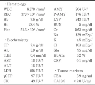
の 腫 癌 を 指 摘 さ れ 同 部 に 圧 痛 を 認 め た . 上 部 内視鏡検査で十二指腸内腔に突出する隆起性病変を 認め,十二指腸腫蕩が疑われ当院紹介となった Table 1 Laboratorydata on firstvisit . Hematology WBC 8,270/mm3 AMY 204UII RBC 373X 104 Imm3 P-AMY 176IUII Hb 7.6g/dl LYP 243IUII Ht 28.4% BUN 5 mg/dl Plat 51.3 X 10⁴ Imm3 Cr 0.62 mg/dl Na 139mEqll . Biochemistry K 4.5 mEql1 TP 7.4g/dl Cl 103 mEql1 Alb 3.9 g/dl Glu 95 mg/dl T.Bil 0.4mg/dl HbA1c 5.2% AST 18IUII CRP 0.1mgl出 ALT 18IUII LDH 158IUII . Tumor markers yGTP 97IUII CEA 3.9ng/ml CK 49IUIl CA19-9 <2.0 U/ml 生 活 歴 : (喫煙J
2
0
本/日x30
年間.(飲酒〕ウイスキー 2
0
0m
l
/
日
x25
年間. 焼 酎3
0
0
m
l
/
日
x1
年間. 来院時現症:腹部は平坦,軟で,右季肋部に圧痛 を認めた. 来院時血液検査(
T
a
b
l
e
1
)
:貧血と勝酵素の上昇 を認めたが,腫蕩マーカーの上昇など他に指摘でき るような異常はなかった. 上部内視鏡検査および上部消化管造影(
F
i
g
.
1
)
:
十二指腸下行脚の膝側には,粘膜下腫蕩を疑わせる 比較的やわらかい多結節状の隆起性病変を認め,内 腔狭窄を来たしていた.被覆粘膜の不整は軽度であ り,粘膜面からの生検では中等度の炎症やそれに伴 う上皮の反応性変化がみられたが,明らかな腫蕩性 変化は認められなかった.CT
およびMRI/MRCP(
F
i
g
.
2
)
:
CT
では十二指 腸下行脚左側を主座とし,内部に石灰化と嚢胞性変 化を伴い,遅延性に造影される最大約7cm
の不整 形腫痛を認めた.MRI
では同腫癌と謄との境界は保 たれていると考えた.MRCP
では主豚管・総胆管に 異常所見は認められず,十二指腸下行脚左側に多発 嚢胞を認めた. 以上の所見から十二指腸粘膜下腫蕩の診断で手術 を施行した 手術所見(
F
i
g
.
3
):
十二指腸下行脚と棒頭部の聞 にー塊となった弾性硬の白色腫癌を認め,周囲には 炎 症 性 変 化 が 著 明 で あ っ た 腫 癌 を 含 め 醇 温 存 十 二 指腸部分切除を試みたが,腫癌と捧の剥離はできなFig. 1 Gastroduodenal endoscopy and gastroduodenography
a) Gastroduodenal endoscopy shows stenosis of the second portion of theduodenum due to multinodularelevatedlesions, suggesting submucosal tumor (*).
b)Gastroduodenography shows stenosis with multinodular elevated lesions on the pancre -atic side of the second portion of the duodenum (arrows).
-E44-かったため,亜全胃温存棒頭十二指腸切除術を施行 した. の所見より GPと診断した. 術後経過は順調で第13病日に退院となった. 考 察 病理学的所見
(
F
i
g
.
4
)
:粘膜面には隆起性変化を 認めたほか,白色腫癌を含めて groove領域と十二 指腸壁には白色の線維性組織を認めた.groove領域 には軽度拡張した副勝管や散在性に嚢胞性変化も認 められた. 組織所見では,膝・ groove領域の著明な線維化を 認めたほか,十二指腸にも粘膜下層主体に線維増生 とリンパ球浸潤がみられ,これら線維化巣には偽嚢 胞が散見された主藤管や副勝管には石灰化を伴う 濃縮した膝液を認めたが,主勝管,副豚管および総 胆管を含めて腫傷性変化は認められなかった以上 GPは 1973年, Beckerら)Jにより十二指腸下行部, 膝頭部および総胆管の聞の溝である groove領域に 限局した慢性膝炎として報告された.1982年, Stole ら幻は慢性勝炎に対し膝頭十二指腸切除術を施行し た1
2
3
例を組織学的に検討し3
0
例(
2
4
.4%)に本症 を認め,全例が男性で,80%
以上が大酒家であった と報告している.発生機序については,長期にわた る過度のアルコール摂取や喫煙が粘調度の高い膝液 分泌を促し,さらに増加したガストリンやコレシス トキニンによる Brunner腺過形成も加わり,副勝管 Fig. 2 CT and MRI/MRCPa) An irregularly shaped mass shows enhancement in the late arterial phase. The mass is 7 cm in diameter, with calcification (arrow) and cystic change (arrowhead). and is present
on the left side of the second portion of the duodenum on CT.
b) The irregularly shaped mass shows enhancement in the equilibrium phase on CT.
c) MRI (T2-weighted) reveals the boundary between the irregularly shaped mass on the left side of the second portion of the duodenum and the pancreas (arrows).
d) MRCP shows no abnormal findings in the main pancreatic duct or common bile duct. Multiple cystic lesions are present on the left side of the second portion of the duodenum (arrows).
-Transverse colon Fig. 3 Operative findings An elastic, hard, white mass isseenbetween the secondportionof theduodenum and the pancreatic head, and marked inflammatory changesare apparent aroundthe mass. 領域での膝液流出障害により炎症を生じると考えら れている.胆道系疾患 消化性潰蕩,副乳頭欠損, 手術および外傷なども成因として挙げられている2)
GP
に特徴的な画像所見として,上部内視鏡検査 における十二指腸下行脚の浮腫性狭窄4)や,CT
での groove領域の腫癌状軟部組織の描出が挙げられる. ダイナミックCT
およびM
RI
での,線維化を反映し た遅延性濃染所見も特徴的とされる制. 組織学的にはgroove領域に慢性捧炎による線維 化がみられ,十二指腸にはBrunner腺の過形成や線 維性増殖による粘膜・粘膜下層の肥厚を伴うことも 多く,捧内,十二指腸壁内の偽嚢胞形成も特徴的と される.石灰化は稀だが病歴が長い症例では認めら れることがある2) また胆道構造が比較的正常に保たれるのも本疾患 に特徴的な所見である7) これらの所見を反映し十 二指腸狭窄に伴う悪心瞳吐や体重減少,腹部の腫癌 触診などを呈する一方で黄痘の頻度は低いとされ る2) 本症例では主捧管および総胆管に異常所見を認め ず,膝頭部に限局した腫癌形成と十二指腸狭窄などGP
を示唆する所見が認められていたにも関わら ず,本症の診断に至らず,十二指腸粘膜下腫蕩を第 ーに考えた.十二指腸粘膜下腫蕩には, gastrointes -tinal stromal tumor(
G
I
S
T
)
, neuroendocrine tumor(NET)
,リンパ腫等の鑑別が挙げられるが,画像所 見上GIST
を 最 も 疑 っ た 消 化 管GIST
は通常境界 明瞭な腫蕩であるが,腫蕩径の増大に伴い消化管外 に突出し腫蕩辺縁は結節状あるいは鋸歯状となり 隣接臓器へ浸潤する.腫蕩内部は不均一な低吸収域 を示し石灰化,壊死,嚢胞形成も出現する8
¥
十二 指腸GIST
においても同様の所見を呈する9
¥
Retrospecti veに見ると,CT
上腫癌の境界が不明 瞭であること,著明な炎症性変化の存在,石灰化や 嚢胞性変化が腫癌内だけでなく膝内にも存在するこ となどGIST
との相違点が挙げられた.鑑別を困難 にしたのは,MRI
上で腫痛と棒組織の聞に連続性を 指摘できなかったことがあるが,それ以上に我々のGP
に対する認識度の低さが要因であったと考え る.本症は発症頻度が低く稀な疾患であるが,その 病態を認識しておくことで鑑別疾患に挙げることが 可能であったと反省させられた. 本疾患に対する治療前診断率はその疾患概念の浸 透に伴い近年向上していると思われるが,一方で膝 癌などの悪性腫蕩との鑑別は依然困難である.2
0
0
3
年 に 定 義 さ れ たgroovepancreatic carcinoma (以 下,GPC)
勺まGP
と臨床的および画像的特徴が類似 しており,両者の混在した病態も存在するl川 5).GPC
は腫癒内に嚢胞性病変は伴わないことが多いが3)GP
で嚢胞性病変が顕著でない例などは両者の鑑別 が困難となる.山j畢ら日)の副醇管原発勝癌本邦報告1
0
例のまとめでも,7
例はGP
を含む炎症性疾患と の鑑別を要し術前に副勝管癌と診断できた症例は なく, groove領域の勝炎および膝癌の術前鑑別診断 の難しさを指摘している.GP
に対する治療は従来外科的治療が行われるこ とが多く仁, ~勝華頭十二指腸切除術あるいは何らかのバ イパス術が施行されてきたl7ト)ト川刊→-2幻一ll の報告も増加しており却→4引μ剖4) この中には内視鏡治療 が有用でで、あつたとする報告も認められる矧→4叫お剖8)¥. 本症 例でで、も神術f
前にGP
の診断が成されていれば禁酒など の生j活舌指導による保存的治療を試みる選択肢があつ たと思われる. しかし保存的治療でも改善が見られ ない場合や,悪性腫蕩との鑑別が困難な場合は依然 手術が必要とされておりl仰 本症例でも十二指腸粘 膜下腫蕩を念頭に置いていたこともあり,術前にGP
を疑った場合でも手術を選択したと考える.GP
を疑う症例に対しバイパス術や保存的治療を -E46-Fig. 4 Histologicalfindings
a)Resectedspecimen. An elevatedlesion isapparent on theduodenalmucosa (*). b)Cut surfaceon line b, showing a whitish, fibrotic lesion inthe groove area.
c) Cut surface onlineC, showing pancreatic tissueand the whitish, fibroticlesionin the groove area.The mildly dilated duct of Santoriniand pseudocystsare also confirmedin the
groove area.
d) Low-power view of the lesionshows marked fibrosisofthegroove area and theduct of
Santorini containsinspissated pancreatic juicewith calcification(hematoxylinand eosin(HE)
staining).
e) Marked fibrosisinthe pancreaticparenchyma and inspissatedpancreaticjuicewith cal -cificationinthemain pancreaticductareidentified (HE staining, X 30).
f)Duodenal wallshows marked thickening with fibrousproliferationand lymphocytic infil
-trationmainly inthe submucosa.The duodenalwall also containspseudocysts (HE stain -ing, X 15). po V, papillaofVater;mdp, minor duodenal papilla;ga, groovearea; dm, duodenalmucosa; pan, pancreas;pc, pseudocyst;cbd, common bile duct;fib, fibrosis;sd, ductofSantorini; mpd, main pancreaticduct. 選択する場合には,悪性との鑑別が極めて困難であ ることから,その可能性が否定できない場合には棒 頭十二指腸切除術を行うことも考慮すべきと考え る. い場合があり,保存的治療が奏功しない症例や悪性 腫蕩が否定できない場合は積極的に手術を検討すべ きである. 盆士 1ロ1・ 墨A E岡 十二指腸粘膜下腫蕩との鑑別が困難であった
GP
の1
例 を 経 験 し た 大 酒 家 に 生 じ た 十二指腸狭窄を 伴う膝頭十二指腸病変ではGP
も念頭におく必要が ある.この場合, 白験例のような十二指腸粘膜下腫 療やGPC
などの腫蕩性変化との臨床的鑑別が難し - E47-開示すべき利益相反はない. 文 献 1)Becker V: Bauchspheicheldruse.InSpezielle Pa -thologische Anastomie, Bd V.
I
Springer, Berlin, Heidelberg, New York (1973)2) Stole M, Weiss W, Volkholz H et al:A special
-tis". Hepatogastroenterology 29: 198-208, 1982
3) Gabata T, Kadoya M, Terayama N et al: Groove pancreatic carcinomas: radiological and pathologi -cal findings. Eur Radioll3: 1679-1684,2003 4) Mohl W, Hero-Gross R, Feifel G et al: Groove pan -creatitis: an important di旺erentialdiagnosis to ma-lignant stenosis of the duodenum. Dig Dis Sci 46: 1034-1038,2001 5) Itoh S, Yamakawa K, Shimamoto K et al: CT findings in groove pancreatitis: correlation with histopathological findings. J Comput Assist To開 mogr 18: 911-915, 1994
6)Ir色H,Honda H, Kuroiwa T et al: MRI of groove pancreatitis. J Comput Assist Tomogr 22: 65ト655, 1998 7)小林道也,荒木京二郎,中村生也ほか:十二指腸狭 窄を呈したgroovepancreatitisの1例.日消外会誌 28 : 2290-2294,1995 8)西 田 俊 朗 :CT' MRIによる臨床診断.IGISTの診 断 と 治 療 実 践 マ ニ ュ ア ルJGIST研究会,pp20-3,1 エルゼピア・ジャパン,東京 (2006)
9) Kim HC, Lee JM, Son KR et al: Gastrointestinal stromal tumors of the duodenum: CT and barium study findings. Am J Roentgenoll83: 415-419, 2004 10) Tan CH, Chow PK, Thng CH et al: Pancreatic
adenocarcinoma that mimics groove pancreatitis: Case report of a diagnostic dilemma. Dig Dis Sci 51: 1294-1296,2006
11) Aimoto T, Uchida E, Nakamura Y et a:lClinico -pathologic study on pancreatic groove carcinoma. Pancreas 33: 255-259, 2006 12)大西佳文,山崎哲,鈴木智哉ほか:Groove膝炎と 鑑別困難であった捧癌の1例.肝胆勝治研誌 6: 41-48,2008 13)目 黒 敬 義 , 山 本 毅 , 西 岡 可 奈 ほ か : 若 年 発 症 したpancreaticgroove carcinomaの1例 . 日 消 誌 105: 1078-1086,2008 14) Ishigami K, Tajima T, Nishie A et al: Differential diagnosis of groove pancreatic carcinomas vs. Groove pancreatitis: Usefulness of the portal ve -nous phase. Eur J Radiol 74: e95-e100, 2010
15) Malde DJ, Oliveira・CunhaM, Smith AM: Pancre
-atic carcinoma masquerading as groove pancreati -tis: Case report and review of literature. JOP 12: 598-602,2011 -E48-16)山津邦宏,寺島裕夫,横畠徳祐ほか:Groove pan -creatitisとの鑑別に苦慮したまれな副醇管領域原 発勝癌の1切除例-自験例を含めた本邦報告10例 の ま と め と 共 に 醇 臓 22 : 65-73,2007 17)宜保淳也,伊藤鉄英,河漫顕ほか:女性に発症し, pure formからsegmentalformに進展したGroove pancreatitisの一例.臨と研 82: 127-131,2005 18)竹中芳冶,春日井尚,宮島伸宜ほか:全胃温存醇頭 十二指腸切除術を施行したgroovepancreatitisの 1例. 日臨外会誌 64 : 2855-2860,2003 19)友松宗史,安井智明,生田真ーほか:門脈閉塞を 伴ったGroovepancreatitisに対し幽門側胃切除, Roux-en Y再 建 術 を 施 行 し た1例.日消外会誌 44 : 991-996, 2011 20)藤島則明,浜口伸正,谷田信行ほか:胃空腸吻合を 行ったgroovepancreatitisの1例.高知医師会医誌 16 : 248-253, 2011