retina: from the initial research on rd mutant mice to zebrafish genetic approaches
Author Maria Iribarne, Ichiro Masai journal or
publication title
Journal of Neurogenetics
volume 31
number 3
page range 88‑101
year 2017‑08‑16
Publisher Informa UK Limited, trading as Taylor &
Francis Group.
Rights (C) 2017 The Author(s).
Author's flag publisher
URL http://id.nii.ac.jp/1394/00000409/
doi: info:doi/10.1080/01677063.2017.1358268
Creative Commons Attribution‑NonCommercial‑NoDerivatives License (http://creativecommons.org/Licenses/by‑nc‑nd/ 4.0/),
Full Terms & Conditions of access and use can be found at
http://www.tandfonline.com/action/journalInformation?journalCode=ineg20
ISSN: 0167-7063 (Print) 1563-5260 (Online) Journal homepage: http://www.tandfonline.com/loi/ineg20
Neurotoxicity of cGMP in the vertebrate retina:
from the initial research on rd mutant mice to zebrafish genetic approaches
Maria Iribarne & Ichiro Masai
To cite this article: Maria Iribarne & Ichiro Masai (2017) Neurotoxicity of cGMP in the vertebrate retina: from the initial research on rd mutant mice to zebrafish genetic approaches, Journal of Neurogenetics, 31:3, 88-101, DOI: 10.1080/01677063.2017.1358268
To link to this article: https://doi.org/10.1080/01677063.2017.1358268
© 2017 The Author(s). Published by Informa UK Limited, trading as Taylor & Francis Group.
Published online: 16 Aug 2017.
Submit your article to this journal
Article views: 473
View Crossmark data
Citing articles: 5 View citing articles
REVIEW ARTICLE
Neurotoxicity of cGMP in the vertebrate retina: from the initial research on rd mutant mice to zebrafish genetic approaches
Maria Iribarne and Ichiro Masai
Okinawa Institute of Science and Technology Graduate University, Onna, Okinawa, Japan
ABSTRACT
Zebrafish are an excellent animal model for research on vertebrate development and human diseases.
Sophisticated genetic tools including large-scale mutagenesis methodology make zebrafish useful for studying neuronal degenerative diseases. Here, we review zebrafish models of inherited ophthalmic dis- eases, focusing on cGMP metabolism in photoreceptors. cGMP is the second messenger of phototrans- duction, and abnormal cGMP levels are associated with photoreceptor death. cGMP concentration represents a balance between cGMP phosphodiesterase 6 (PDE6) and guanylate cyclase (GC) activities in photoreceptors. Various zebrafish cGMP metabolism mutants were used to clarify molecular mecha- nisms by which dysfunctions in this pathway trigger photoreceptor degeneration. Here, we review the history of research on the retinal degeneration (rd) mutant mouse, which carries a genetic mutation of PDE6b, and we also highlight recent research in photoreceptor degeneration using zebrafish models.
Several recent discoveries that provide insight into cGMP toxicity in photoreceptors are discussed.
ARTICLE HISTORY Received 1 April 2017 Accepted 18 July 2017 KEYWORDS
Zebrafish; photoreceptor degeneration; genetic mutants; retina; Aipl1;
Pde6c
Introduction
In vertebrates, the neural retina consists of six major classes of neurons: retinal ganglion cells, which form the innermost layer of the optic cup, three types of interneurons, namely amacrine cells, bipolar cells, and horizontal cells, which form the intermediate nuclear layer, and two types of photorecep- tor neurons, namely rods and cones, which mediate photo- transduction under dim and normal light conditions, respectively (Figure 1(A)). In the last several decades, the neural retina has been used as an excellent platform to study neuronal differentiation in the central nervous system of ver- tebrates. In addition, the neural retina provides a good model for studying neuronal degeneration and regeneration.
Here, we focus on photoreceptor degeneration caused by genetic mutations of a central component of phototransduc- tion molecule, cGMP phosphodiesterase 6 (PDE6) by review- ing previous mouse and human studies and zebrafish research, and identify its underlying mechanism, cGMP tox- icity, which causes photoreceptor cell death.
Photoreceptor structure and the phototransduction pathway
Cone and rod photoreceptors mediate phototransduction in the retina under dim and normal light conditions, respect- ively. Because zebrafish are diurnal, they have retinas rich in cones as well as rods, making them a convenient model to
study cone pathologies (Fadool & Dowling, 2008). Humans have red-, green-, and blue-sensitive cones, while zebrafish have four types, which express red, green, blue, and UV opsins, respectively, and which employ a planar pattern called the row mosaic (Raymond, Barthel, Rounsifer, Sullivan, & Knight,1993).
Photoreceptors have highly specialized functional struc- tures, formed by an outer segment (OS), an inner segment containing a mitochondria-rich ellipsoid, a nucleus, and a synaptic terminal (Figure 1(B)). The OS consists of photo- receptive membranes that form multiple stacked membrane disks, like piles of coins. These accommodate a large number of phototransduction molecules, such as rhodopsin and opsins (Goldberg, Moritz, & Williams,2016), indicating that phototransduction is initiated in the OS. The transport of phototransduction molecules and membrane components from the inner segment to the OS is mediated through a specialized primary cilium called the connecting cilium (Wang & Deretic,2014).
The molecular network for the vertebrate phototransduc- tion cascade is shown inFigure 2. In vertebrate photorecep- tors, cyclic nucleotide-gated (CNG) channels determine membrane potential by promoting Naþ/Ca2þ ion flow into or out of cells. Under dark conditions, a high level of cGMP maintains CNG channels in the open state, maintaining a steady influx of Naþ, and Ca2þ. Once the photoreceptors are exposed to light, opsins or rhodopsin activates a G-protein, transducin, which subsequently activates PDE6. Activated
CONTACTMaria Iribarne [email protected]; Ichiro Masai [email protected] Okinawa Institute of Science and Technology Graduate University, 1919-1 Tancha, Onna, Okinawa 904-0495, Japan
ß2017 The Author(s). Published by Informa UK Limited, trading as Taylor & Francis Group.
This is an Open Access article distributed under the terms of the Creative Commons Attribution-NonCommercial-NoDerivatives License (http://creativecommons.org/Licenses/by-nc-nd/
4.0/), which permits non-commercial re-use, distribution, and reproduction in any medium, provided the original work is properly cited, and is not altered, transformed, or built upon in any way.
https://doi.org/10.1080/01677063.2017.1358268
PDE6 hydrolyses cGMP. The reduced intracellular concen- tration of cGMP causes cGMP to dissociate from the CNG channel, which closes the channel. This subsequently reduces the ion influx that hyperpolarizes photoreceptors. Guanylate cyclase-activating proteins (GCAPs) function as Ca2þ sen- sors. When Ca2þ ions bind to GCAPs, GCAPs inhibit gua- nylate cyclase (GC) activity. On the other hand, when Ca2þ ions dissociate from GCAPs, this activates GC activity. After light stimuli close the CNG channel, resulting in decreased Ca2þ influx, GCAPs activate GC to recover cGMP levels.
Thus, in the phototransduction process, PDE6 and GC are functionally linked by intracellular Ca2þ levels and regulate the cGMP concentration in photoreceptors.
The phototransduction pathway is shared by both rod and cone photoreceptors; however, phototransduction com- ponents differ in rods and cones. For example, rods possess the visual pigment, rhodopsin, while opsins are present in
cones. Furthermore, rod and cone transducin, PDE6, GC, GCAP and CNG channels differ in their subunit composi- tions (Table 1) (Larhammar, Nordstrom, & Larsson, 2009).
In rods, PDE6 is a hetero-tetramer consisting of two closely related aand b subunits and two identicalc subunits, while cone PDE6 consists of two homodimers of a0 and c0 subu- nits (Lagman, Franzen, Eggert, Larhammar, & Abalo, 2016).
PDE6 a, b, a0, c, and c0 subunits are also designated as PDE6a, PDE6b, PDE6c, PDE6g, and PDE6h, respectively.
Phototransduction molecules are transported from the ER and Golgi bodies to the OS. Rhodopsin constitutes the majority of OS-resident proteins and belongs to the G- protein-coupled receptor family, which has seven transmem- brane domains. The last four amino acids comprising the VXPX motif at the C-terminus of rhodopsin are important for trafficking to the OS and are targeted by a small GTPase, Arf4, which interacts with Rab11 and Rab8 to sort rhodopsin
Figure 1. Zebrafish retina. (A) Section of a 7 dpf zebrafish retina (left) and a schematic drawing of the retinal neural circuit, which comprises six major classes of ret- inal neurons (right). (B) Electron microscopic (EM) image (left) and schematic drawing (right) of rod and cone photoreceptors. Photoreceptors have specialized struc- tures: the outer segment (OS), the inner segment, and a synaptic terminus. The inner segment contains the mitochondria-rich ellipsoid. The connecting cilium extends from the inner segment to the OS. The OS consists of multiple, stacked, photoreceptive membrane disks and accommodates a large number of phototrans- duction molecules, including opsins, transducins, and Pde6.
into transport vesicles budding from the trans-Golgi network and delivering them to the OS (Pearring, Salinas, Baker, &
Arshavsky, 2013). Many other proteins transported to the OS are associated with membranes via lipid modifications.
Transducin a and c subunits, GCAPs, and opsin kinase GRK1 are lipidated by farnesylation, acylation, and myristoy- lation (Pearring et al., 2013). Rod PDE6 is farnesylated and
geranylgeranylated on its a and b subunits (Anant et al., 1992; Qin, Pittler, & Baehr, 1992), respectively, while cone PDE6 is likely geranylgeranylated on both of its a0 subunits (Wensel, 2008). Following prenylation of the C-terminal cysteine of PDE6, which ends in a CAAX domain, the last three amino acids are removed by the endoprotease RAS- converting enzyme 1 (RCE1) (Christiansen, Kolandaivelu,
Figure 2. Photoreceptors and the phototransduction pathway. (A) Diagram of cone and rod photoreceptors showing the different compartments, the outer (OS) and inner segment (IS), the nucleus, and the synapsis (Syn). (B) Schematic representation of the phototransduction pathway. Photon absorption and activation of opsin lead to dissociation of transducinasubunit fromb/csubunits, which subsequently activates PDE6. Activated PDE6 then catalyzes cGMP hydrolysis. Reduction of cGMP closes CNG channels on the plasma membrane and reduces intracellular Ca2þlevels. Then, hyperpolarization of the photoreceptor ceases neurotransmitter release. Reduced [Ca2þ] activates GCAP, which stimulates GC to synthesize cGMP, opening CNG channels again.
Table 1. Nomenclature of rod- and cone-specific subunits of phototransduction molecules in human and zebrafish. In zebrafish, some of the subunits have extra paralogs caused by gene duplication in teleost fish.
Bergo, & Ramamurthy, 2011) and subsequently prenylcys- teine is methylated by isoprenylcysteine carboxyl methyl- transferase (IMCT) (Christiansen et al., 2016). These two modifications are critical for trafficking functional PDE6 to the OS. Photoreceptor-specific GCs, retGC1 and retGC2 (also designated as GUCY2D and GUCY2F), are trans-mem- brane proteins. Double knockdown of retGC1 and retGC2 impairs protein levels of GCAP1, transducins, PDE6, and GRK1, as well as their delivery to the OS, suggesting that these lipidated proteins are transported to the OS via GC- bearing transport vesicles (Baehret al.,2007). These findings indicate that intracellular protein modification and transport tightly regulate functional integrity of phototransduction molecules.
Retinal degeneration diseases in humans
Retinal degeneration is a devastating condition leading to blindness. Here, we will focus on the inherited retinal degen- erative diseases, such as retinitis pigmentosa, cone-rod dys- trophy, achromatopsia, and Leber congenital amaurosis (LCA) among others. Retinitis pigmentosa is characterized by night blindness and loss of mid-peripheral visual field in its early stages. Later, as the disease progresses, loss of far peripheral visual field and central vision occur, suggesting rod-cone dystrophy in which primarily rods degenerate, fol- lowed by the second loss of cones (Hartong, Berson, &
Dryja,2006). Cone-rod dystrophy is a distinct type of retinal disease causing initial cone degeneration followed by the loss of rods (Hamel, 2007). Achromatopsia is a retinopathy, which presents loss of cone functions, leading to color blind- ness, poor visual acuity, photophobia, and nystagmus.
Characteristically, it appears at birth or infancy of patients (Aboshiha, Dubis, Carroll, Hardcastle, & Michaelides, 2016).
LCA is a severe disease with congenital loss of rods and cones at early stages (den Hollander, Roepman, Koenekoop,
& Cremers,2008). Over 238 genes are known to participate in these retinal diseases (RetNet, http://www.sph.uth.tmc.
edu/RetNet). These genes are roughly classified into several distinct functional groups, which are classified depending upon whether they impact phototransduction (Rattner, Sun,
& Nathans, 1999), retinoid metabolism (Saari, 2012), intra- cellular transport through the connecting cilium (Rachel, Li,
& Swaroop, 2012), or establishment of the OS (Goldberg et al.,2016). However, because of the large number of genes associated with retinal degeneration, it is difficult to develop an efficient treatment. So far, only partial positive results have been obtained through treatment with neuroprotective compounds, gene therapy, cell transplantation, or artificial devices (Cuencaet al.,2014).
cGMP metabolism in photoreceptor degeneration As mentioned above, genetic mutations of a variety of genes are associated with photoreceptor degeneration in humans.
Hereafter, we focus on cGMP metabolism in photoreceptor degeneration. A good starting point is an early finding in a spontaneous mouse mutant, retinal degeneration (rd)
(Keeler, 1924). The rd mutation induced photoreceptor degeneration in accordance with Mendelian inheritance. In 1974, Farber and Lolley reported that cGMP concentration is elevated in degenerating photoreceptors of the rd mutant mouse (Farber & Lolley, 1974). Following this study, it was reported that high concentrations of cGMP are linked to photoreceptor degeneration in frog (Farber & Lolley, 1977) and dog retinas (Aquirre, Farber, Lolley, Fletcher, & Chader, 1978). These reports raised the possibility that abnormal accumulations of cGMP cause photoreceptor degeneration in the vertebrate retina.
Indeed, cGMP is the key messenger in phototransduction in vertebrate photoreceptors, and the rd mutant gene enco- des a rod-specific subunit of PDE6, PDE6b (Bowes et al., 1990). Soon after, genetic mutations in rod-specific subunits of PDE6, PDE6a, and PDE6b were identified in human patients suffering from retinitis pigmentosa (Huang et al., 1995; McLaughlin, Sandberg, Berson, & Dryja,1993), further suggesting that cGMP metabolic dysfunctions are important for understanding human genetic retinal diseases. In parallel, a genetic mutation was found in rod transducin a-subunit (also called GNAT1) in human patients with the Nougaret form of congenital stationary night blindness (Dryja, Hahn, Reboul, & Arnaud,1996). Mutations in the cone transducin a-subunit (also designated GNAT2) were identified in human patients with Achromatopsia, which shows total lack of cone functions (Kohl et al., 2002). Gnat1 mutant mice show that rods fail to show the response to flash light by single-cell recording, however display a very mild retinal degeneration (Calvert et al., 2000). gnat2 mutant zebrafish show no detectable level of cone a transducin and relatively normal retinal morphology (Brockerhoff et al., 2003). These data suggest that a blockade of phototransduction upstream of PDE6 in both rods and cones does not induce photo- receptor degeneration to the level of the rd mutant mice.
These observations also suggest that cGMP metabolic dysfunction is more directly linked to photoreceptor degeneration.
cGMP levels are maintained by a balance between PDE6 and GC activities. In humans and mice, there are two GCs, retGC-1 and retGC-2. retGC1 is expressed in both rods and cones, whereas retGC2 is expressed only in rods (Table 1) (Yang, Foster, Garbers, & Fulle, 1995). Gain-of-function mutations of retGC-1 are likely to increase cGMP concentra- tion and to cause autosomal dominant cone-rod dystrophy (Tuckeret al.,1999; Wilkieet al.,2000). Furthermore, a gen- etic mutation, Y99C, in GCAP1 disrupts the 3rd EF hand motif, thereby preventing Ca2þ binding to GCAP1. Thus, GCAP1 is released from Ca2þ-mediated inhibition and per- manently activates retGC1 (Payne et al., 1998), resulting in autosomal dominant cone dystrophy (Sokal et al., 1998).
These observations also support the idea that elevated cGMP levels trigger photoreceptor degeneration in humans and mice. Conversely, loss-of-function mutations of retGC-1 are expected to decrease cGMP production and reported to induce LCA in human (Perrault et al., 1996). It was also reported that cGMP levels are significantly reduced in chick GC1mutant (Semple-Rowland, Lee, Van Hooser, Palczewski,
& Baehr, 1998), suggesting that lower cGMP levels link to
photoreceptor degeneration. As we mentioned before, double knockdown of retGC-1 and retGC-2 markedly decreases the level of PDE6 in mice (Baehr et al., 2007), indicating some interdependence between PDE6 and GC activity. These find- ings show that dysfunction on cGMP metabolism results in abnormal cGMP levels, which are toxic to photoreceptors.
Functional CNG channels in photoreceptors are hetero- tetramers consisting of a subunits (also designated as CNGA) and b subunit (also designated as CNGB) (Larhammar et al.,2009). Rods and cones use distinct subu- nits. Rods have CNGA1 and CNGB1, whereas cone channels consist of CNGA3 and CNGB3 (Table 1). The hetero-tetra- mer channel consists of three CNGA1 and one CNGB1 in rods (Zheng, Trudeau, & Zagotta, 2002), and two copies of CNGA3 and CNGB3 in cones (Peng, Rich, & Varnum, 2004). Mutations of CNGA3 and CNGB3 were reported in patients with achromatopsia (Kohl et al., 1998; Kohl et al.,
2000). InCnga3 mutant mice, accumulation of cGMP causes photoreceptor degeneration (Xuet al., 2013), supporting the correlation between high levels of cGMP and photoreceptor cell death.
Mechanisms underlying photoreceptor cell death triggered by cGMP accumulation
During the past twenty years, many studies have elucidated the molecular mechanism underlying photoreceptor degen- eration caused by defects in PDE6 function (Figure 3(A–B)). In the rd mouse, mutations of the Pde6b gene are likely to cause accumulation of cGMP in rods. Consistently, rods first degenerate, followed by cones. Since PDE6b is expressed exclusively in rods, cone degeneration is triggered by loss of rods, probably through the bystander effect
Figure 3.Possible cone cell death pathways in response to cGMP accumulation. (A) Diagram of the phototransduction pathway in wild-type photoreceptors.
Intracellular cGMP levels are regulated by a balance between PDE6 and GC activity. Ca2þinflux through CNG channels inhibits GCAPs, so the closure of CNG channels decreases intracellular Ca2þlevels, which subsequently activate GCAP and then increase GC activity, resulting in cGMP production. (B) Apoptotic pathways inPde6b mutant mice. The absence of PDE6b activity induces the accumulation of cGMP. High levels of cGMP chronically open CNG channels, which subsequently enhance a series of Ca2þ-dependent cellular processes, including the activation of calpain. Calpain activates AIF to translocate from mitochondria to nuclei, in which AIF pro- motes chromatin degradation. Calpain also activates caspase 12 to promote apoptosis. (C) The apoptotic pathway ofCnga3/Cngb3mutant mice. The absence of cone CNG channels decreases Ca2þinflux, which may cause persistent activation of GCAPs and retGC1, resulting in elevated cGMP. Highly elevated cGMP activates PKG and enhances ER stress response regulators, including ATF6 and CHOP. Activation of calpain, AIF, and caspase 12 is also observed.
(Frasson et al., 1999; Narayan, Wood, Chidlow, & Casson, 2016). Thus, two questions arise. First, what molecular mechanism mediates rod death in the rd mouse? Second, what is the molecular mechanism that induces cone death in response to rod loss in the rd mouse? The first question also raises a series of other questions. If cone pde6 gene, pde6c, is mutated, do these mutant cones also accumulate cGMP and undergo degeneration? In this case, does loss of pde6c mutant cones affect survival of genetically healthy rods?
Early studies on therdmutant mouse focused on the first question, the molecular mechanism underlying rod cell death. In rd mice, rods undergo a common mode of cell death, apoptosis (Chang, Hao, & Wong, 1993; Portera- Cailliau, Sung, Nathans, & Adler, 1994). However, overex- pression of anti-apoptotic factors Bcl2 or BclXL showed con- flicting results. Joseph and Li reported that overexpression of Bcl2 or BclXL did not inhibit photoreceptor degeneration in rd mice (Joseph & Li, 1996); however, Chen et al. found that overexpression of Bcl2 rescued photoreceptor cell death inrdmice (Chenet al.,1996). Furthermore, the involvement of caspases is controversial. Activation of caspases and release of cytochrome C were reported in therdmouse pho- toreceptors (Jomary, Neal, & Jones, 2001), whereas caspase activation was not observed in rd mice (Doonan, Donovan,
& Cotter, 2003). Yoshizawa et al. reported that caspase inhibition mildly rescued photoreceptor degeneration in rd mice (Yoshizawa et al., 2002), whereas Zeiss et al. showed that photoreceptor degeneration proceeded even in rd cas- pase3-/- double mutant mice (Zeiss, Neal, & Johnson, 2004).
Although photoreceptor death is associated with apoptotic traits such as DNA fragmentation, it is likely that the cas- pase-independent pathway mediates rod degeneration in rd mutant mice.
Accumulation of intracellular cGMP is expected to maintain high levels of Ca2þ via CNG channels. It was reported that application of a Ca ion channel blocker res- cues photoreceptor degeneration and visual functions in rd mice (Frasson et al., 1999), although there were conflicting reports (Pawlyk, Li, Scimeca, Sandberg, & Berson, 2002;
Pearce-Kelling et al., 2001). Knockdown of rod-specific sub- units of CNG channels, CNGA1 and CNGB1, strongly res- cues rod photoreceptor degeneration in rd mice (Paquet- Durand et al., 2011; Tosi et al., 2011), suggesting that dele- terious Ca2þ influx through CNG channels causes rod cell death in the absence of PDE6b. Consistently, it was reported that calcium-activated proteases, calpains, are highly activated in degenerating rd mutant photoreceptors (Paquet-Durand et al., 2006). One calpain substrate is apoptosis-inducing factor (AIF). AIF was discovered as the first protein that mediates caspase-independent apoptosis (Susin et al., 1999). AIF is a flavoprotein located in the mitochondrial intermembrane space and is involved in energy and redox metabolism (Hangen, Blomgren, Benit, Kroemer, & Modjtahedi, 2010; Modjtahedi, Giordanetto, Madeo, & Kroemer, 2006). Under stress conditions, AIF is cleaved and translocates to the nucleus, leading to chroma- tinolysis in a caspase-independent manner. AIF release from mitochondria is stimulated by not only calpain-
mediated cleavage, but also by reactive oxygen species (ROS) (Churbanova & Sevrioukova, 2008) and poly(ADP- ribose) (PAR) polymer (PARP) (Yu et al., 2002; Yu et al., 2006), which is produced by PAR polymerase 1, a family of proteins involved in DNA repair and genome instability, suggesting that oxidative stress and subsequent DNA dam- age modify calpain-mediated cleavage of AIF. Retinas of rd mice exhibit increased activity of calpains and PARP, which mediate mitochondrial AIF release (Paquet-Durand et al., 2007; Sanges, Comitato, Tammaro, & Marigo, 2006).
Blockade of the calpain or PARP pathway inhibits nuclear translocation of AIF and rod cell death in rd mice (Murakami, Ikeda, et al., 2012; Sahaboglu et al., 2010;
Sanges et al., 2006). These data suggest that AIF and its regulatory mechanisms induce rod photoreceptor cell death in the absence of PDE6b. In addition to the Ca2þ/calpain/
AIF pathway, cGMP-dependent protein kinases (PKG) are activated and regulate photoreceptor cell death in rd mice (Paquet-Durand, Hauck, van Veen, Ueffing, & Ekstrom, 2009). Very recently, it was also reported that microglia eliminate photoreceptors in rd mice (Zhao et al., 2015), suggesting that microglial phagocytosis contributes to rod cell elimination in the absence of PDE6b.
Zebrafish forward genetics generate visual mutants Zebrafish are widely used as a vertebrate model in develop- mental genetics, because of their high fertilization rate, large numbers of eggs per female, rapid embryonic development (Kimmel, Ballard, Kimmel, Ullmann, & Schilling, 1995), and an established chromosome linkage map (Geisler et al.,1999;
Knapik et al., 1998; Postlethwait et al., 1994). Moreover, zebrafish embryos are transparent, so a series of excellent fate-map studies (Kimmel, Warga, & Schilling,1990; Woo &
Fraser, 1995) and high-resolution time-lapse live imaging techniques have been applied to early stage of development (Olivier et al., 2010) and also combined with an anatomical atlas documenting gene expression in the nervous system (Ronnebergeret al.,2012).
The pioneering work of Streisinger showed the utility of this genetic model to study neuronal development using mutant strains (Grunwald & Streisinger, 1992; Streisinger, Walker, Dower, Knauber, & Singer, 1981). Soon after, sev- eral laboratories carried out large-scale, forward-genetic screening with the mutagenic agent, ethyl nitrosourea (ENU) (Driever et al., 1996; Haffter et al., 1996), or viral insertion to identify recessive mutants in zebrafish (Amsterdam et al., 1999). Because zebrafish depend heavily upon vision, they are especially useful for research on the visual system (Fadool & Dowling, 2008). Different behav- ioral tests have been established to evaluate visual perform- ance, such as the optomotor response (OMR) (Neuhauss et al., 1999) and the optokinetic response (OKR) (Brockerhoff et al., 1995). Visual impairment can be eval- uated as soon as 5 days post-fertilization (dpf), when cone photoreceptors start to function (Rinner, Rick, &
Neuhauss, 2005). Several types of screening focusing on the visual system were performed. Mutants were identified based upon OKR, OMR, or electroretinogram (ERG)
responses (Brockerhoff et al., 1995; Muto et al., 2005;
Neuhauss et al., 1999; Nishiwaki et al., 2008) and retinal morphology (Gross et al., 2005; Malicki et al., 1996; Masai et al., 2003). This mutant collection is an essential resource for various studies of retinal developmental biology and functions.
Zebrafish cone-specific subunit of PDE6, PDE6c, mutants
As mentioned above, genetic mutations of rod-specific subu- nits of PDE6, PDE6a and PDE6b, cause retinitis pigmentosa in humans (Huang et al., 1995; McLaughlin et al., 1993).
Furthermore, the rd mutant gene encodes PDE6b (Bowes et al.,1990). In both cases, rods initially degenerate, followed by a progressive degeneration of cones. Because cGMP con- centration is elevated in degenerating photoreceptors of rd mutant mice (Farber & Lolley, 1974), these findings suggest that chronically high levels of cGMP trigger rod photorecep- tor degeneration in mice and humans. One interesting ques- tion is whether a similar situation occurs in cones. Does knockdown of cone-specific subunits of PDE6 cause cone degeneration? Until 2009, no genetic mutation of cone-spe- cific subunit of PDE6,a’ subunit (also designated as PDE6c), had been reported in mice or humans. Another interesting issue is the interdependence of rod and cone maintenance.
Since PDE6b is a rod-specific subunit, cone degeneration in rd mouse mutants is likely to depend on the initial loss of rods, suggesting a ‘bystander effect’, in which healthy neu- rons undergo cell death when they are neighbor to dead or dying neurons (Frasson et al., 1999). Several possibilities were proposed (Narayan et al., 2016): (1) degenerating rods may release toxic materials to cones, (2) rods may secrete factors that support cone survival, (3) rod loss may affect delivery of nutrients to cones via blood vessels and glia, or (4) rod loss may decrease oxygen consumption, leading to increase of reactive oxygen species, which cause cone dam- age by oxidative stress. However, it was not clear whether rods show a similar bystander effect in response to cone loss. Interestingly, knockdown of retGC1 in mice shows cone-specific dystrophy, but rods do not degenerate despite a reduced response in rod-mediated ERG (Yanget al.,1999), suggesting that the interdependence between rod and cone maintenance is not mutual. Thus, the interdependence of rod and cone maintenance is interesting to understand homeostasis of retinal functions and also important from a clinical point of view.
A cue to understand rod survival in the cone-degenerat- ing environment came from zebrafish mutagenesis. A non- sense allele of zebrafish pde6c mutants was isolated by Brockerhoff’s lab (Stearns, Evangelista, Fadool, &
Brockerhoff, 2007). In this allele, loss of Pde6c activity indu- ces rapid cone degeneration at embryonic stages. Some rods also undergo degeneration, although rod degeneration is lim- ited to areas with a low rod density. Concurrently, we also isolated a hypomorphic mutant allele ofpde6c,namedeclipse (els) (Nishiwaki et al., 2008). Theels mutant has a missense mutation in thepde6cgene, causing an amino acid substitu- tion in a conserved regulatory domain called GAF-A. This
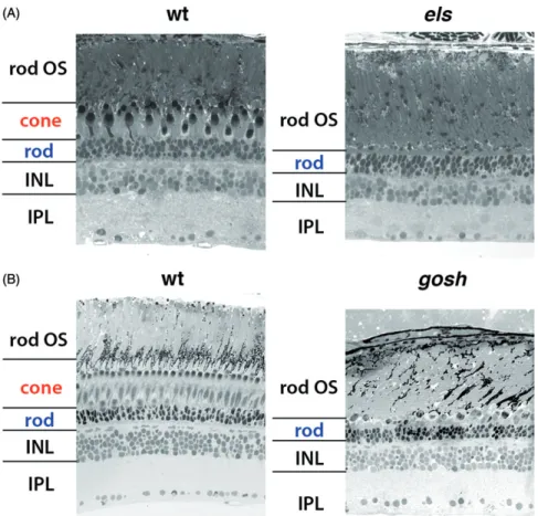
allele causes cell death in cones and probably rods at an early embryonic stage (Nishiwaki et al., 2008). In later developmental stages, cones undergo slow, but progressive degeneration, and are completely eliminated by 6 months post-fertilization (mpf), but rods are retained to form the outer nuclear layer (Figure 4(A)), suggesting that cones are eventually eliminated in the absence of cone Pde6. It was reported that transplanted wild-type cones persist in zebra- fish null pde6c mutant retinas (Lewis, Williams, Lawrence, Wong, & Brockerhoff, 2010). These findings indicate that as in the case of rod PDE6, cone PDE6 is required for cone maintenance in a cell-autonomous manner.
Zebrafishpde6cmutants provide an interesting insight on the interdependence between rod and cone maintenance. In both null and hypomorphic alleles of zebrafish pde6c mutants, some rods seem to undergo cell death at embryonic stages from 6 to 9 dpf (Nishiwakiet al.,2008; Stearnset al., 2007). Since the maturation of rods proceeds until 3 weeks post-fertilization (wpf) in zebrafish (Branchek & Bremiller, 1984), it is likely that cone cell death influences the survival of maturing rods. Interestingly, we observed a slow but nor- mal OKR under dim light conditions in zebrafishelsmutant larvae at 3 wpf, suggesting rod-mediated scotopic vision (Nishiwaki et al.,2008). Inels mutants, almost all cone cells are eliminated, but rods are retained with normal subcellular morphology at 6 mpf (Figure 4(A)) (Nishiwaki et al., 2008).
Thus, although rods are deformed and some of them are likely to undergo cell death in embryonic stages, the els mutant larvae maintain scotopic vision. From these observa- tions, loss of cones does not compromise scotopic vision and rod maintenance in adult zebrafish. After zebrafish pde6c mutants were reported, human PDE6c mutations were reported and linked to autosomal recessive achromatopsia (Chang et al., 2009; Thiadens et al., 2009). These findings suggest that, like zebrafish pde6c mutants, cone-specific degeneration occurs in humanPDE6cpatients.
However, it remains unknown why the bystander effect of rods in the cone-degenerating environment is weak. In rd10 mutant mice, a missense mutation occurs in exon 13 of the Pde6b gene (Chang et al.,2002). Rod photoreceptor death is characterized by apoptotic features, but receptor-interacting protein (RIP) kinase mediates necrotic cone cell death in rd10mutant mice (Murakami, Matsumoto,et al.,2012), sug- gesting that programmed necrosis, called necroptosis, plays a critical role in non-cell-autonomous cone cell death in the absence of rod PDE6. These data suggest that bystander effects of cones in the absence of rod PDE6 are linked to necroptosis. A weak bystander effect of rods in the cone- degenerating environment may be explained if rods do not have molecular machinery to receive death signals for nec- roptosis or if the cone-degenerating environment does not release signals for necroptosis. Second, in zebrafish els mutants, rod numbers recover from embryonic to adult stages, and this recovery depends on retinal regeneration triggered by M€uller cells (Nishiwaki et al., 2008). If rod regeneration is active enough to mask non-cell-autonomous rod degeneration in zebrafish pde6c mutants, it is expected that rods would gradually recover during development.
However, as M€uller cell-mediated neuronal regeneration is
active in fish but not in mammals (Wan & Goldman, 2016;
Wilken & Reh, 2016), it is hard to imagine that a similar regeneration mechanism supports rod functions in human achromatopsia patients carrying a PDE6c mutation. It is interesting to investigate whether rod cell number and func- tions are maintained throughout development in human achromatopsia.
Zebrafish aryl hydrocarbon receptor-interacting protein-like 1 (Aipl1) mutant
As previously mentioned, PDE6 is synthesized and anchored to the ER membrane by prenylation of a C-terminal cysteine located in the CAAX motif. Further modification of the CAAX motif is required for the stability of PDE6 subunits and their transport to the OS (Christiansen et al., 2011;
Christiansenet al., 2016). In addition to these modifications, the maturation and functional integrity of PDE6 depends on aryl hydrocarbon receptor-interacting protein-like 1 (AIPL1).
AIPL1 mutation was originally identified as the cause of human LCA4 (Jacobson et al., 2011; Sohocki et al., 2000).
Retinal phenotypes in mice with an Aipl1 mutation were studied as an animal model for human LCA4. Aipl1 null mutant mice show rapid degeneration of both rods and cones in early post-natal stages (Dyeret al.,2004; Liu et al., 2004; Ramamurthy, Niemi, Reh, & Hurley, 2004). Analyses of Aipl1 mutant mice revealed that AIPL1 is required for
maintenance of rod-specific subunits of PDE6, PDE6a, and PDE6b. A transcription factor, neural retinal leucine zipper (NRL), promotes rod differentiation by suppressing cone specification (Mearset al.,2001). InAipl1;Nrldouble mutant mice, almost all retinal photoreceptors are specified as cones, but cones fail to mediate phototransduction and undergo degeneration through a loss of PDE6c (Kolandaivelu, Singh,
& Ramamurthy, 2014). Furthermore, Aipl1 mutant mice expressing human AIPL1 under the control of rod-specific Nrlpromoter showed defects in cone phototransduction and maintenance (Kirschman et al., 2010). Even though both cases employed artificially cone-dominated environments, these data exclude the possibility that cone loss of Aipl1 mutant mice results entirely from the loss of rods. Instead, the authors suggested that AIPL1 is cell autonomously required for cone function and survival.
Biochemical analyses revealed that in the absence of AIPL1, rod and cone PDE6 subunits are synthesized nor- mally, but are degraded through the ubiquitin–proteasome system (Kolandaivelu, Huang, Hurley, & Ramamurthy,2009;
Kolandaivelu et al., 2014). Thus, AIPL1 is not implicated in synthesis of PDE6, but helps to achieve proper functioning, by assisting correct folding, prenylation, and assembly of PDE6 subunits (Kolandaivelu et al., 2009). Recently, it was proposed that AIPL1 also interacts with the inhibitory sub- unit, PDE6g, after PDE6 assembly (Yadav, Majumder, Gakhar, & Artemyev, 2015). Thus, AIPL1 is an important
Figure 4. Retinal phenotypes of zebrafishpde6candaipl1bmutants. (A) Semi-thin sections of zebrafish wild-type andpde6c(els) mutant retinas at 6 mpf. Cone pho- toreceptors are exclusively eliminated, but rods are retained, suggesting cone-specific cell death. Adapted, with permission, from Nishiwakiet al. (2008). (B) Semi- thin sections of zebrafish wild-type andaipl1b(gosh) mutant retinas at 3 mpf. Thegoshmutant shows cone-specific cell death very similar to that ofelsmutants.
Adapted, with permission, from Iribarneet al. (2017).
regulator for rod and cone PDE6 function in mammalian photoreceptors.
Very recently, we reported that the zebrafish gold rush (gosh) mutant harbors a mutation in aipl1 (Iribarne et al., 2017). The zebrafish gosh mutant was originally identified as an OMR-defective mutant (Mutoet al.,2005). We examined retinal phenotypes of thegoshmutant and found that they are very similar to those of the zebrafish hypomorphic pde6c mutant, els. The gosh mutant does not show OKR, and dis- plays abnormal photoreceptor morphology, such that some photoreceptors undergo cell death during the embryonic stage. At 3 mpf, cones are eliminated, but rods are retained in the outer nuclear layer in the gosh mutant retina (Figure 4(B)). However, a complementation test between thegoshand els mutations indicated normal photoreceptor morphology and OKR in trans-heterozygous embryos, suggesting that the gosh mutant gene is not pde6c. We cloned the gosh mutant gene and found that it encodes Aipl1. There are two aipl1 homologous genes, namely aipl1a and aipl1b, annotated on the zebrafish genomic database. aipl1a is expressed in rods and UV cones, whereasaipl1bis expressed in all four types of cones. The gosh gene encodes Aipl1b, a cone-specific Aipl1.
We found that cone Pde6c is markedly reduced in the gosh mutant, and that thegosh mutation genetically interacts with the zebrafish pde6c mutation, els. These data suggest that Aipl1 is required for Pde6c stability and function in zebrafish.
The requirement of AIPL1 for PDE6 is conserved across ver- tebrate species from fish to humans.
The zebrafish conegc (gc3)mutant,zatoichi
Mammalian photoreceptors express two GCs. retGC1 is expressed in rods and cones, whereas retGC2 is expressed only in rods. In zebrafish, there are three retGCs, gc1–3.gc3 is expressed in all cones, whereas gc1–2 are expressed in rods and UV cones (Ratscho, Scholten, & Koch, 2009). gc3 mutants have been isolated by the Baier lab and named zatoichi(zats125 andzats376) (Muto et al.,2005).zat mutants show abnormal OKR and OMR, although their retinal morphology is normal during larval stages. However, retinal phenotypes in the zat mutant have not been studied in detail. We knocked downgc3with morpholino antisense oli- gos and found that, like zat, retinal morphology, including photoreceptors is normal at 4–5 dpf (Iribarne et al., 2017).
In addition, it was reported that PDE6c activity is reduced or absent in retGC1 knockdown mice (Baehr et al., 2007).
We found that Pde6c activity was drastically reduced in gc3 morphants (Iribarneet al.,2017). Furthermore, interestingly, Gc3 levels are also drastically reduced in zebrafish els and gosh mutants at the embryonic stage before cones undergo degeneration. Thus, stability of Aipl1, Pde6c, and Gc3 is reciprocally interdependent in zebrafish cone photoreceptors.
Since it was reported that PDE6c fails to be transported to the cone OS in retGc1 mutant mice (Baehr et al., 2007;
Karan, Zhang, Li, Frederick, & Baehr, 2008), the coupling mechanism between cGMP metabolic enzymes may be con- served between fish and mice.
At present, the molecular basis of this coupling mechan- ism is unknown. One possibility is that Gc3 and Pde6c are
co-transported from the ER to the OS in the same vesicles.
However, there is conflicting evidence. Retinal degeneration 3 (RD3) mutations cause photoreceptor degeneration known as LCA12 in human. In mice, RD3 knockdown affects trans- port of retGC1/2 to the OS but not that of PDE6c (Azadi, Molday, & Molday, 2010), indicating that transport of PDE6c and retGC1 does not always coincide. Another possi- bility is that some negative feedback mechanism may keep an appropriate balance between Gc3 and Pde6c activity to prevent fluctuation of cGMP concentration. High cGMP lev- els lead to constitutive activation of CNG channels, resulting in high Ca2þinflux, which subsequently inhibits GCAPs and also cGMP production. If this negative feedback is not enough to prevent cGMP accumulation, Gc3 may be degraded by some unknown mechanism. These observations suggest that regulation of cGMP metabolic enzymes is more complex than we expected.
Mechanism of cone degeneration in the absence of cone PDE6
Previous mouse studies showed that cGMP accumulation correlates with rod photoreceptor degeneration. However, decreased cGMP concentrations were also reported in Aipl1 mutant mice, in which rod and cone PDE6s are not main- tained (Kolandaivelu et al., 2014; Liu et al., 2004). In add- ition, the GC1 mutant chick shows low cGMP levels in photoreceptors, which were around 10–20% of those of wild-type sibling, prior to photoreceptor degeneration (Semple-Rowland et al., 1998). Our analyses on zebrafish cone-specific cGMP metabolic enzymes suggest reciprocal interdependence of the stability of Pde6c, Aipl1b, and Gc3.
Therefore, it is important to investigate cGMP levels in degenerating cones. Anti-cGMP antibody has been used for monitoring cGMP levels in photoreceptors (Michalakis, Xu, Biel, & Ding, 2013). Immunohistochemical analyses using the anti-cGMP antibody revealed that cGMP level is increased in photoreceptors of the zebrafish pde6c null mutant, pde6cw59, at 4 dpf (Viringipurampeer et al., 2014).
However, we found that cGMP level is not elevated in pho- toreceptors in either els or gosh mutants at 4 and 7 dpf, although there are a few cells with higher cGMP levels (Iribarneet al.,2017). It is possible that the anti-cGMP anti- body does not have enough sensitivity to detect subtle increases in cGMP levels, which possibly occur in gosh and els mutants. It is also interesting that higher cGMP level is only observed in the severe pde6c allele, which show rapid cone degeneration, but not in the weak pde6c allele, els, which shows slow cone degeneration. These observations suggest that intracellular cGMP levels correlate with the severity or speed of cone degeneration.
No accumulation of cGMP inels andgoshmutant photo- receptors raises the possibility that some negative feedback mechanism prevents cGMP accumulation. Since Gc3 level is reduced in els and gosh mutants at 7 dpf, the loss of Gc3 activity may prevent chronic elevation of cGMP in these mutants (Iribarneet al.,2017). InAipl1;Nrl double knockout mice, PDE6c was not maintained, and cGMP level was reduced under dark-adapted conditions (Kolandaivelu et al.,
2014). Interestingly, the retGC1 level is also reduced in these double mutant mice. Thus, the absence of PDE6c activity links to instability of GC proteins throughout vertebrate ani- mals. If this is the case, such a homeostatic mechanism may function as a safeguard to protect photoreceptors against fluctuating cGMP levels. An acute reduction of Pde6 activity may occur at early embryonic stages in the zebrafish pde6c null mutant, pde6cw59, elevating cGMP levels despite such homeostatic mechanisms. On the other hand, in the weak allele ofpde6c mutants, els, suppression of Gc3 activity may more effectively prevent cGMP accumulation, resulting in slower cone degeneration. Further study will be necessary to unravel the molecular basis for the coupling of PDE6 and GC activity.
The next important question is what molecular mechan- ism induces cone degeneration in the absence of PDE6c activity. Although a failure of Pde6c-mediated GMP hydroly- sis must start cone degeneration process, we did not observe accumulation of cGMP in els or gosh mutants at early embryonic stages (Iribarne et al., 2017). At the moment, we do not know whether cGMP levels of els or gosh mutant cones are kept in the normal physiological range or chronic- ally in lower levels. Abnormal low cGMP levels are thought to chronically maintain lower Ca2þ levels by closing the CNG channels, which is toxic to neuronal cells (Fain, 2006).
Thus, lower cGMP levels mimic the situation where constant light illumination triggers photoreceptor degenerations, which was proposed as ‘the equivalent light hypothesis’.
(Fain & Lisman,1993). However, the photoreceptor degener- ation mechanism coupled with low cGMP levels is still unclarified. In mouse Pde6b mutants, rod cell death is induced by apoptosis in a cell-autonomous manner, whereas cone cell death is induced cell non-autonomously by a new programed type of necrosis, called necroptosis, which depends on receptor-interacting protein kinases 1 and 3 (RIP1 and RIP3) (Murakami, Matsumoto, et al., 2012).
Recently, it was reported that photoreceptor cell death in the zebrafish pde6c null mutant, pde6cw59, depends on Rip3 (Viringipurampeer et al., 2014). The authors showed that Rip3 knockdown rescues cell death and visual responses in zebrafishpde6cmutants. They speculated that rod Pde6 sub- units may be artificially expressed in pde6c mutant cones, where they mediate cone phototransduction even in the absence of Pde6c. However, we did not observe OKR in zebrafish elsor gosh mutant embryos injected with morpho- lino rip3 antisense oligos (data not shown). Furthermore, it was reported that cone cell death in zebrafish pde6cw59 mutants is induced cell autonomously (Lewiset al.,2010), in contrast to the cell-non-autonomous necroptotic cone cell death in Pde6b mutant mice (Murakami et al., 2015). Thus, it is still unknown whether necroptosis is a major mechan- ism of cone degeneration in zebrafishpde6cmutants.
In humans and mice, mutations of PDE6c cause achro- matopsia, in which cones degenerate, but rods are main- tained (Changet al.,2009; Thiadenset al.,2009). In addition to PDE6c, genetic mutations of cone-specific subunits of CNG channels, CNGA3 and CNGB3, cause achromatopsia (Kohlet al.,1998; Kohl et al.,2000). In Cnga3mutant mice, accumulation of cGMP causes cone photoreceptor
degeneration (Xuet al.,2013), confirming that high levels of cGMP cause cone cell death. Since this cone cell death is res- cued by knockdown of retGC1, the authors suggested that the absence of CNGA3 results in lower [Ca2þ], which indu- ces persistent activation of GCAPs so that retGC1 increases cGMP levels. These data suggest a cone cell death mechan- ism caused by accumulation of cGMP without excessive Ca2þ influx (Figure 3(C)). Expression of ER stress response markers, Grp78/Bip, phosphor-elF2a, and CHOP as well as Ca2þrelease from ER, is enhanced in cone photoreceptors of CNGA3 and CNGB3 mutant mice (Thapa et al., 2012), sug- gesting that abnormal Ca2þ homeostasis activates ER stress responses in cones. Furthermore, calpain, caspase 12, and AIF are also activated in these CNG mutants, implying that the ER stress response activates calpain-mediated caspase 12 and AIF pathways. Recently, it was reported that blockade of the cGMP/cGMP-dependent protein kinase (PKG) signaling pathway reduces ER stress response and cone cell death in Cnga3 mutant mice (Ma et al., 2015), suggesting that PKG mediates cone cell death, probably through the ER stress response. Interestingly, it was reported that genetic muta- tions of the ER stress response regulator, ATF6A, cause ach- romatopsia associated with no photopic vision and severe cone loss, especially in the fovea in humans (Ansar et al., 2015; Kohl et al., 2015). This indicates that the ER stress response is important for cone survival in humans. Since opsins are frequently mislocalized outside the OS in both els and gosh mutant photoreceptors (Iribarne et al., 2017;
Nishiwaki et al., 2008), it will be interesting to examine whether the ER stress response contributes to cone degener- ation in zebrafish pde6c and aipl1b mutants, and whether zebrafish Cnga3 or Cngb3 knockdown induces cone cell death, as in zebrafish pde6c and aipl1b mutants. Further studies will be required to elucidate the mechanism underly- ing cone cell death in response to cGMP metabolic dysfunctions.
Conclusions
Studies ofrdmutant mice have contributed to understanding the pathological process of human retinitis pigmentosa caused by genetic defects in phototransduction. In the cur- rent model, accumulation of cGMP triggers rod cell death, probably through excessive Ca2þ influx, and activation of calpain and AIF. rd mutant mice also provided a good model to study the mechanism underlying bystander cone cell death in the absence of rod PDE6 activity. Zebrafish mutant screening identified many visual mutants. They include photoreceptor degeneration mutants, in which cone- specific cGMP metabolic enzymes, including Pde6c, Aipl1b, and Gc3, are mutated. In this review, we showed that these mutants provide a good model for studying mechanisms underlying cone cell death in the absence of Pde6c activity.
At present, we are still investigating how cGMP levels are modulated in cones of zebrafish pde6c/aipl1b mutants, and also whether dysfunctions of cGMP metabolism activate the similar or different cell death pathways between rods of Pde6b mutant mice and cones of zebrafish pde6c/aipl1b mutants. Zebrafish are suitable for recently developed
genome editing technology, such as CRISPR/Cas9 and live imaging techniques, so we believe that zebrafish genetic approaches will further contribute to understanding molecu- lar and cellular mechanisms of cGMP toxicity to retinal pho- toreceptors, and also therapy development for human patients suffering from retinitis pigmentosa and achromatopsia.
Acknowledgements
We thank OIST Graduate University for its generous support of the Developmental Neurobiology Unit, and we thank Steven D. Aird for editing the manuscript.
Disclosure statement
The authors have no competing interests.
Funding
This work was supported by an internal grant from OIST to IM.
ORCID
Maria Iribarne http://orcid.org/0000-0002-9576-8922 Ichiro Masai http://orcid.org/0000-0002-6626-6595
References
Aboshiha, J., Dubis, A.M., Carroll, J., Hardcastle, A.J., &
Michaelides, M. (2016). The cone dysfunction syndromes. British Journal of Ophthalmology,100, 115–121. doi: 10.1136/bjophthalmol- 2014-306505
Amsterdam, A., Burgess, S., Golling, G., Chen, W., Sun, Z., Townsend, K., …Hopkins, N.(1999). A large-scale insertional muta- genesis screen in zebrafish.Genes and Development,13, 2713–2724.
Anant, J.S., Ong, O.C., Xie, H.Y., Clarke, S., O'brien, P.J., & Fung, B.K.
(1992). In vivo differential prenylation of retinal cyclic GMP phosphodiesterase catalytic subunits. The Journal of Biological Chemistry,267, 687–690.
Ansar, M., Santos-Cortez, R.L., Saqib, M.A., Zulfiqar, F., Lee, K., Ashraf, N.M., …Leal, S.M. (2015). Mutation of ATF6 causes auto- somal recessive achromatopsia.Human Genetics,134, 941–950.
Aquirre, G., Farber, D., Lolley, R., Fletcher, R.T., & Chader, G.J. (1978).
Rod-cone dysplasia in Irish setters: A defect in cyclic GMP metabol- ism in visual cells. Science, 201, 1133–1134. doi: 10.1126/science.
210508
Azadi, S., Molday, L.L., & Molday, R.S. (2010). RD3, the protein associ- ated with Leber congenital amaurosis type 12, is required for guany- late cyclase trafficking in photoreceptor cells. Proceedings of the National Academy of Sciences of the United States of America, 107, 21158–21163. doi: 10.1073/pnas.1010460107
Baehr, W., Karan, S., Maeda, T., Luo, D.G., Li, S., Bronson, J.D., … Palczewski, K. (2007). The function of guanylate cyclase 1 and gua- nylate cyclase 2 in rod and cone photoreceptors. The Journal of Biological Chemistry,282, 8837–8847.
Bowes, C., Li, T., Danciger, M., Baxter, L.C., Applebury, M.L., &
Farber, D.B. (1990). Retinal degeneration in the rd mouse is caused by a defect in the beta subunit of rod cGMP-phosphodiesterase.
Nature,347, 677–680. doi: 10.1038/347677a0
Branchek, T., & Bremiller, R. (1984). The development of photorecep- tors in the zebrafish, Brachydanio rerio. I. Structure. Journal of Comparative Neurology,224, 107–115. doi: 10.1002/cne.902240109
Brockerhoff, S.E., Hurley, J.B., Janssen-Bienhold, U., Neuhauss, S.C., Driever, W., & Dowling, J.E. (1995). A behavioral screen for isolating zebrafish mutants with visual system defects. Proceedings of the National Academy of Sciences of the United States of America, 92, 10545–10549. doi: 10.1073/pnas.92.23.10545
Brockerhoff, S.E., Rieke, F., Matthews, H.R., Taylor, M.R., Kennedy, B., Ankoudinova, I., … Hurley, J.B. (2003). Light stimulates a transdu- cin-independent increase of cytoplasmic Ca2þ and suppression of current in cones from the zebrafish mutant nof. Journal of Neuroscience,23, 470–480.
Calvert, P.D., Krasnoperova, N.V., Lyubarsky, A.L., Isayama, T., Nicolo, M., Kosaras, B., … Lem, J. (2000). Phototransduction in transgenic mice after targeted deletion of the rod transducin alpha -subunit. Proceedings of the National Academy of Sciences of the United States of America,97, 13913–13918.
Chang, B., Grau, T., Dangel, S., Hurd, R., Jurklies, B., Sener, E.C., … Wissinger, B. (2009). A homologous genetic basis of the murine cpfl1 mutant and human achromatopsia linked to mutations in the PDE6C gene.Proceedings of the National Academy of Sciences of the United States of America,106, 19581–19586.
Chang, B., Hawes, N.L., Hurd, R.E., Davisson, M.T., Nusinowitz, S., &
Heckenlively, J.R. (2002). Retinal degeneration mutants in the mouse. Vision Research, 42, 517–525. doi: 10.1016/S0042- 6989(01)00146-8
Chang, G.Q., Hao, Y., & Wong, F. (1993). Apoptosis: Final common pathway of photoreceptor death in rd, rds, and rhodopsin mutant mice.Neuron,11, 595–605. doi: 10.1016/0896-6273(93)90072-Y Chen, J., Flannery, J.G., LaVail, M.M., Steinberg, R.H., Xu, J., & Simon,
M.I. (1996). bcl-2 overexpression reduces apoptotic photoreceptor cell death in three different retinal degenerations.Proceedings of the National Academy of Sciences of the United States of America, 93, 7042–7047. doi: 10.1073/pnas.93.14.7042
Christiansen, J.R., Kolandaivelu, S., Bergo, M.O., & Ramamurthy, V.
(2011). RAS-converting enzyme 1-mediated endoproteolysis is required for trafficking of rod phosphodiesterase 6 to photoreceptor outer segments. Proceedings of the National Academy of Sciences of the United States of America, 108, 8862–8866. doi: 10.1073/pnas.
1103627108
Christiansen, J.R., Pendse, N.D., Kolandaivelu, S., Bergo, M.O., Young, S.G., & Ramamurthy, V. (2016). Deficiency of isoprenylcysteine carboxyl methyltransferase (ICMT) leads to progressive loss of photoreceptor function.Journal of Neuroscience,36, 5107–5114. doi:
10.1523/JNEUROSCI.0176-16.2016
Churbanova, I.Y., & Sevrioukova, I.F. (2008). Redox-dependent changes in molecular properties of mitochondrial apoptosis-inducing factor.
The Journal of Biological Chemistry, 283, 5622–5631. doi: 10.1074/
jbc.M709147200
Cuenca, N., Fernandez-Sanchez, L., Campello, L., Maneu, V., De la Villa, P., Lax, P., & Pinilla, I. (2014). Cellular responses following retinal injuries and therapeutic approaches for neurodegenerative diseases. Progress in Retinal and Eye Research,43, 17–75. doi: 10.
1016/j.preteyeres.2014.07.001
den Hollander, A.I., Roepman, R., Koenekoop, R.K., & Cremers, F.P.
(2008). Leber congenital amaurosis: Genes, proteins and disease mechanisms.Progress in Retinal and Eye Research,27, 391–419. doi:
10.1016/j.preteyeres.2008.05.003
Doonan, F., Donovan, M., & Cotter, T.G. (2003). Caspase-independent photoreceptor apoptosis in mouse models of retinal degeneration.
Journal of Neuroscience,23, 5723–5731.
Driever, W., Solnica-Krezel, L., Schier, A.F., Neuhauss, S.C., Malicki, J., Stemple, D.L., … Boggs, C. (1996). A genetic screen for mutations affecting embryogenesis in zebrafish.Development,123, 37–46.
Dryja, T.P., Hahn, L.B., Reboul, T., & Arnaud, B. (1996). Missense mutation in the gene encoding the alpha subunit of rod transducin in the Nougaret form of congenital stationary night blindness.
Nature Genetics,13, 358–360. doi: 10.1038/ng0796-358
Dyer, M.A., Donovan, S.L., Zhang, J., Gray, J., Ortiz, A., Tenney, R.,
… Sohocki, M.M. (2004). Retinal degeneration in Aipl1-deficient mice: A new genetic model of Leber congenital amaurosis. Brain Research. Molecular Brain Research,132, 208–220.