Introduction
Gastrointestinal nematodosis caused by trichos- trongylids (Trichostrongyloidea: Trichostron- gylidae) has been recognized as an important disease of captive wild ruminants[10, 14, 23
]. However, despite the importance of this disease few studies on nematode parasites of captive wild ungulates have been reported
[27]. This may be in part due to the lack of expertise available to assist in the identification of wild animal para- sites[14
]. Although there are now molecular assays for the identification of economically important gastrointestinal nematodes of cattle
[38]
, the identification of nematode parasites of wild animals still relies upon morphological examination
[6, 30]because to date few molecu- lar studies of wild animal nematode parasites have been published. The present study was therefore designed to add to our knowledge of nematode parasites of captive Chinese Water Deer ( Hydropotes inermis)focussing in particular upon two ostertagiine species that exhibit polymorphism in domestic ruminants. The over-
all objectives of the present study were to:
(i) identify the genera and species of abomasal nematodes recovered at post mortem from Chinese Water Deer at Whipsnade Wild Animal Park;
(ii) compare the relative numbers of Ostertagia leptospicularis and Skrjabinagia kolchida in each host;and
(iii) evaluate the measurement of the proconus as an aid to the identification of O. le- ptospicularis from O. ostertagi.
Materials and Methods
The study was conductd at Whipsnade Wild Animal Park (WWAP), opened in 1931 by the Zoological Society of London. WWAP which is a 265 hectare zoological collection in Bedfordshire (at 51.8°N, 0.5° W), UK, specializing in exhibiting and breeding herd animals, especially ruminants, in large grass enclosures and has been highly successful in breeding many, often endangered,
species. Gastrointestinal parasitism of the rumi- nants is an important cause of disease and mortal- ity at the Park and has been the subject of a number of investigations
[7, 9, 11, 16, 17, 22, 27,29] .
The nematodes examined in this study were recovered during the post mortem examination of Chinese Water Deer,that had been found recently dead or that had been euthanased at WWAP between January 2000 and February 2001, by the veterinary officer Dr. E. J. Flach. The present study included 12 Chinese Water Deer details of which are given in Table 1.
Nematodes were collected by opening the abomasum of individual Chinese Water Deer along its greater curvature, collecting the con- tents in a bucket and washing the mucosa under a stream of tap water to facilitate the recovery of adult stages. Abomasal washings were made up to ca.10 L,an aliquot (2%)withdrawn and formal-
dehyde added to give a final concentration of 5%
prior to storage and subsequent examination
[9]. Abomasal samples were examined in a petri dish under a dissecting microscope and all the nematodes were removed and counted.
J. Rakuno Gakuen Univ.,34(2):223
Department of Pathobiology, School of Veterinary Medicine, Rakuno Gakuen University, Ebetsu, Hokkaido 069‑8501, Japan
Mitsuhiko A
SAKAWA(Accepted 22 December 2009)
Morphological Observations on Male Nematodes of the Subfamily Ostertagiinae in Capive Chinese Water Deer ( :
Artiodactyla:Mammalia) at Whipsnade Wild Animal Park, UK
★ 字 取 り あ り
★
All mature male nematodes were isolated from fixed samples,mounted on microscope slides and were cleared in lactoglycerole. A coverslip was then placed on top of each slide in prior to detailed examination. The nematodes were then identified microscopically using standard tax- onomical keys[30
]and photographed where appropriate. After identification, the following measurements were made for each male Oster- tagia and Skrjabinagia worm:body length,spicule length, oesophageal length, proconus (ventral swelling of the genital cone) height, Sjobergʼ s organ (where present) and bursa (Fig.1). These features were measured using an eyepiece graticule mounted in the 10X eye piece and 4X, 10X,and 40X objectives,calibrated initially using a stage micrometer. Each small division of the eyepiece graticule represented 12.5μm using the X4 objective, 5μm using the X10 objective and 1.25μm using the X40 objective lens.
Results
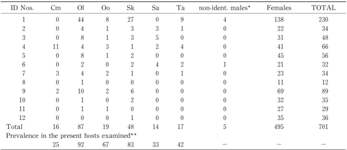
Nematode genera and species. A total of 701 nematodes recovered from Chinese Water Deer were examined in this survey. These belonged to six species,i.e. Camelostrongylus mentulatus (ab-
breviated to Cm in Table 2), Ostertagia le- ptospicularis (Ol), O. ostertagi (Oo), Skrjabinagia kolchida (Sk), Spiculopteragia asymmetrica (Sa)
and Trichostrongylus axei (Ta).
Number of worms. The number of male worms examined from each deer is shown in the Table 2. Among the worms, Ostertagia le- ptospicularis and Skrjabinagia kolchida were not
only the most numerous (total 87 and 48 individ- uals, respectively) but also the most prevalent (92% and 83%, respectively) nematode species recovered. Behind the result,there are intensity
(total 19 individuals) and prevalence (67%) of O.
ostertagi. Number and prevalence of other para- site were between 14 and 17, and between 25%
and 42%, respectively.
Ratio of Ostertagia leptospicularis :Skrjabinagia kolchida . The number of O. leptospicularis and S.
kolchida obrained from each Chinese Water Deer and thence the ratio of the two species are given in Table 3. According to the table, prevalence figures were more evenly balance at 64.6% O.
leptospicularis:35.4% S. kolchida.
Table 1 Chinese Water Deer examined at Whipsnade Wild Animal Park:background information.
ID No.
Clinical
No. Age Weight
(kg) Sex Found dead PM
Date remarks
1 XR47 old mature 7.6 male 28/Sep./2000 28/Sep./2000 found dead 2 XS7 young adult 6.1 male 3/Jan./2001 4/Jan./2001 found dead
3 XS16 adult 5.8 male Eu. 6 /Feb./2001 8/Feb./2001 found collapsed and very thin, euthanased 4 XS18 adult 7.5 male 12/Feb./2001 13 /Feb./2001 ⎜⎜
5 XR2 adult 8.0 male 12/Jan./2000 13/Jan./2000 found dead
6 XS6 adult 6.6 female 3/Jan./2001 4/Jan./2001 found dead with very poor condition 7 XR52 subadult 5.7 male 23/Nov. /2000 23/Nov./2000 found dead with poor condition 8 Data not available (Institute of Zoology reference no.18)
9 Data not available (Institute of Zoology reference no.19) 10 Data not available (Institute of Zoology reference no.23) 11 Data not available (Institute of Zoology reference no.25) 12 Data not available (Institute of Zoology reference no.27)
Fig.1 Diagram showing proconus (or genital cone) and Sjoberg organ in a male Skrjabinagia kolchida. Note: Sjoberʼ s organ is not presnt in Ostertagia species.
M easurements of Ostertagia spp. and Skrjabinagia kolchida . In the present study,
approximately 17.9% of male Ostertagia spp.
nematodes observed were identified as O. oster- tagi. Two Ostertagia species recorded in the present study may be differentiated by examina- tion of the proconus which is well-developed (with an acute-angled bursa; Fig.2) in the case of O.
leptospicularis and less well developed (with a
rounded bursa;Fig.5) in the case of O. ostertagi (see Fig.9). However,in this study,bursal shape and proconal size was somewhat variable. For example, eg., presences of individuals of O. le- ptospicularis with slightly lower proconus within well-developed bursa (Fig.3),or with proconus of the “ leptospicularis” type within slightly rounded bursa (Fig.4). On the other hand, there are sev- eral individuals of O. ostertagi with relatively
Table 2 Nematode examined from individual of Chinese Water Deer.
ID Nos. Cm Ol Oo Sk Sa Ta non-ident. males Females TOTAL
1 0 44 8 27 0 9 4 138 230
2 0 4 1 3 3 1 0 22 34
3 0 8 1 3 5 0 0 31 48
4 11 4 3 1 2 4 0 41 66
5 0 8 1 2 0 0 0 45 56
6 0 2 0 2 4 2 1 21 32
7 3 4 2 1 0 1 0 23 34
8 0 1 0 0 0 0 0 11 12
9 2 10 2 6 0 0 0 69 89
10 0 1 0 2 0 0 0 32 35
11 0 1 1 0 0 0 0 27 29
12 0 0 0 1 0 0 0 35 36
Total 16 87 19 48 14 17 5 495 701
Prevalence in the present hosts examined
25 92 67 83 33 42 − − −
:Ostertagia sp. including L5.
:Nematode positive number of the deer/Total number of the deer examined X 100 (%).
Abbreviations. Cm:Camelostrongylus mentulatus, Ol:Ostertagia leptospicularis, Oo:O. ostertagi, Sk:Skrjabinagia kolchida, Sa:
Spiculopteragia asymmetrica, Ta:Trichostrongylus axei.
Table 3 Ratio of Ostertagia leptospicularis and Skrjabinagia kolchida obtained from individual Chinese Water Deer.
ID Nos. Ol Sk Ol+Sk
1 44 (62.0%) 27 (38.0%) 71
2 4 (57.1%) 3 (42.9%) 7
3 8 (72.7%) 3 (27.3%) 11
4 4 (80.0%) 1 (20.0%) 5
5 8 (80.0%) 2 (20.0%) 10
6 2 (50.0%) 2 (50.0%) 4
7 4 (80.0%) 1 (20.0%) 5
8 1 (100.0%) 0 (0.0%) 1
9 10 (62.5%) 6 (37.5%) 16
10 1 (33.3%) 2 (66.7%) 3
11 1 (100.0%) 0 (0.0%) 1
12 0 (0.0%) 1 (100.0%) 1
Range 0-44 0-27
Mean 7.3 (64.6%) 4.0 (35.4%) 11.3 (100%)
SD 12.01 7.42
SEM 12.01 /3.46=3.47 7.42/3.46=2.14 95%CI 0.5-14.1 -0.19-8.19
developed proconus (Fig.6) and
/or bursal lobe (Fig.7). Hence, the measurements of height of proconus and bursa with body, spicule and oe- sophagus of Ostertagia spp.including Skrjabinagia kolchida ,which is considered as morphotype of O.
leptospicularis were done.
Each measurement was shown in appendixes 1 and 2,and its statistical comparisons and relation- ship between the data were made as shown in the Table 4 and Figures 10
‑14. In general, height of proconus of O. leptospicularis was larger than one of O. ostertagia ,although there is overlap between both ranges (Tab.4, Figs.10 and 15). Bursal height is almost constant in Ostertagia spp., although bursa of Skrjabinagia kolchida is slightly
larger than ones of Ostertagia spp. (Tab.4, Fig.
12). Skrjabinagia kolchida is easily differenciated from Ostertagia spp. because of presence of Sjobergʼ s organ (Figs.1 and 8). And, there is a remarkable variation of the heights of genital cone and Sjobergʼ s organ. On the other hand, there is also no evident differences in the values of the measurements of body,spicule and oesoph- agus between Ostertagia spp.(Tab.4,Figs.11 and 13). However, it was cleared that the values of the measurements, especially oesophagus length and bursal height, of S. kolchida is longer and
/or larger than ones of Ostertagia spp. (Tab.4;Figs.
11
‑13).
Fig.2 Posterior extremity of male of Ostertagia le- ptospicularis,left-lateral view,showing a well- developed procpnus and acute-angled bursa ( ). Nematode ID.-No.44 (cf. appendix 1).
Fig.4 Posterior extremity of male of Ostertagia le- ptospicularis, right-lateral view, showing a well-developed procpnus and slightly rounded bursa ( ). Nematode ID.-No.3 (cf. appendix 1).
Fig.3 Posterior extremity of male of Ostertagia le- ptospicularis, right-lateral view, showing a slightly lower proconus and acute-angled bursa ( ). Nematode ID.-No.95 (cf.appendix 1).
Fig.5 Posterior extremity of male of Ostertagia ostertagi, left-lateral view, showing lower and rounded proconus, and more rounded bursa ( ). Nematode ID.-No.85 (cf. appendix 1).
Discussion
Chinese Water deer, the only species found in the genus Hydropotes, occurs in Korea and in eastern China
[26]. The head and body length is approximately 850 mm, tail length 70 mm, shoul- der height 500 mm and body weight 30kg. This cervid species lives among tall reeds and rushes along rivers and also frequents tall grass areas on mountains and cultivated fields. In the wild Chinese Water Deer have been hunted for their colostrum used in folk medicine and also because they are considered to be an agricultural pest.
This has resulted in the species being classified as near threatened by the IUCN. Fortunately,
Chinese Water Deer have also been bred success- fully in captivity,including the herd established at Whipsnade Wild Animal Park. There have been few reports describing the parasites found in Chinese Water Deer. The trematode families, Dicrocoeliidae and Paramphistomatidae, were reported in wild Chinese Water Deer in China
[35,37
]. In Britain,Corbet and Harris
[4]and Ohira et al.[27
]described finding the nematodes Camelostrongylus mentulatus, Ostertagia le-
ptospicularis, O. ostertagi, Skrjabinagia (
=Oster-tagia) kolchida, Spiculopteragia asymmetrica and Trichostrongylus axei in the abomasum, T. colu-
briformis in the small intestine and Oesophagos- tomum venulosum in the large intestine. Previ- ous work
[27]at WWAP revealed the prevalence of abomasal nematodes in 14 Chinese Water Deer to be Ostertagia leptospicularis and Skrjabinagia kolchida (43%), O. ostertagi (14%), Camelostron- gylus mentulatus, Spiculopteragia asymmetrica and Trichostrongylus axei (7%). In general,members of the genus Trichostrongylus are found in a wide range of domestic and
/or wild ungulates and
Fig.7 Posterior extremity of male of Ostertagia ostertagi,left-lateral (slightly sub-dorsal)view, showing slightly longer bursa ( ). Nematode ID.-No.6 (cf. appendix 1).
Fig.9 Comparative morphology of caudal bursas (ventral views) of Ostertagia ostertagi(a),O.
leptospicularis(c), and their hybrid (b)[34]. Fig.8 Posterior extremity of male of Skrjabinagia
kolchida, sub-ventral dorsa view. Nematode ID.-No.102 (cf. appendix 2).
Fig.6 Posterior extremity of male of of Ostertagia ostertagi, left-lateral (slightly sub-dorsal)view showing slightly developed proconus.
Nematode ID.-No.2 (cf. appendix 1).
lagomorphs (accidentally, rodents)[1, 30, 32
]though tend to be of low pathogenecity in temper- ate regions
[36].
With the exception of T. axei, the nematodes found in Chinese Water Deer belong to the sub-
family Ostertagiinae, The taxonomy and system- atics of the Ostertagiinae,which includes between 7 and 17 genera
[14]depending upon the statusafforded to strains, polymorphism, species com- plexes and hybridization
[15],is still been subject to much debate. Among them, the polymor- phism hypothesis was based on the following observations
[14, 15, 18]
: (i) pairs of male mor- photypes consistently occur together, with one constituting a “major”proportion and the other a
“minor”proportion of the combined population;
and (ii) consistent structural differences allow recognition of each of the morphological types.
In the past this led to the recognition of separate genera and species for major and minor mor- photypes. The proposal for polymorphism has been corroborated based on morphological, bio-
chemical, and molecular ground
[19‑21
].
Despite this debate, the species belonging to this subfamily seem to fall naturally into one of two groups
[6](i)Ostertagia sens.lat.,parasites of Bovidae, originating from parasites of lagomor- phs;and (ii)Spiculopteragia sens. lat.,parasites of Cervidae, originating from parasites of suids and tragulids. The ostertagiines are characterized by having a reduced buccal capsule and a well-
developed copulatory bursa in the male. Cer- vical papillae are prominent,and the synlophe
[6]is composed of a large number of cuticular ridges that are perpendicular to the body surface. The genital cone, especially when swollen, is called a
Table 4 Summary of morphological measurements of Ostertagia spp. and Skrjabinagia kolchida obtained from Chinese Water Deer.
O. ostertagi O. leptospicularis S. kolchida Body Range 4.6mm-6.9mm (n=18) 3.0mm-7.3mm (n=84) 3.8mm-7.8mm (n=47)
Mean 5.81 6.02 6.35
SD 0.826 0.911 0.995
SEM 0.19 0.10 0.15
95%CI 5.44-6.18 5.82-6.22 6.06-6.64
Spicule Range 145um-225um (n=19) 145um-190um (n=85) 140um-225um (n=48)
Mean 166.0 164.5 169.5
SD 19.55 12.90 20.4
SEM 4.40 1.40 2.94
95%CI 157.4-174.6 161.8-167.2 163.74-175.3
Oesophagus Range 500um-825um (n=18) 525um-975um (n=84) 500um-913um (n=45)
Mean 722.9 762.5 783.8
SD 102.65 73.38 78.63
SEM 24.20 8.00 11.72
95%CI 675.5-770.3 746.8-778.2 760.8-806.8
Proconus or genital coneRange 19um-33um (n=19) 28um-56um (n=85) 13um-69um (n=24)
Mean 26.9 39.3 24.4
SD 4.03 4.66 11.64
SEM 0.92 0.51 2.99
95%CI 25.1-28.7 38.3-40.3 18.5-30.26
Sjobergʼs organ Range ⎜⎜ ⎜⎜ 38um-138um (n=48)
Mean ⎜⎜ ⎜⎜ 84.5
SD ⎜⎜ ⎜⎜ 25.11
SEM ⎜⎜ ⎜⎜ 3.62
95%CI ⎜⎜ ⎜⎜ 81.4-87.6
Bursa Range 125um-175um (n=14) 125um-190um (n=79) 135um-250um (n=43)
Mean 148.5 151.5 180.5
SD 15.35 15.70 26.90
SEM 3.62 1.77 4.10
95%CI 141.4-155.6 148.0-155.0 172.5-188.5
“proconus”
[15, 21]. The lateral rays of the bursa are in a pattern of 2
‑1
‑2 or 2
‑2
‑1[6
]. Ientification of this nematode group is therefore based on the structure of the bursa,genital cone,
and spicules in males and dimensions of the oeso- phageal valve and the configuration of the synlo- phe
[6, 15, 30]in the female.
The life cycles of the ostertagiines is direct,i.e.
adult worms reside in the abomasum,embryonat- ed eggs are passed in feces, and the first to third larval stages are free-living. The infective third- stage is ensheathed, and parasitic development and the prepatent period require between 2 and 3 weeks. Early fourth-stage larvae may be retained in the abomasal mucosa for extended periods of time prior to resuming maturation to the adult stage, a phenomenon known as hypobiosis
[14].
The ostertagiines are among the most path-
ogenic of the strongyles known in ruminants
[14], the most marked changes occurring as the fifth larval stage (or young adult) emerges from the abomasal glands
[2, 31, 36]
.
Of the nematodes recorded from the Chinese Water deer, the genus Ostertagia is the most pathogenic
[2, 25, 31, 36]
. Skrjabinagia kolchida is considered a minor pathogen of cattle
[36]and,
in addition, regarded as the “minor morphotype”
of Ostertagia leptospicularis[14, 18
]and so itsoccurrence in captive or wild ruminants is of potential significance from the epidemiological point of view.
Ostergiinae other than Ostertagia and Skrjabinagia in Chinese Water deer include the following two nematodes:
(i)
.
Although this nematode has been associated with a wide range of artiodactyle hosts,especialy the Camelidae and Bovidae
[8,27,29],it has been reported in several species of Cervidae including red deer
[7, 12, 27, 29]. The nematode causes ostertagid-like lesions in the abomasum of sheep
[13]
and has been a major cause of mortality in Thomsonʼ s gazelles
[17]and blackbuck[10]in
Fig.10 Relationship between proconus (or genital cone) and bursa of Ostertagia spp. and Skrjabinagia kolchida obtained from Chinese Water Deer.
Fig.11 Relationship between body and spicule of Ostertagia spp. and Skrjabinagia kolchida obtained from Chinese Water Deer.
zoos.
(ii) .
The genus Spiculopteragia is more prevalent in wild red, fallow and sika deer although little is
known of its life-cycle and pathogenecity
[25]. S. asymmetrica has been recovered from farmed red deer in East Anglia and may cause a Type II ostertagiosis
[3].
The present study revealed that same six
Fig.12 Relationship between body and bursa of Ostertagia spp. and Skrjabinagia kolchida obtained from Chinese Water Deer.
Fig.13 Relationship between body and oesophagus of Ostertagia spp. and Skrjabinagia kolchida
obtained from Chinese Water Deer. Fig.15 Graph showing distribution of proconus height in Ostertagia ostertagia and O. le- ptospicularis from Chinese Water Deer.
Fig.14 Relationship between Sjobergʼs organ and genital cone or bursa of Skrjabinagia kolchida obtained from Chinese Water Deer.
species of nematode were recovered from the abomasa of Chinese Water Deer that were report- ed by Ohira et al.
[27]also working with Chinese Water Deer at WWAP. It is likely that O. le-
ptospicularis and its minor morphotype, S. kol- chida, were derived from other ruminabnts graz- ing at Whipsnade, rather than China, since these two parasites are confined to the western Palear- ctic and occur in cervids and bovids
[14]. Whilst O. ostertagi was identified from animals in the present study, the “minor morphotype”of this parasite, Skrjabinagia (Ostertagia) lyrata[14, 18
]was not. This may be because O. ostertagi nor- mally occurs at low levels,in both numerical and prevalence terms in wild bovid and cervid hosts
[14]or that Chinese Water Deer are not the
parasiteʼ s primary host. Other species obtained from the Chinese Water Deer in this study on, Camelostrongylus mentulatus and Trichostrongylus axei, have been associated with a wide range of not only wild but also domestic camelids and
/or bovids
[29, 30, 32]. Since both these nematodes were recorded in an earlier at WWAP by Ohira et al.
[27],both appear to be well-established in the Chinese Water Deer. Although Spiculopteragia asymmetrica was recovered by both Ohira et al.
[27]
and the present author,it was not possible to determine whether this parasite was a primary parasite of Chinese Water Deer or merely a secon-
dary (or accidental)parasite of this host species.
This parasite is regarded as a primary parasite of Palearctic cervids, such as Cervus elaphus, C.
dama, C. nippon and Capreolus capreolus by Skrjabin et al.
[30]and Hoberg et al.
[14].
In the present study, Ostertagia leptospicularis and Skrjabinagia kolchida were not only the most numerous but also the most prevalent nematode species recovered from Chinese Water Deer.
This is unlike the situation in domestic cattle( Bos taurus) where S. kolchida tends to be much less common. For example, Mulrooney et al.
[24]found S. kolchida in only 18% of Oregon calves and that the mean burden was two worms. In contrast, the mean burden and prevalence of O.
leptospicularis were 88 and 73%,respectively. S.
kolchida has therefore tended to be regarded as the minor “minor morphotype”of Ostertagia le-
ptospicularis
[14, 18]. It is remarkable that the prevalence of S. kolchida is relatively high in Chinese Water Deer in the present study;indeed,
one animal harbouring S. kolchida had no O.
leptospicularis (deer ID No.12) (Table 2). The prevalence of O. ostertagi in this study(67%)was higher than the 14% reported by Ohira et al.
[27]thouygh less than the prevalence in domestic cattle (100%)
[24]. The number and prevalence of other parasite species suggests that they are minor nematode species of the abomasum in Chinese Water Deer.
Previous work reported by Mulrooney et al.
[24]indicated that the mean prevalence of two
polymorphic species pairs was 96.1% O. le-
ptospicularis:3.9% S. kolchida and 99.7% O. oster- tagi : 0.3%. However, in this study, the corre- sponding prevalence figures were more evenly balance at 64.6% O. leptospicularis: 35.4% S.
kolchida . Clearly, the not only the intensity but also the prevalence of S. kolchida in Chinese Water Deer at WWAP is remarkable high. The factors responsible for this phenomenon require further investigation.
In the present study, approximately 17.9% of male Ostertagia spp. nematodes observed were identified as O. ostertagi according to standard identification keys
[30,34]
(Fig.9). According to these keys,the two Ostertagia species recorded in the present study may be differentiated by exami-
nation of the proconus which is well-developed (with an acute-angled bursa;Fig.2)in the case of O. leptospicularis and less well developed (with a rounded bursa;Fig.5)in the case of O. ostertagi.
However,in this study,bursal shape and proconal size of the ostertagid nematodes appeared to be somewhat variable. For example, some O. le- ptospicularis had a slightly smaller proconus contained within a well-developed bursa (Fig.3) while others had a well-developed proconus within a slightly rounded bursa (Fig.4). Con-
versely,some individual O. ostertagi had a reason-
ably well-developed proconus (Fig.6) in a more
acute-angled bursa (Fig.7). Therefore,a number
of morphological measurements were made in-
cluding the height of the proconus and the length
of the bursa,the body,spicule and oesophagus for
both Ostertagia spp.and Skrjabinagia kolchida ,the minor morphotype of O. leptospicularis
[18,19],in order to evaluate the relative value of each as identification criteria.
Proconus. According to the 95% CI,this study confirmed that the height of the proconus of O.
leptospicularis was larger than that of O. ostertagi , although there was some overlap in the range of values measured for the two worm species (Table 4, Figs.10 and 15). This morphological feature may therefore be used as an aid to the identifica-
tion of O. leptospicularis and O. ostertagi.
Bursa. Bursal length was found to be fairly constant amongst the Ostertagia spp.,though that of Skrjabinagia kolchida was slightly larger (Tab.
4,Fig.12)because there was a gap in the 95% CI between Ostertagia spp. and S. kolchida . There- fore,bursal size is of no value as an identification criterion for distinguishing between the Ostertagia species and Skrjabinagia kolchida .
Sjobergʼ s organ. Skrjabinagia kolchida was easily differentiated from the Ostertagia spp. by the presence of Sjobergʼ s organ (Figs.1 and 8)and the absence of a well-developed proconus.
Furthermore, it was evident that there was remarkable variation in the height of the genital cone and Sjobergʼ s organ within this species because the SDs were 11.64μm in the genital cone and 26.90μm in Sjobergʼ s organ, respectively
(Tab.4).
Other measurements. Conversely, there were no obvious differences in the length of the body, oesophagus or spicules between the two Ostertagia spp.becaues there were overlap between the 95%
CI in these measurements between both species (Tab.4, Figs.11 and 13). However, oesophageal and bursal length of S. kolchida were significantly longer than the corresponding values for O. le- ptospicularis and O. ostertagi.
Conclusions
Whilst the six nematode species reported from the abomasum of Chinese Water Deer were the same as those listed by Ohira et al.
[27], this is the first report of the relative numbers of each of these species in this host. Ostertagia le-
ptospicularis and Skrjabinagia kolchida were the dominant species in Chinese Water Deer both in numerical and prevalence terms. It was also remarkable that the prevalence and intensity of infection with S. kolchida was relatively high in Chinese Water Deer (64.6% O. leptospicularis : 35.4% S. kolchida) compared with the relative numbers that have been reported from domestic cattle. Precisely why there were such large num- bers is not known. This study also confirmed that measurement of the proconus could be used as a valuable criterion in the identification of O.
leptospicularis and O. ostertagi.
However, before using criterion, more investi- gation about the other host species and worms populations including experimental hybrids
[34].
The values of the other measurements,eg.,bursa, body, spicule and oesophagus between Ostertagia spp.are almost constant,but ones of Skrjabinagia kolchida were larger than ones of Ostertagia spp..
This phenomenon is interesting because S. kol- chida is the minor morphotype of O. leptospicular- is. It is remarkable that there is variation of the heights of genital cone and Sjobergʼ s organ in S.
kolchida. Both phenomena should be re- investigated with regard to several matters, eg., nematode growth, hybridizations, host species, environments and so on, in future.
Acknowledgements
I would like to thank to Drs.Mark T.Fox and Lynda M. Gibbons, Department of Infectious Disease and Pathology, the Royal Veterinary College, for their help and support during the conduct of this research project. The author wishes to thank Dr.Edmund J.Flach,Veterinary Science Group, Whipsnade Wild Animal Park, Zoological Society of London,for the donation of the present samples and post mortem records,Mr.
Terry Dennet, the Institute of Zoology, London, for assistance with photo-micrography. Mr.
Andrew Mackie, the Royal Veterinary College,
for laboratory assistance, Dr. Anthony W. Sains-
bury, London Zoo, Veterinary Science Group,
Institute of Zoology, for support during my stay
at the Institute and Prof. D. Jacobs, Department
of Infectious Disease and Pathology, the Royal
Veterinary College, for, for helping my activities at the in the Royal Veterinary College Hawk- shead Campus. I am very grateful to Rakuno Gakuen University, for providing me with the funding to stay in London between 2000 and 2001,
and to all of my family for their encouragements.
The present publication was supported by a Grant-in-Aid (Nos.18510205, 20380163) of the Ministry of the Education, Science and Culture, Japan.
References
1. Asakawa, M. and Uchikawa, K.1991.A new host and locality record for Trichostrongylus retortaeformis (Zeder, 1800) (Nematoda: Tri- chostrongyloidea: Trichostrongylidae) from the Japanese grass vole, Microtus montebelli (Milne-Edwards) (Rodentia: Microtidae) in Nagano Prefecture, Japan. J. Rakuno Ga-
kuen Univ., Nat. Sci., 16:15
‑20.
2. Bisset,S.A.,Kleinjan,E.D.and Vlassoff,A.
1984. Development of Ostertagia leptospicular- is in cattle, and the differentiation of in- fective larvae and female adults from those of O. ostertagia. Vet. Parasitol., 16:23
‑33.
3. Connan, R. M. 1991. Type II ostertagiasis in farmed red deer. Vet. Rec., 128:233
‑235.
4. Corbet, G. B.and Harris,S.1991.The Hand- book of British Mammals.3rd Ed.,Blackwell Scientific Publications, UK.
5. Durette-Desset, M.-C. 1981. A hypothesis on the systematic position of the Ostertagiinae within the Trichostrongyloidea. Par-
asitology, 82:175
‑177.
6. Durette-Desset, M.-C. 1983. The superfamily Trichostrongyloidea. (Eds. Anderson, R. C., Chabaud, A. G. and Willmott,S.).Key to the Nematodes of Vertebrates, No.10.Common- wealth Institute of Helminthology, Farnham Royal, CAB, UK.
7. Dutton, C. 1995. Gastrointestinal nematodes in exotic ungulates from the “Passage through Asia” exhibit at Whipsnade Wild Animal Park.Project Report of MSc in Wild Animal Health, Royal Veterinary College,
London.
8. Flach,E.J.1986.Gastro-intestinal parasitism
in ungulates at Edinburgh Zoo with particular reference to Camelostrongylus mentulatus infection in blackbuck. MSc thesis, Univer-
sity of Edinburgh.
9. Flach,E.J.1997.Investigation and control of gastrointestinal parasitism in zoo ungulates.
Verh. ber. Erkrg. Zootiere, 38:359‑ 365.
10. Flach, E. J. and Sewell, M. M. H. 1987.
Gastro-intestinal nematodiasis in blackbuck ( Antelope cervicapra ) at Edinburgh Zoo. J.
Zoo Anim. Med., 18:56
‑61.
11. Hastings, B. E. and Kock, R. A. 1988. A rationale for the control of nematode en- doparasitism in Bactrian camels ( Camelus bactrianus). Internatl. Symp. Dis. Zoo Wild Anim., 30:125
‑134.
12. Hernandez, S., Martinez, F., Calero, R., Moreno, T. and Navarette,I.1980.Parasitos del ciervo ( Cervua elaphus ) en Cordoba. 1.
Primera relacion. Rev. Iberica Parasitol., 40:
93
‑106.
13. Hilton,R.J.,Barker,I.K.and Rickard,M.D.
1978. Distribution and pathogenecity during development of Camelostrongylus mentulatus in the abomasum of sheep.Vet.Parasitol.,4:
231
‑242.
14. Hoberg, E. P., Kocan, A. A., and Rickard,L.
G. 2001. Gastrointestinal strongyles in wild ruminants.In (Eds.Samuel,W.M.,Pybus,M.
J., and Kocan, A. A.) Parasitic Diseases of Wild Mammals.Iowa State University,USA:
209‑ 213.
15. Jansen, J. and Gibbons, L. M. (Eds.). 1981.
Workshop no.14.Systematics and biology of Ostertagia sens. lat. (Nematoda:Trichostron-
gylidae). Parasitology, 82:175
‑189.
16. Kock, R. A. 1984. Review of nematode para- sites and anthelmintic usage in ungulates at Whipsnade Zoo, the Zoological Society of London. Bri. Vet. Zool. Soc. Newsletter and Summary of papers, Mar. 1984, 17:7
‑11.
17. Kock, R. A. 1986. Enteric nematode infesta- tions in Thomsonʼ s gazelles, Gazella thom- soni, at Whipsnade Park, the Zoological Society of London.J.Zoo Anim.Med.,17:61
‑64.
18. Lancaster, M. B., Hong, C. and Michel, J. F.
1983. Polymorphism in the Trichostron- gylidae.In (Eds.Stone,A.R.,Platt,H.M.and Khalil, L. F.)Concepts in Namatode System-
atics, Systematics Association Special Vol- ume, No.22, Academic Press, UK:293
‑302.
19. Lichtenfels,J.R.and Hoberg,E.P.1993.The systematics of nematodes that cause oster- tagiasis in domestic and wild ruminants in North America: an update and a key to species. Vet. Parasitol., 46:33
‑53.
20. Lichtenfels,J.R.,Hoberg,E.P.and Zarlenga, D. S. 1997. Systematics of gastrointestinal nematodes of domestic ruminants: advances between 1992 and 1995 and proposals for future research. Vet. Parasitol., 72:225
‑238.
21. Lichtenfels,J.R.,Pilitt,P.A.and Lancaster, M. B. 1988. Systematics of the nematodes that cause ostertagiasis in cattle, sheep and goats in North America. Vet. Parasitol., 27:
3
‑12.
22. Manton, V. J. A. 1971. Some problems in the control of intestinal parasites. Internatl.
Symp. Dis. Zoo Wild Anim., 13:159‑ 162.
23. Mikolon, A. B., Boyce, W. M., Allen, J. L., Gardner, I. A. and Elliott, L. F. 1994.
Epidemiology and control of nematode para- sites in a collection of captive ungulates. J.
Zoo Wildl. Med., 25:500
‑510.
24. Mulrooney,D.M.,Bishop,J.K.and Zimmer- man, G. L. 1991. First report of Ostertagia leptospicularis (Nematoda: Trichostron- gyloidea) in calves ( Bos taurus) from North America. J. Helminthol. Soc. Wash., 58:260
‑262.
25. Munro, R. 1994. Gastro-intestinal parasites.
In (Eds. Alexander, T. L. and Buxton, D) Management and Diseases of Deer, 2nd Ed., A Veterinary Deer Society Publication, UK:
126
‑128.
26. Nowak, R. 1999. Chinese Water Deer. In Walkers Mammals of the World, Vol.2. 6th ed.. The John Hopkins University Press,
USA:1093
‑1094.
27. Ohira, H., Flach, E. J., Fox, M. T. and Gob- bons,L.M.1997.Observations on gastrointes- tinal nematode burdens of exotic ruminants at Whipsnade Wild Animal Park. Merck,
Sharp and Dohme Report:1
‑37.
28. Rickard, L. G. and Zimmerman, G. L. 1986.
First report of Ostertagia kolchida (Nematoda: Trichostrongyloidea) from North America. Proc. Helminthol. Soc.
Wash., 53:136
‑138.
29. Shiferaw Desta, F.1999.Observations on the morphology of male Camelostrongylus mentulatus in exotic ungulates at Whipsnade Wild Animal Park.Project Report of MSc in Wild Animal Health, Royal Veterinary Col-
lege, London.
30. Skrjabin, K. I., Shikhobalova, N. P., and Schultz,R.S.1954.Essentials of Nematology.
Vol.3.Trichostrongylids of animals and man.
(Ed. Skrjabin, K. I.)Academy of Sciences of the USSR,Moscow.(Translated by the Israel Programme for Scientific Translations,
1960):1
‑704.
31. Soulsby, E. J. L. 1965. Text Book of Veteri- nary Clinical Parasitology. Vol.,1, Hel- minths.Blackwell Scientific Publication,UK.
32. Soulsby,E.J.L.1982.Helminths,Arthropods and Protozoa of Domesticated Animals. 7th ed., Lea & Febiger, USA.
33. Stevenson, L. A., Gasser, R. B. and Chilton, N. B. 1996. The ITS-2rDNA of Teladorsagia circumcincta, T. trifurcata and T. davtiani (Nematoda: Trichostrongylidae) indicates that these taxa are one species. Internatl. J.
Parasitol., 26:1123
‑1126.
34. Suarez, V. H., Durette-Desset, M.-C. and Cabaret, J. 1993. Description of Ostertagia ostertagi and Ostertagia leptospicularis hybrids in experimentally infection sheep. J. Par-
asitol., 79:874
‑878.
35. Tang, C. and Jiang, C. F. 1986.On three new species of dicrocoeliid trematodes from Hubei Province,China.Acta Zootaxonomica Sin., 11:337
‑343.
36. Urquhart, G. M., Armour, J., Duncan, J. L., Dunn,A.M.and Jennings,F.W.1987.Veteri- nary Parasitology. Longman Scientific &
Technical, UK.
37. Wang, X. Y. 1979. Systematic studies on amphistomatous trematodes from China. 2.
Paramphistomatidae
[Paramphistomidae]:
Paramphistominae and Gastrothylacinae, with notes on some new species.Acta Zootax- onomica Sin., 4:327
‑338.
38. Zarlenga, D. S., Barry, C. M, Gasbarre, L. C.
and Boyd P. C.2001.A multiplex PCR assay for differentiating economically important gastrointestinal nematodes of cattle. Vet.
Parasitol., 97:201
‑211.
要 旨
英国ウイップスネード野生動物公園内に生息するシ カ科動物キバノロ(Hydropotes inermis)オステルタ ジア亜科雄線虫の形態学的検討
シカ科動物キ バ ノ ロ(Hydropotes inermis)は 中 国・朝鮮半島の湿原地帯に自然分布するが,ロンドン 動物学会のウイップスネード野生動物公園(ロンド ン北方約 40
km
のイングラント地方に所在)内にも 半野生下の状態で多数生息する。本研究はこのキバ ノロの第4胃に寄生するオステルタジア亜科線虫(家畜で病原性の高い種を含む)について検討した。
検 討 項 目 は 全 般 的 な 線 虫 相,
Ostertagia le- ptospicularis
とSkrjabinagia kolchidaの 各 宿 主 個
体における出現数比率,交接嚢およびそのほかの雄 生殖器(proconusなど)の形態的多型を応用したオ ス テ ル ターグ 胃 虫Ostertagia ostertagi
とO. le-
ptospicularis
との簡便な鑑別法であった。今回の検討では6種(Camelostrongylus mentulatus, Oster-
tagia leptospicularis, O. ostertagi, Skrjabinagia kol-
chida, Spiculopteragia asymmetrica, Trichostron- gylus axei
),計 701個体の線虫が得られた。これらに うちO. leptospicularis
とS. kolchida
とが寄生率お よび寄生数とも他種を凌駕した。これらに次いで,オ ステルターグ胃虫が検出されたので,反芻類家畜な どへの本線虫種の媒介という側面からキバノロは警 戒すべきであることが指摘された。出現比O. le- ptospicularis:S. kolchida
は 64.6:35.4で,ほかの 反芻類における値に比して後種の出現比が高かった 点も注目された。また,Ostertagia
属2種およびS.
kolchida
(こ の 種 はO. leptospicularis
の 一mor- photypeとされる)で交接嚢など雄生殖器に大きさ
や形態に若干の変異が認められた。そこで,今後の疫 学調査において簡便な鑑別方法を確立する目的で形 態情報の整理を行うため,proconus高,交接嚢高,生
殖円錐高,Sjobergʼ s organ
高,体長,交接刺長,食道 長について測定した。これらのうち,proconus高に
関してはO. leptospicularis
の方がオステルターグ 胃虫のものより発達している傾向が認められたの で,この性質は鑑別指標としての候補となりうると 考 え ら れ た。S. kolchida
の 生 殖 円 錐 とSjobergʼ s
organ
は個体間で変異が著しいことが明らかにされた。本 論 文 は,2001年 9 月,著 者 が ロ ン ド ン 大 学
Royal Veterinary Collegeとロンドン動物学会とが
共同開講する専門職大学院Master of Science in Wild Animal Health
(野生動物医学)に在学中,学位認定のため
Royal Veterinary Collegeに提出され
たProject Report
を一部改稿したものである。Summary
This study was designed to add to our knowledge of ostertagiines of captive Chinese Water Deer ( Hydropotes inermis) focussing in particular upon two species that exhibit polymorphism in domestic ruminants because there has not been record from the host species. The objectives of the present study were to:-identify the genera and species of abomasal nematodes recovered at post mortem from Chinese Water Deer at Whipsnade Wild Animal Park,;-compare the relative numbers of Ostertagia leptospicularis and Skrjabinagia kolchida in each host;and -evaluate the measurement of the proconus as an aid to the identifica- tion of Ostertagia leptospicularis from O. ostertagi. A total of 701 nematodes recovered were examined in this survey,and these belonged to six species,i.e. Camelostrongylus mentulatus, Ostertagia leptospicularis, O.
ostertagi, Skrjabinagia kolchida, Spiculopteragia asymmetrica and Trichostrongylus axei. O. leptospicularis and
S. kolchida were not only the most numerous but also the most prevalent. Behind this,there are intensity
and prevalence of O. ostertagi. Ratio of Ostertagia leptospicularis :Skrjabinagia kolchida was more evenly
balance at 64.6% O. leptospicularis:35.4% S. kolchida . It is remarkable that the prevalence of S. kolchida
is relatively high in Chinese Water Deer. The not only the intensity but also the prevalence of S. kolchida
i n Chinese Water Deer at Whipsnade Wild Animal Park is remarkable high. The factors responsible for
this phenomenon require further investigation. In this study,bursal shape and proconal size was somewhat
variable. Hence, the measurements of height of proconus and bursa with body,spicule and oesophagus of