緒 言
解離性脳動脈瘤に関する報告は,本疾患に対する関心 の高まりと診断技術の向上に伴い増加している3,4).そ の多くは椎骨動脈の解離性動脈瘤に関する報告である.
椎骨脳底動脈以外の末梢部に発生した解離性脳動脈瘤の 報告は少なく,特にくも膜下出血(subarachnoid hemor rhage;SAH)で発症した上小脳動脈(superior cerebel lar artery;SCA)解離の報告は少ない.今回,我々は 出血源として特定困難であった破裂末梢性解離性上小脳 動脈瘤の1例を経験したので報告する.
症例呈示
患者は55歳,男性.突然の意識障害で発症し当院へ 救急搬送された.既往歴,家族歴は特記事項なし.意識 レベルは Glasgow Coma Scale(GCS)E3V2M5,命令に 従わず,四肢を非合目的に動かしていた.単純頭部 CT で脳幹前面に強い SAH および急性水頭症を認めた(Fig.
1).Hunt & Kosnik grade Ⅲ,World Federation of Neurosurgical Societies(WFNS)grade Ⅳ,Fisher group 3と診断した.引き続き施行した脳血管造影検査 では,明らかな嚢状動脈瘤を認めなかった.
症例報告
JNET 7:323-329, 2013出血源として特定困難であった破裂末梢性解離性 上小脳動脈瘤の 1 例
武井 淳1,2) 西村健吾1) 石橋敏寛1) 村山雄一1) 結城一郎1)
荒川秀樹1) 加藤正高1) 見崎孝一1) 大橋洋輝2) 常喜達裕2) 阿部俊昭2)
A case of ruptured peripheral dissecting superior cerebellar artery aneurysm for which the source of bleeding was difficult to confirm
Jun TAKEI1,2) Kengo NISHIMURA1) Toshihiro ISHIBASHI1) Yuichi MURAYAMA1) Ichiro YUKI1) Hideki ARAKAWA1) Masataka KATO1) Koichi MISAKI1) Hiroki OHASHI2) Tatsuhiro JOKI2) Toshiaki ABE2)
1)Division of Endovascular Neurosurgery, Department of Neurosurgery, Tokyo Jikei University School of Medicine 2)Department of Neurosurgery, Tokyo Jikei University School of Medicine
●Abstract●
Objective: We report a case of ruptured peripheral dissecting superior cerebellar artery (SCA) aneurysm as a rare and difficult to confirm source of subarachnoid hemorrhage (SAH).
Case presentation: A 55yearold male patient was admitted to our hospital with impaired level of consciousness. Although the patient's CT showed SAH, no intracranial arterial saccular aneurysm was seen on DSA. However, there was a suspicion of morphological changes in the lateral pontomesencephalic segment of the left SCA. An MRI 4 days after onset showed cerebellar infarction in the left SCA territory.
This raised reasonable suspicion that the SAH was caused by a ruptured dissecting SCA. On day 6 after onset, a parent artery occlusion of the left SCA was performed. After the operation a transient right conjugate gaze deviation was observed.
Conclusion: A dissecting SCA may be the possible cause of SAH. We expect that parent artery occlusion can be an effective method of management, as it is in other locations for this type of pathology.
●Key Words●
dissecting aneurysm, subarachnoid hemorrhage (SAH), superior cerebellar artery (SCA) 1)東京慈恵会医科大学付属病院 脳神経外科 脳血管内治療部
2) 同 脳神経外科
<連絡先:武井 淳 〒105-0003 東京都港区西新橋3-25-8 Email: jun.takei1986@gmail.com>
(Received July 15, 2013:Accepted September 30, 2013)
右椎骨動脈造影で左 SCA の lateral pontomesencephalic segment に径3 mm の紡錘状の拡張所見を認めたが,こ の時点で破裂部位の確定診断に至らず,急性水頭症に対 して脳室ドレナージのみを施行し待機的観察とした
(Fig. 2).第3病日に再度施行した脳血管造影検査でも,
同様の所見であった.第4病日に施行した頭部 MRI 検 査で左 SCA 領域に diffusionweighted image(DWI)で 高信号領域を認めた(Fig. 3).この時点での神経症状は,
意識レベル GCS E3V5M6,構音障害,下肢に優意な左 片側の運動失調を認めた.
以上の経過から,左 SCA の紡錘状の拡張所見は出血 および虚血を来した解離性上小脳動脈瘤と診断し,第6 病日に動脈瘤塞栓および親血管閉塞を目的とした血管内 手術を施行した.全身麻酔下に右椎骨動脈に5Fr Shuttle Sheath(Cook Medical, Bloomington, IN, USA)を留置し た.HyperForm(eV3 Covidien, Irvine, CA, USA)4 mm
×7 mm を脳底動脈に待機させ,microcatheter(Excelsior SL-10; Stryker, Kalamazoo, MI, USA)を左 SCA に誘導 した.解離部より遠位側へ microcatheter 先端を留置し たのち,Target coil 2 mm ×3 cm(Stryker)をアンカ D
A R
B
C Fig. 1
CT on admission showing subarachnoid hemorrhage predominant in the perimesencephalic cisterns.
Takei J, et al
ーとして遠位側へ留置し解離部近位側まで合計6本使用 し閉塞した(Fig. 4).血管造影で解離部より先が造影 されないことを確認し手技を終了した.術後に一過性の 右共同偏視,左方視時の水平性眼振を認めたほかは,新 たな神経脱落症状は認めなかった.第7病日に施行した 頭部 MRI で左 SCA 領域の DWI での高信号領域の拡大 を認めた(Fig. 5).
術前より認めた構音障害,左上下肢運動失調のためリ ハ ビ リ テ ー シ ョ ン を 行 い, 発 症44日 目 に modified Rankin Scale(mRS)4で転院した.発症90日の時点で は mRS 3まで改善した.
考 察
上小脳動脈瘤は全動脈瘤の1.7% といわれ,その中で も 末 梢 性 の 上 小 脳 動 脈 瘤 は0.2% と い わ れ る.Ana RodríguezHernández らの報告18)では,末梢性上小脳 動脈瘤は2789例中2例と極めて稀である18).また,末 梢性上小脳動脈瘤の形状は嚢状動脈瘤の報告例が多
い12,16).末梢性解離性上小脳動脈瘤は,脳虚血症状で発
症する例と出血形式で発症する症例とに大別されるが,
A B
Fig. 2
Vertebral angiography demonstrating dilation and stenosis of the lateral pontomesencephalic segment of the left superior cerebellar artery (SCA).
Fig. 3
An axial diffusionweighted image(DWI) MRI 4 days after onset showing infarcts in the territory of the left SCA.
今回我々が経験した SAH で発症した末梢性上小脳動脈 の解離性動脈瘤は渉猟した限り10例と極めて稀な症例 と考えられる1,6,9,10,14,15).
くも膜下出血の15-20% は初回脳血管撮影にて出血源 が特定されない17).Dalyai らは,初回脳血管撮影で出血 源が同定できなかった254例中10例(3.9%)は1週間 後の脳血管撮影で破裂脳動脈瘤が発見されたと報告して いる2).これら10例のうち末梢性脳動脈瘤は3例であ る.本症例では SAH 発症当日に施行した DSA で上小 脳動脈末梢部に pearl and string 様の血管壁不整を認め
た.後方視的には入院時の頭部 CT で左迂回槽に優位な SAH を認めるが,その時点では責任病変の確診に至ら ず待機的観察とした.発症3日目に施行した DSA でも 同様の形態異常を認めた.発症4日目に施行した頭部 MRI にて左小脳脚での高信号領域を認め,左 SCA 領域 の虚血性変化を伴っていることから,解離性上小脳動脈 瘤破裂による SAH との診断に至った.出血源不詳のく も膜下出血の要因として,本例のような末梢性解離性脳 動脈瘤を一考することは重要と考える.
上小脳動脈領域梗塞は構音障害,下肢優位の運動失調 D
A B
C Fig. 4
A:Left Vertebral angiogram (lateral view) showing distal aneurysm of the left SCA.
B:Intraaneurysmal contrast injection showing the aneurysm and the distal branches.
C, D:Lateral views after parent artery occlusion.
Takei J, et al
を特徴としており11),本症例でも同様の症状を認めた.
治療時期については,本症例では発症6日目に手術を施 行したが,自験例を含めた11例のうち発症2日以内に 手術に至ったのは3例で15),5例は発症3-10日に手術 が施行されている.このように SAH で発症した SCA 末梢部解離性動脈瘤は出血部位の確定診断が難しく,治 療が遅れる傾向にあると言える.
一般的に出血発症の解離性脳動脈瘤は急性期での再出 血の確率が高く,その場合の予後は悪いため外科的治療 が推奨されている13,19).外科的治療には開頭術による trapping,proximal clipping や血管内治療による endo vascular trapping などがある.Proximal clipping 後の術 後再出血や trapping 後の小脳および脳幹梗塞のリスク もまた報告されている4,13,19,20).今回我々は動脈瘤を含め た上小脳動脈のコイル塞栓術を選択した.
SCA hemispheric branch および marginal branch の動 脈瘤治療に関してはさまざまな報告がある.一般的に trapping および proximal ligation を施行しても神経学的 脱落症状の出現は少ないとされる5,8).その理由として,
hemispheric branch の末梢支配域と vermian branch ま たは marginal branch の末梢支配域の重複や,marginal branch と前下小脳動脈末梢との間に anastomosis の存 在7),SCA 末梢支配域に silent area が比較的多いこと8)
が考えられる.しかしながら,本症例では術後 MRI で SCA 末梢支配域に脳虚血拡大を示唆する所見を認めた.
これは SCA caudal trunk より塞栓せざるを得なかった こと,および左前下小脳動脈がやや低形成であったこと が原因の一つではないかと推測される.
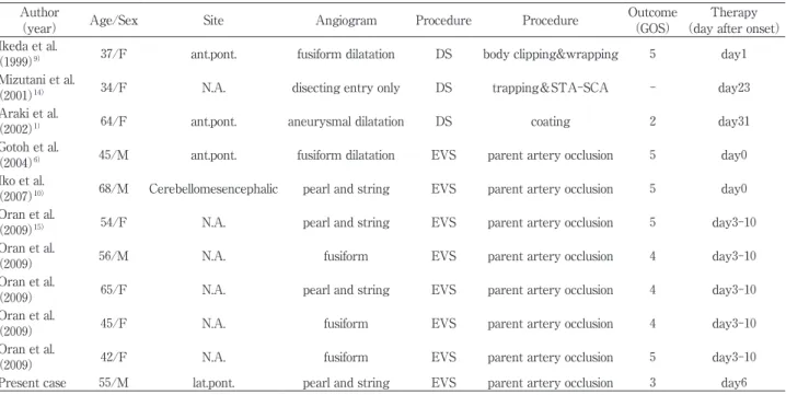
SAH で発症した上小脳動脈解離性動脈瘤の報告に関 し治療方法,予後について比較検討した(Table 1).
平均年齢は53.2歳,性別は男性4名,女性7名であった.
治療方法は直達手術例が3例,血管内治療例が8例であ った.直達手術の内訳は trapping and STA-SCA bypass 1例,body clipping and wrapping 1例,coating 1例であ った.血管内治療はすべて parent artery occlusion が行 われた.予後は Glasgow Outcome Scale(GOS)で評価 を行った.直達手術群および血管内治療群の2群に分け,
その傾向を示す(Table 2).直達手術群と比較して,
血管内治療群の予後は劣らない成績であった.本動脈瘤 に対する血管内治療の利点とは,SAH 急性期の脳腫脹 に手術手技が影響を受けないことがあげられる.一方で,
脳幹部穿通枝の確認ができないことは血管内治療の欠点 である.SCA は主幹部および rostral,caudal trunk それ ぞれの分岐部より近位に穿通枝の分枝が存在する.主幹 部では平均2本,rostral trunk からは平均5本,caudal trunk からは平均2本分岐しているとされる7).血管内 治療による parent artery occlusion の際には,これら脳 幹部穿通枝の温存のためには可能な限り末梢のみを閉塞 する工夫が必要である.
末梢部 SCA の解離性脳動脈瘤破裂による SAH に対 しては,報告例が少ないため定型的な治療方針は定まっ ていない10).現在までの報告では直達手術群および血 管内治療群ともに神経学的予後に明らかな差を認めず,
本症例のように親血管を含む動脈瘤塞栓術は有効な治療 であると考えられた.
結 語
1. 出血源として特定困難であった破裂末梢性上小脳動 脈瘤の1例を報告した.
2. 血管内治療による親血管閉塞術は本動脈瘤治療の有 用な選択肢である.
本論文に関して,開示すべき利益相反状態は存在しない.
Fig. 5
An axial postoperative DWI MRI showing expansion of infarcts in the territory of the left SCA.
文 献
1) Araki T, Fujiwara H, Murata H, et al: Subarachnoid hemorrhage due to ruptured dissecting aneurysm of peripheral superior cerebellar artery.
30:1345-1351, 2002.
2) Dalyai R, Chalouhi N, Theofanis T, et al: Subarachnoid hemorrhage with negative initial catheter angiography: a review of 254 cases evaluating patient clinical outcome and efficacy of short and Longterm repeat angiography.
72:646-652, 2013.
3) Fransen P, De Tribolet N: Dissecting aneurysm of the posterior inferior cerebellar artery. 8:381- 386, 1994.
4) Friedman AH, Drake CG: Subarachnoid hemorrhage from intracranial dissecting aneurysm. 60:325-334, 1984.
5) Fukui S, Minamida Y, Kubota T, et al: A case of peripheral, fusiform type aneurysm originating from the superior cerebellar artery. 26:163-167, 1998. 6) Gotoh H, Takahashi T, Shimizu H, et al: Dissection of the
superior cerebellar artery: a report of two cases and review of the literature. 11:196-199, 2004.
7) Hardy DG, Peace DA, Rhoton AL Jr: Microsurgical anatomy of the superior cerebellar artery.
6:10-28, 1980.
8) Hidaka M, Shimoda M, Shibuya N, et al:Peripheral Superior Cerebellar Artery Aneurysm.
( ) 29:533-537, 1989.
9) Ikeda K, Shoin K, Taguchi H, et al: Postpartum dissecting aneurysm of the superior cerebellar arterycase report.
( ) 39:852-857, 1999.
10) Iko M, Kazekawa K, Aikawa H, et al: Case of ruptured superior cerebellar artery dissection treated by endovascular embolization. 59:72-75, 2007.
11) 桑原 聡,平山恵造,小島重幸,他:上小脳動脈領域梗塞の
臨床的特徴.脳卒中 14:159-165,1992.
12) Mabuchi S, Kamiyama H, Abe H: Distal aneurysms of the superior cerebellar artery and posterior inferior cerebellar artery feeding an associated arteriovenous malformation:
case report. 30:284-287, 1992.
13) Mizutani T, Aruga T, Kirino T, et al: Recurrent subarachnoid hemorrhage from untreated ruptured vertebrobasilar dissecting aneurysms.
36:905-913, 1995.
14) Mizutani T, Kojima H, Asamoto S, et al: Pathological mechanism and threedimensional structure of cerebral dissecting aneurysms. 94:712-717, 2001. 15) Oran I, Cinar C, Yagci B, et al: Ruptured dissecting
aneurysms arising from nonvertebral arteries of the posterior circulation: endovascular treatment perspective.
15:159-165, 2009.
16) Papo I, Caruselli G, Salvolini U: Aneurysm of the superior Table 1 Clinical characteristics of eleven patients with dissecting aneurysms.
Author
(year) Age/Sex Site Angiogram Procedure Procedure Outcome
(GOS) Therapy
(day after onset)
Ikeda et al.
(1999)9) 37/F ant.pont. fusiform dilatation DS body clipping&wrapping 5 day1 Mizutani et al.
(2001)14) 34/F N.A. disecting entry only DS trapping&STA-SCA - day23
Araki et al.
(2002)1) 64/F ant.pont. aneurysmal dilatation DS coating 2 day31
Gotoh et al.
(2004)6) 45/M ant.pont. fusiform dilatation EVS parent artery occlusion 5 day0 Iko et al.
(2007)10) 68/M Cerebellomesencephalic pearl and string EVS parent artery occlusion 5 day0 Oran et al.
(2009)15) 54/F N.A. pearl and string EVS parent artery occlusion 5 day3-10
Oran et al.
(2009) 56/M N.A. fusiform EVS parent artery occlusion 4 day3-10
Oran et al.
(2009) 65/F N.A. pearl and string EVS parent artery occlusion 4 day3-10
Oran et al.
(2009) 45/F N.A. fusiform EVS parent artery occlusion 4 day3-10
Oran et al.
(2009) 42/F N.A. fusiform EVS parent artery occlusion 5 day3-10
Present case 55/M lat.pont. pearl and string EVS parent artery occlusion 3 day6
ant.pont.: anterior pontomesencephalic segment, DS: direct surgery, EVS: endovascular surgery, GOS: Glasgow Outcome Scale lat.pont.: lateral pontmesencephalic segment, N.A.: not applicable, SCA: superior cerebellar artery, STA: superficial temporal artery
Table 2 Summary of reported SCA aneurysm patients.
Direct surgery Endovascular surgery
No. of cases 3 8
Age 45.0 53.8
Sex Male 0, Female 3 Male 4, Female 4 GOS (mean) 3.5 4.4 GOS: Glasgow Outcome Scale
Takei J, et al
cerebellar artery. 7:15-17, 1977.
17) Rinkel GJ, van Gijn J, Wijdicks EF: Subarachnoid hemorrhage without detectable aneurysm. A review of the causes. 24:1403-1409, 1993.
18) RodríguezHernández A, Zador Z, Rodríguez Mena R, et al: Distal aneurysms of intracranial arteries: application of numerical nomenclature, predilection for cerebellar arteries, and results of surgical management.
80:103-112, 2012.
19) 山浦 晶,吉本高志,橋本信夫,他:非外傷性頭蓋内解離性
動脈病変の全国調査(第2報).脳卒中の外科 26:87-95, 1998.
20) Yamaura I, Tani E, Yokota M, et al: Endovascular treatment of ruptured dissecting aneurysms aimed at occlusion of the dissected site by using Guglielmi detachable coils. 90:853-856, 1999.
JNET 7:323-329, 2013
要 旨
【目的】出血源として特定困難であった破裂末梢性解離性上小脳動脈瘤の1例を認めたので報告する.【症例】55歳,
男性.意識障害で発症し,当院へ救急搬送となった.頭部 CT ではくも膜下出血を認めたが,脳血管造影検査で 主幹動脈に明らかな嚢状動脈瘤を認めなかった.左上小脳動脈末梢部に形状変化を認めたが,確定診断に至らず 待機的観察となった.発症3日後に施行した3D-DSA でも左上小脳動脈末梢部の形状変化を認め,MRI で左上小
脳動脈領域に梗塞を認めることから,左上小脳動脈解離性脳動脈瘤破裂によるくも膜下出血と判断した.発症6
日後,動脈瘤を含めた上小脳動脈の閉塞術を施行した.術後,画像上に左上小脳動脈領域脳梗塞の拡大と一過性
の右共同偏視を認めた.【結語】後頭蓋窩のくも膜下出血の原因として上小脳動脈解離を考慮する必要がある.破
裂解離性上小脳動脈瘤に対する治療として親血管を含む動脈瘤塞栓術は有効な治療であると考えられた.