MINERALOGICAL PROPERTIES OF BULALA FLINT CLAY,
CAMARINES NORTE, PHILIPPINES
著者
Mamaril-Diegor E.J., Tomita K
journal or
publication title
鹿児島大学理学部紀要. 地学・生物学
volume
22
page range
11-22
別言語のタイトル
フィリッピン, カマリネス ノルテ, ブララ産フリ
ントクレーの鉱物学的性質
URL
http://hdl.handle.net/10232/5977
MINERALOGICAL PROPERTIES OF BULALA FLINT CLAY,
CAMARINES NORTE, PHILIPPINES
著者
Mamaril-Diegor E.J., Tomita K
journal or
publication title
鹿児島大学理学部紀要. 地学・生物学
volume
22
page range
11-22
別言語のタイトル
フィリッピン, カマリネス ノルテ, ブララ産フリ
ントクレーの鉱物学的性質
URL
http://hdl.handle.net/10232/00006945
Rep. Fac. Sci., Kagoshima Univ.,(Earth Sci. & Biol.) No. 22, p.1ト22, 1989.
MINERALOGICAL PROPERTIES OF BULALA FLINT CLAY,
CAMARINES NORTE, PHILIPPINES
EJ. Mamaril-Diegor and K. Tomita*
(Received September 5, 1989)
ABSTRA CT
Mineralogical studies by means of X-ray Diffraction Analysis (XRD) , Scanning Electron Microscopy (SEM) , Infrared Absorption Spectroscopy (IR) and Differential Thermal Analysis (DTA) have been conducted on a flint clay deposit found in Barrio Bulala, Camarines Norte, Philippines. Results were obtained from raw specimen as well as on specimens heated at 550,
650, 900, 1000, 1100, 1200, 1300℃.
Based on X-ray diffraction analysis, the Bulala flint clay is mainly composed of●
welトordered kaolinite with minor amounts of alunite. SEM micrographs, DTA curves and IR spectra show that the clay is readily transformed to metakaohnite, γ -alumina, mullite and cnstobalite by heat treatment.
INTRODUCTION
Several workers have conducted studies on a flint clay deposit found in Barrio BuiAIa, Capalonga, Camarines Norte, Philippines. Miranda first reported its occurrence in 1966
during a regional mapping of Camarines Norte. A follow-up detailed geologic study was subsequently conducted by Zepeda in 1967, and he discussed the probable origin of this clay deposit (Zepeda, 1968). Caleon (1974) also undertook a geological investigation with particular interest on its geologic reserves upon a paid request by Firestone Ceramics, Inc. These previous works, however, were mainly concerned with the geology, occurrence, origin
●
and reserves with some reference on the mineralogical and chemical composition of this clay
●
deposit. In this paper, we present the recent results of x-ray powder diffraction (XRD), differential thermal analysis (DTA) , infrared absorption spectroscopy (IR) and scanning electron microscopy (SEM) studies on the changes in the mineralogical properties of this flint clay upon heat treatment. These findings are significant because they represent the first study on the thermal behavior of this type of clay which is the first of its kind in the
Philippines.
●
GEOLOGIC SETTING
The flint clay sample used in this study came from Barrio Bulala, Capalonga, Camarines
12 E.J. Mamaril-Diegor and K. Tomita
Norte, Philippines. The general geology of the Bulala area was described in detail by Zepeda (1968), Caleon (1974) and the UNDTCD (1987). The principalrock types consist of serpentinized peridotites, greenschists and agglomerate pyroclastics with intercalated tuffaceous sedimentary rocks and chert. The flint clay deposit was reported to be a product of hydrothermal alteration of the volcanic and sedimentary rocks (Zepeda, 1968). It occurs as solution fillings along fractures and alteration products derived from the aluminuGus materials of the host rocks. Veinlets of flint clay vary in thickness from a millimeter to about 40cms.
METHODOLOGY
Chips (1×lcm) and powdered specimens of the flint clay were fired using an electric furnace at temperatures of 550, 650, 900, 1000, 1100, 1200, 1300, and 1400℃ and were kept constant at their respective temperatures for at least 1 hr. An oriented specimen was also
prepared by the usual sedimentation method and was heated to 550℃ in order to determine
the effect of heat treatment on an oriented specimen of clay mineral, i.e. kaolinite. Raw and
heated powdered and oriented specimens were analyzed on a Rigaku (Geigerflex) X-ray
diffractometer using a Ni-filtered CuKa radiation at 30 kV and 15 mA. DTA curves were
obtained with a Rigaku Thermoflex apparatus at a heating rate of 10℃ per minute up t0
℃ Raw and heated chips newly broken to expose fresh, natural fracture surfaces
were used for SEM analysis in order to observe the morphological changes that took place
during the heat treatment. SEM studies were carried out on a JEOL JSM-25 scanning
electron microscope, after placing the samples in brass holders and coating them with sputtered Pd-Au for good electrical conductivity. IR analysis was conducted on a Ninon Bunko infrared spectrometer employing the potassium bromide tablet method of sample●
preparation in which a small amount of powdered specimens was mixed and ground with 300mg of potassium bromide and pressed into a disc.
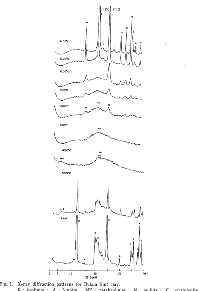
RESULTS AND DISCUSSION X-RAY DIFFRACTION DATA
X-ray diffraction patterns of raw and heated specimens of the flint clay are shown in Fig. 1. The flint clay consists dominantly of relatively well-crystallized kaolinite with minor amounts of alunite. Petrographic and x-ray studies of samples collected by Zepeda (1968) and the UNDP Group (UNDTCD, 1987) showed that other accessory minerals include quartz, magnetite, iron oxide (goethites? ) and pyrite.
Generally, kaolinite when heated t0 550℃ showed collapsed x-ray reflections. In the
case of the flint clay, heatingto this temperature indicated that only the intensity of the basal and related reflections of kaolinite were slightly reduced. Oriented specimen exhibited similar XRD pattern implying that the effect of heat treatment to 550℃ is not dependent on the orientation of the clay. At 650℃, these reflections collapsed completely and werereplaced by weak asymmetrical peaks (14A and 4Å) broadeningtoward high angle O's; the 4A peak, however, persisted to higher temperature (1200). Some strong peaks, which are
Mineralogical Properties of Bulala Flint Clay, Camarines Norte, Philippines 13
20 30 40 c 29(CuKdU
Fig. 1. x-ray diffraction patterns for Bulala flint clay.
14 E.J. Mamari卜Diegor and K. Tomita
indicated in Fig. 2, appeared at l000℃ and were determined mainly to belong to mullite.
Cristobalite, as indicated by its strongest peak (4.04A) , appeared at 1300℃. Weiトdefined
x-ray patterns for mullite and cristobalite were observed at ℃.
11000C
10000C
15 20 25 30 35 40 45 50 20(CuKa)
Fig. 2. X-ray diffraction patterns for specimens heated to 1000℃ showing mullite
(M) and γ-alumina (A) d-spacings in A.
Based on the results of the XRD studies, the behavior of kaolinite to heat treatment could be attributed to its continuous transformation from one mineral phase to another.
The slight decrease in intensity of kaolinite reflection at 550℃ suggested ・that a loss of
weight due to small amount of dehydroxylation must have occurred. Removal of water
molecules and OH ions in kaolinite was completed at 650℃ and caused a collapse in its crystal structure as reflected by the asymmetrical broad peaks at 14A and 4A. A similar observation was made by Brindley and Nakahira (1959) in their study of kaolinite. They attributed the unusual occurrence of a longer-order spacing (14Å) after heat treatment to the crystal size of and expulsion of water in kaolinite. Likewise, Hill (1955) recorded the
same result from the kaolin polymorph dickite when it was heated to 700℃. However, he
Mineralogical Properties of Bulala Flint Clay, Camarines Norte, Philippines 15
failed to obtain similar effect with kaolinite and ascribed this result to the different structure of dickite.
In his study of kaolinite and nacrite, Rinne (1924) found that the dehydration products
(metakaolin and metanacrite, respectively) above 550℃ gave weak patterns and that these
products were amorphous or mixtures of amorphous substances. By applying single crystal X-ray techniques, Brindley and Nakahira (1958) showed that kaolinite is first transformed to metakaolin and subsequently to a cubic spineトtype phase by a process of
●
orderly crystallization. According to these authors, when kaolinite is subjected to heat, the reactions are represented as :
A1203 2SiO2 2H20
2(A1203 - 2SiO2)
2A1203 3SiO23(Al203 - SiO2
925℃
℃1200℃
A1203 2SiO2 + 2H20 , metakaohn 2A1203 3SiO2 + SiO2 ● spinel phase2(A1203 SiO2) + SiO2
1 I 1 mullite cristobalite
3 A1203 2SiO2 + SiO2
3 : 2 mullite cristobalite
Tsuzuki (1961) otherwise pointed out that the metakaolin is transformed into γ -alumina having a deficient spinel structure, in lieu of a cubic A卜Si spinel phase.
● ● ∫
In the present study, neither the A卜Si spinel nor the γ -alumina were clearly observed as only x-ray powder diffraction technique was used. It is quite interesting to note here, however, that a broad peak at 1.98A (Fig. 2) was observed when the sample was heated t0
℃, which could indicate the formation of γ-alumina (Torillo et a/, 1982). Further-more, some indistinct peaks of mullite began to appear at this temperature and became enhanced at advanced temperatures. These observations could be explained by the fact that kaolinite is transformed first into metakaolin which is not wholly amorphous but still retains some crystallinity (Brindley and Nakahira, 1959). According to Tsuzuki (1961),
the packing of oxygen layers is loose but the contraction of these layers advances as the temperature rises and this probably gives impetus to the formation of a framework of oxygen
●
for 7 -alumina. Although the regular arrangement of the oxygen of the 7 -alumina follows that of metakaolin, the regularity of the atomic arrangement is still not sufficient to yield x-ray diffraction. Generally, cations migrate more easily than oxygen ions, and therefore, they are more numerous in the most suitable positions in the oxygen framework at such higher temperatures. As further migration and rearrangement of cations proceeds, almost all aluminum ions are consumed in the formation of γ-alumina, in the case of poorly crystallized kaolin minerals. But in a welトcrystallized kaolin minerals such as the kaolinite present in the flint clay, aluminum ions remain because the rearrangement and migration of ions is more difficult, and they readily combined with silicon and oxygen ions
●
forming mullite. Thus, the ratio of mullite to γ -alumina tends to increase with an increase in crystallinity of the original kaolin mineral. These conclusions made by Tsuzuki agreed with the observations of the present authors in which the 7 -alumina, as indicated by the
16 E.J. Mamari卜Diegor and K. Tomita
weak peak at 1.98A, occurs only in a relative minimal amount between 1000 and 1100℃ as
compared to that of mullite. On the other hand, the residual silicon and oxygen ions form amorphous silica (or "diffused" silica by Brindley and Nakahira) as shown by the persistence of the broad peak at 4Å. As the temperature increases, a corresponding increase in the concentration of silica occurs that facilitates the formation of cristobahte. At still higher temperature, the crystallization of mullite and cristobahte improves remarkably and is accompanied by the disappearance of the amorphous pattern. The double reflections of the former, (120) and (210), are also resolved (Fig. 1.)DIFFERENTIAL THERMAL DATA
DTA curves (Fig. 3) of the flint clay further support the observations based on the XRD results. Raw sample showed double endothermic peaks at 598℃ and 624℃. The
authors attribute this to the presence of two types of kaolin minerals. However, further
studies are needed. The small exothermic peak at 701℃ indicates the presence of alunite.
The rapid transformation of the 7 -alumina to mullite was indicated by the sharp
exothermic peak at around 977℃ and 983-985℃ for the raw and heated samples,
respectively. The difference in the exothermic peaks of the raw and heated samples could be due to the presence of impurities, i.e. alunite, that lowers the exothermic peak of the
former. Moreover, the K ions released from the destroyed alunite probably markedly
reduced the rate of formation of cristobalite from kaolinite (Holdridge, 1957).
Unfortun-ately, this could not be confirmed since the formation of cristobalite occurs above 1000℃
which is beyond the temperature range of the differential thermal analyzer used in the present study.INFRARED ABSORPTION SPECTROSCOPY DATA
The infrared spectrum of the flint clay (Fig. 4) is dominated by the absorption bands of kaolinite at 3675, 3650, 1100, 1000, 940, 905, 795, 530, 430cm 1 (Table 1). The sharp doublet at 3675 and 3650cm are assigned to OH-stretchingmodes whereas a weak band at 1625cm"1,totheOH-vibratingmode. The bands at940, 905cm arise from the vibrations of inner and inner surface OH groups, respectively (Russell, 1987). The strong broad absorption near llOOcm x and supporting bands between 400 and 800cm are due to
Si-0ト(A卜0) modes.
Heat treatment produced a number of significant changes in the spectrum. Increasing
the temperature to 550℃ brought no distinguishable changes except that the intensity of the
absortion bands was slightly reduced and the 1000cm shifted to higher wavenumber. At
650C, dehydroxylation occurred as reflected by the disappearance of all OH vibrational
modes. Furthermore, the (Si-0ト(A卜0) bands in the region between 1000-1100cm i
merged and became a broad peak′at llOOcm" ㌔ The relatively sharp peaks between400-800cm , likewise assigned to (Si-0ト(A1-0), disappeared and were replaced by a
peak at 470cm . These results suggest that the crystalline structure of the kaolinite was largely destroyed and was being replaced by a partially amorphous phase of metakaolinite.Mineralogical Properties of Bulala Flint Clay, Camarines Norte, Philippines 17
」 二.二__二_∴_.山L i- 1 6_写_旦Iー_ I . I J 100 200 300 400 500 600 700 800 900 1000
TEMPERATURE (-C)
Fig. 3. DTA curves for Bulala flint clay.
Aside from the sharpening and shifting of peaks to higher wavenumbers, a similar spectrum is observed at 900℃ As the temperature was raised to 1000℃ new broad peaks at 550
and 730cm began to appear and were enhanced at further heating, indicating that mullite
was being formed at this point. The single band at 470cm can be assigned to (A卜O卜
and (Si-0トbending modes in a random arrangement of (AIO^トand (SiO4トtetrahedra
(Freund, 1974). As the crystallization of mullite advances, silica and Al/Si ordering
causes the band to split into separate bands (470, 550 and 730cm ) which shift apart and gain sharpness at about 1300-C. A weak shoulder peak of the llOOcm absorption band
眉 目 署 日 月 ガ 日 掛 - 刀 7 割 朋 -〟 -弘 D n 止 別 訂 判 別 東 屋 一 室 u 古 川 y l 1400-C 13000C 12000C
18 E.J. Mamaril-Diegor and K. Tomita
「∵二一∴二
^ ^ ^ ^ ^ ^ ^ ^ ^ ^ ^ ^ 蝣 ^ ^ ^ ^ ^ ^ ^ ^ ^ ^ ^ ^ ^ ^ ^ ^ ^ ^ ^ ^ ^ ^ 」 ^ ^ ^ ^ ^ ^ ^ ^ ^ ^ ^ ^ ^ ^ ^ ^ ^ ^ ^ ! ^ ^ ^ ^ ^ ^ ^ ^ ^ ^ ^ ^ ^ ^ ^ ^ ^ ^ ^ ^ ^ ^ ^ ^ ^ ^ ^ ^ ^ ^ ^ ^ ^ ^ 5 ^ ^ ^ ^ ^ ^ ^ ^ ^ ^ ^ ^ ^ ^ ^ h ^ ^ ^ ^ ^ ^ ^ ^ ^ ^ ^ ^ ^ ^ ^ ^ ^ ^ ^ ^ ^ ^ ^ ^ ^ ^ ^ ^ ^ b ^ ^ ︰.日日..日日‖ 一 44 36 28 20 18 16 14 12 10 8 × 100 Wayenumbor (cm'M
Mineralogical Properties of Bulala Flint Clay, Camarines Norte, Philippines 19
Table 1. Infrared absorption data (in cm"1) of raw and heated specimens of Bulala flint clay.
Raw 3690 3650 1100 1000 940 905 795 758 700 530 470 430 550℃ 3700 1100 1040 920 795 695 540 470 430
650℃ 1100
900℃ 1100
1000℃ 1100
1100℃ 1100
1200℃ 1100
1300℃ 1170 1100
800 810 810 820 730 910-820 730 900-830 730 540 470 470 560 470 560 470 560 470 560 470 1400℃ 1170 1100 920-880 800 740 620 560 470 J(1170cm ) also started to show and was determined to be part of the spectrum of cristobalite. Additional absorption bands (620, 800cm ) of cristobalite were finally obtained at 1400℃ The results of the IR analysis confirmed those that were obtained from X-ray diffraction.
SCANNING ELECTRON MICROSCOPY DATA
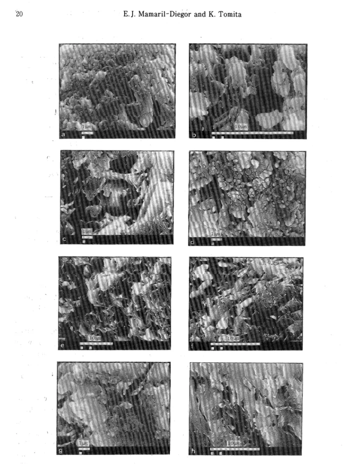
Scanning electron micrographs show that kaolinite occurs as interlocking spherical
crystals (Fig. 5a) and as hexagonal plates (Fig. 5b). By heatingto 650℃ minute fractures
within the crytals were developed that smaller, irregular grains were also noted (Fig. 5d). At increasing temperature, the subsequent loss of interlayer OH in the structure of kaolinite brought about a gradual change in morphology from individual crystals into a coalescensed mass (Fig. 5 e-h) ; but retention of some original flaky shapes of the kaolinite crystals were
still clearly visible at 1200℃ The initial development of a mosaic structure was observed at 1100℃ (Fig. 5g) which became pronounced at 1300℃ with the appearance of some rounded particles (Fig. 6). At 1400-C, a few needle-like crytals occurred in random orientation as shown in Fig. 7.
The above-observed changes in morphology with increasing temperature could be attributed to the formation of different crystal phases which was confirmed by the results of X-ray analysis. The change from kaolinite to metakaolin due to removal of structural water brought about the coalescense of individual crystals. Although the transformation of the latter to γ -alumina was not clear, it was probably indicated by the later development of
the mosaic texture. Mullite and cristobalite were hardly distinguished at lower
temperatures but rounded particles probably of cristobalite and a network of needle-like20 E.J. Mamari卜Diegor and K. Tomita
Fig. 5. Scanning electron micrographs for Bulala flint clay heated at different temperatures.
a:rawl; b:raw2; c:550℃; d:650℃; e:900℃; f:1000℃; g:1100℃;
h:1200℃.
Mineralogical Properties of Bulala Flint Clay, Camarines Norte, Philippines 21
Fig. 6. Scanning electron micrographs for Fig. 7. Scanning electron micrographs for specimen of Bulala flint clay heated specimen of Bulala flint clay heated
to 1300℃ to 1400℃ under different
magnifica-tions.
CONCLUSION
Based on the above discussion, the flint clay is composed dominantly of
welトcrystal-●
lized kaolinite with small amount of alunite (and some accessory minerals) , that responded
to heat treatment by continuously changing into new mineral phases, i.e. ka
●
metakaolinーγ -alumina + mulliteーmullite + cristobalite, as shown by XRD,
oH;
ite ー
A and
IR analyses. On the other hand, scanning electron microscopy revealed that these transformations were accompanied with corresponding changes in morphology from individuaf spherical and platy particles of kaolinite into masses of metakaolin and γ -alumina, and in turn into well-crystallized mullite and cristobalite crystals.
22 E.J. Mamaril-Diegor and K. Tomita
ACKNOWLEDGMENT
The authors wish to express their thanks to the Bureau of Mines and Geo-Sciences, Philippines for allowing us to use their facilities during the sample collection and providing
● ●
us some of the unpublished reports ; and to Conrado Miranda, Lorna Vicente, Lydia Rajas
and Bella Pandi for accomanyingthe senior author in the field. We are also grateful to the
staff of the Institute of Earth Sciences, Kagoshima University for allowing us to use their
laboratory equipment ; to Mr. Tetsuo Kobayashi of the same Institute and Professor N.
Miyauchi of the Faculty of-Agriculture for the use of the computer of the Volcanology
●
Laboratory for word processing and the infrared absorption spectrometer for analysis.
● ●
The moral support and valuable comments of Mr. Wilfredo G. Diegor, Mr. Constante B. Belandres and Mr. Laurence P. James are also gratefully acknowledged.
REFERENCES
Bates, T.F. (1971) The Kaolin Minerals : in Electron-Optical Investigation of Clays, J.A. Gard, ed., Mineralogical Society, London, 109-158.
Brindley, G.W. and Nakahira, M. (1958) New Concept of the Transformation Sequence of Kaolinite to Mullite, Nature, 181, 133-34.
ln二元「(1959) The Kaolin-Mullite Reaction Series : I, II, and HI, /. Am. Ceram. Soc., 42,
Caleon, P.C. (1975) Geologic Reserves of the Flint Clay Property of Firestone Ceramics, Inc. at Bulala, Capalonga, Camarines Norte : Unpublished Report, Bureau of Mines, Philippines, 13pp. Freud, F. (1974) Ceramics and Thermal Transformations of Minerals : in The Infrared Spectra of
Minerals, V.C. Farmer, ed., Mineralogical Society, London, 465-482.
Hill, R.D. (1955) 14 A Spacings in Kaolin Minerals Acta Crystallographies 8, 120.
Holdridge, D.A.. and Vaughan, F. (1957) The Kaolin Minerals (Kandites) : in The Differential Thermal Investigation of Clays, R.C. Mackenzie, ed., Mineralogical Society, London, 99-139.
Llave, C.A. (1971) Geological Investigation of the 13 Mining Claims (PLA-4912-D to PLA-4924-D) in Barrio Bulala, CApalonga, Camarines Norte for Firestone Cer畠mics, Incorporated : Unpublished Report, Bureau of Mines, Philippines, 7pp.
Rinne F. (1924) Rontgenographische Diagnostik beim Brennen von Kalkstein, Dolomit, Kaolin, und Glimmer Z. Krist, 61, 113-124.
Russell, J.D. (1987) Infrared Methods : in A Handbook ofDeterminative Methods in Clay Mineralogy, MJ. Wilson, ed., Blackie and Sons Ltd., London, 133-173.
Torillo, A.R. et al. (1982) Kaolin-rich Clays of the Philippines : A Critical Study of its Behavior on Heating /. Geol. Soc. Phil, 36, 32-46.
Tsuzuki, Y. (1962) Mechanism of the 980-C exotherm of kaolin minerals/. Earth Set., Nagoya Univ., 9, 305-344.
United Nations Department of Technical Cooperation for Development (1987) Geology and Mineralization in the Panganiban - Tabas and Bulala Areas, Camarines Norte, Manila,
DP/UN/PHI-85-001/1, 43pp.
Zepeda, Z.C. (1968) The Geology of the Flint Clay Deposits in Bulala, Capalonga, Camarines Norte Unpublished Report, Bureau of Mines, Philippines, 12pp.
●