九州大学学術情報リポジトリ
Kyushu University Institutional Repository
Stage IA 肺腺癌における上皮間葉転換の意義および 予後への影響について、傾向スコアマッチングを用 いての検討
松原, 太一
https://doi.org/10.15017/2556288
出版情報:Kyushu University, 2019, 博士(医学), 課程博士 バージョン:
権利関係:
Original Study
Clinical and Prognostic Signi fi cance of the Epithelial e Mesenchymal Transition in Stage IA
Lung Adenocarcinoma: A Propensity Score e Matched Analysis
Taichi Matsubara,
1Tetsuzo Tagawa,
1Kazuki Takada,
1Gouji Toyokawa,
1Mototsugu Shimokawa,
2Yuka Kozuma,
1Takaki Akamine,
1Akira Haro,
1Atsushi Osoegawa,
1Masaki Mori
1Abstract
We aimed to analyze the clinical significance of the epithelialemesenchymal transition (EMT) in stage IA lung adenocarcinoma. Tumors with the EMT phenotype were identified in 43.1% of patients with a high ratio of consolidation to tumor diameter. Propensity score matching indicated a significant association of the EMT with shorter survival.
Background:The epithelialemesenchymal transition (EMT) describes the process through which cells lose epithelial characteristics and gain a mesenchymal phenotype. The EMT contributes to tumor invasion and cancer progression, and is associated with metastasis and poor survival of patients with nonesmall-cell lung cancer. However, little is known about the relationships between the EMT and the clinicopathologic characteristics of patients with stage IA lung adenocarcinoma.Patients and Methods:We conducted immunohistochemical analysis of the expression of the EMT markers E-cadherin and vimentin of specimens acquired from 183 consecutive patients with stage IA lung adenocarcinoma. The clinicopathologic significance of the association of the EMT status with E-cadherin and vimentin expression was analyzed after propensity score matching.Results:E-cadherin and vimentin were detected in 68.3%
and 18.6% of stage IA lung adenocarcinomas, respectively. The presence of cells with EMT conversion was asso- ciated with older patient age. A propensity scoreematched cohort (128 patients) was used for further analyses.
Computed tomography revealed that tumors with EMT conversion showed solid-dominant nodules compared to those without conversion. Survival analysis after propensity score matching showed that the EMT correlated with poor disease-free survival (hazard ratio¼2.57,P¼.0451) and overall survival (hazard ratio¼4.23,P¼.0471). Multivariate analysis revealed that the EMT was an independent predictor of shorter disease-free survival.Conclusion:The EMT was a significant predictor of poor prognosis of patients with stage IA lung adenocarcinoma. The EMT status may serve as an indicator for administering adjuvant therapy.
Clinical Lung Cancer,Vol. 20, No. 4, e504-13ª2019 Elsevier Inc. All rights reserved.
Keywords: Early stage, E-cadherin, Lung cancer, Prognosis, Vimentin
Introduction
Lung cancer is the leading cause of cancer-related death world- wide, and the most common histologic lung cancer subtype is adenocarcinoma. The recent development of a multidisciplinary approach for treating lung cancer that includes surgery, radio- therapy, and chemotherapy has improved outcomes of patients with nonesmall-cell lung cancer (NSCLC). Surgical resection is the standard treatment for early-stage adenocarcinomas, particularly
1Department of Surgery and Science, Graduate School of Medical Sciences, Kyushu University, Fukuoka, Japan
2Clinical Research Institute, National Kyushu Cancer Center, Fukuoka, Japan Submitted: Nov 14, 2018; Revised: Mar 5, 2019; Accepted: Apr 12, 2019; Epub: Apr 19, 2019
Address for correspondence: Tetsuzo Tagawa, MD, PhD, Department of Surgery and Science, Graduate School of Medical Sciences, Kyushu University, Fukuoka 812-8582, Japan
Fax:þ81-92-642-5482; e-mail contact:[email protected]
e504 - Clinical Lung Cancer July 2019 1525-7304/$ - see frontmatterª2019 Elsevier Inc. All rights reserved.
https://doi.org/10.1016/j.cllc.2019.04.006
pathologic stage I lung cancer.1 Patients with pathologic stage IA NSCLC experience longer survival after complete resection2,3; however, the survival rate after recurrence is poor.4,5Therefore, it is important to identify survival-associated molecular markers for stage IA NSCLC patients.
The epithelialemesenchymal transition (EMT) describes the process through which cells down-regulate their epithelial charac- teristics and acquire a mesenchymal phenotype. The EMT is inte- gral to development, wound healing, and stem-cell behaviors and contributes tofibrosis, tumor invasion, and metastatic spread, which drive cancer progression.6,7During the EMT, epithelial cells lose junctions, reorganize their cytoskeletons, and reprogram their gene expression profiles to acquire motility an invasive phenotype.8 E-cadherin is an epithelial marker that maintains cellecell adhe- sion and inhibits cell mobility and invasiveness.9Vimentin is a type III intermediate filament that is a marker of the mesenchymal phenotype of the EMT. The down-regulation of E-cadherin expression and concomitant up-regulation of vimentin expression provide the best indicators of the EMT in carcinomas.10Although evidence indicates that the EMT is associated with metastasis and poor prognosis of certain malignant tumors, including lung can- cer,11-13 the clinical significance of EMT phenotypes of stage IA lung adenocarcinoma is unknown.
Here we investigated the expression of EMT markers of stage IA lung adenocarcinoma and applied propensity scoreematched anal- ysis to evaluate the clinical and prognostic significance of EMT status.
Patients and Methods
PatientsWe retrospectively analyzed data of consecutive patients who underwent surgical resection for primary lung adenocarcinomas at the Department of Surgery and Science, Graduate School of Medical Sciences, Kyushu University, between January 2003 and December 2012. We ultimately enrolled 183 patients who were pathologically diagnosed with stage IA lung adenocarcinomas and had available paraffin-embedded specimens. Pathologic tumor stage was defined according to the criteria of the 7th edition of the In- ternational Association for the Study of Lung Cancer staging sys- tem. We examined the clinicopathologic features as follows: age at surgical resection, sex, smoking history, histologic growth type of adenocarcinoma, lymphovascular invasion, and epidermal growth factor receptor (EGFR) mutation status. Furthermore, we reviewed radiologicfindings acquired using computed tomography (CT) and positron-emission tomography/computed tomography (PET/CT) upon surgical resection. After surgical resection, clinical examina- tions (physical examination, blood tests including serum tumor markers, and chest radiograph) were performed at 3-month intervals for the first 3 years and twice a year thereafter. Chest and upper abdominal CT was performed twice a year for thefirst 3 years and annually thereafter.
We measured the greatest consolidation diameter and the entire tumor diameter, including ground glass opacity, using 2- dimensional CT scanning of 1 mm or 2 mm sections, and calculated the ratio of consolidation diameter to tumor diameter
(C/T). We acquired the standardized maximum uptake value (SUVmax) of tumors from PET/CT images to the extent possible.
Written informed consent was obtained from each patient before study inclusion. The institutional review board of Kyushu Univer- sity approved our study (No. 30-41).
Immunohistochemistry
Formalin-fixed, paraffin-embedded specimens were sectioned into 4mm thick sections, deparaffinized in xylene, and rehydrated through a graded ethanol series. Different immunohistochemical (IHC) techniques were used to detect the expression of E-cadherin and vimentin. The E-cadherin IHC protocol was as follows: (1) Target Retrieval Solution (Dako, Glostrup, Denmark) was used for antigen retrieval at 115C for 15 minutes after inhibiting endog- enous peroxidase activity with 3% hydrogen peroxidase in meth- anol. (2) Sections were incubated with an antieE-cadherin monoclonal antibody (HECD-1, 1:1000; Takara, Shiga, Japan) at 4 C overnight. (3) Immunostaining was performed using the Envision Detection System (Dako). (4) Hematoxylin was used as a counterstain.
The vimentin IHC protocol was as follows: (1) Sections were incubated for 30 minutes in 3% hydrogen peroxidase in methanol without antigen retrieval. (2) Sections were incubated with an anti- vimentin monoclonal antibody (V-9, 1:25; Dako) at room tem- perature for 60 minutes. (3) Immunostaining was performed using the Envision Detection System (Dako). (4) Hematoxylin was as a counterstain.
Evaluation of IHC Staining
The levels of E-cadherin and vimentin were independently evaluated by at least two investigators. Vimentin expression was evaluated according the cytoplasmic staining of tumor cells (TCs) because the intensity of vimentin expression appeared to be evenly distributed throughout. In this study, the positive proportion score of vimentin expression was defined as3% of stained TCs because nonspecific staining might be also considered as positive if positivity was defined as1% of TCs. E-cadherin expression was evaluated using staining intensity and the proportion of TCs as previously reported.14-16 E-cadherin staining intensities were classified as follows: 1¼weak staining, 2 ¼moderate staining, 3¼strong staining. The staining of positive TCs was expressed as percentages, and scores were assigned follows: 0¼0,þ1¼1%- 20%,þ2¼21%-40%,þ3¼41%-60%, andþ4¼61%-100%.
The scores were then multiplied to calculate a final E-cadherin staining score. Final scores of 8 were judged positive for E-cadherin expression according to the receiver operating char- acteristic curve. If the independent assessments did not agree, then the stained slides were reviewed by another investigator to achieve consensus.
Patients were then divided into subgroups according to E-cadherin and vimentin expression as follows: (1) negative for vimentin and positive for E-cadherin, (2) positive for both vimentin and E-cadherin, (3) negative for both for vimentin and E-cadherin, and (4) positive for vimentin and negative for E-cadherin. We classified the staining patterns into groups as
-
follows: E, epithelial ¼ 1; I, intermediate ¼ 2 and 3; and M, mesenchymal¼4 (Table 1).
Statistical Analysis
Continuous variables are expressed as the meanstandard de- viation, and categorical variables are expressed as numbers. We performed statistical evaluations using JMP 13 software (SAS
Institute, Cary, NC). For continuous variables, differences were evaluated using a 2-sided Student ttest. For categorical variables, statistical differences between groups were evaluated by the chi- square test or the Fisher exact test. Univariate analysis of the asso- ciations between the EMT and clinical factors was performed using logistic regression analysis. Disease-free survival (DFS) was defined as the interval between the date of surgery and the date of recurrence or death, and was censored at last follow-up. Overall survival (OS) was defined as the interval between the date of surgery and the date of death, and was censored at last follow-up. DFS and OS rates were estimated by the Kaplan-Meier method with the log-rank test.
Univariate and multivariate analyses were used to estimate the hazard ratios (HRs) for independent prognostic values via Cox proportional hazards regression models with the backward elimi- nation method. All factors assessed in the univariate analysis were included in the multivariate analysis.P< .05 was considered sta- tistically significant.
Propensity score matching was performed to reduce the bias inherent in a retrospective study. A logistic regression model was Table 1 Analysis of Expression of E-Cadherin and Vimentin
Vimentin Expression Undetected (N[149, 81.4%)
Vimentin Expression Detected (N[34, 18.6%) E-cadherin
Expression Negative (N[58, 31.7%)
I: 44 (24.0%) M: 14 (7.7%)
E-cadherin Expression Positive (N[125, 68.3%)
E: 105 (57.4%) I: 20 (10.9%)
Abbreviations: E¼epithelial; I¼intermediate; M¼mesenchymal.
Figure 1 Vimentin and E-Cadherin Expression in Lung Adenocarcinoma Specimens. (A) Results of IHC that did not Detect Vimentin Expression. (B) IHC Showing Increased Vimentin Expression, Mainly in Cytoplasm of Tumor Cells. (C) IHC Showing Reduced E-Cadherin Expression. (D) IHC Showing That E-Cadherin Expression was Mainly Detected on Surface and in Cytoplasm of Tumor Cells
Abbreviation: IHC¼immunohistochemistry.
EMT in Lung Adenocarcinoma
e506 - Clinical Lung Cancer July 2019
used to calculate the propensity score using the following variables:
age, sex, predominant histologic growth type, and lymphovascular invasion. A difference in propensity score of 0.20 was adopted as the maximum caliper width.
Results
Patient Characteristics
The clinicopathologic characteristics of the 183 enrolled patients with pathologic stage IA lung adenocarcinoma who underwent surgical resection are shown inSupplemental Table 1in the online version. The median follow-up time was 60.3 months (range, 1.5- 139.2 months). The median age of patients was 68 years (range, 34- 85 years), 83 patients (45.3%) were men, and 88 patients (48.1%) had a history of smoking. One hundred twenty patients (65.6%) underwent lobectomy with standard dissection of the hilar and mediastinal lymph nodes.EGFRmutation status was available for 125 patients; of these, 57 (45.6%) were wild type and 68 (54.4%) harboredEGFRmutations, mainly exon 19 deletions or the L858R point mutation.
IHC analysis of TCs localized E-cadherin to the cell membrane or the cytoplasm (Figure 1AandB), and vimentin was detected in the cytoplasm (Figure 1Cand D). E-cadherin was not detectably expressed in 58 samples (31.7%), and vimentin expression was detected in 34 samples (18.6%; Table 1). We classified 105
(57.4%), 64 (34.9%), and 14 (7.7%) patients into the E, I, and M groups, respectively. The I and M groups were regarded as the EMT conversion group (I/M).
Association Between EMT Conversion and Clinicopathologic Factors of Stage IA Lung Adenocarcinoma
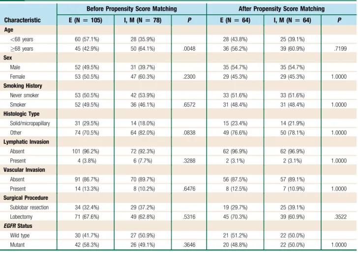
A comparison of clinicopathologic features according to EMT status is shown in Table 2. The EMT conversion occurred more often in patients68 years of age (64.1 vs. 35.9%, P¼.0048).
There were no significant differences between the EMT and other clinicopathologic factors.
Propensity score matching was performed to create a balanced cohort. The 128 matched patients were included in the propensity scoreematched analysis (64 and 64 patients from groups E and I/M, respectively). After matching, the distribution of patients’character- istics among the groups was well balanced (Table 2). There were no significant differences in sex, smoking history, histologic type, lym- phovascular invasion, andEGFRmutation status. The most variable clinical factor was the surgical procedure (P¼.3522). We used this propensity scoreematched population for further analyses.
We investigated the radiologic features of tumors associated with the EMT conversion in the propensity scoreematched cohort (Figure 2). A significantly greater C/T ratio was observed in tumors
Table 2 Associations Between EMT Phenotype and Patient Clinicopathologic Characteristics
Characteristic
Before Propensity Score Matching After Propensity Score Matching
E (N[105) I, M (N[78) P E (N[64) I, M (N[64) P
Age
<68 years 60 (57.1%) 28 (35.9%) 28 (43.8%) 25 (39.1%)
68 years 45 (42.9%) 50 (64.1%) .0048 36 (56.2%) 39 (60.9%) .7199
Sex
Male 52 (49.5%) 31 (39.7%) 35 (54.7%) 35 (54.7%)
Female 53 (50.5%) 47 (60.3%) .2300 29 (45.3%) 29 (45.3%) 1.0000
Smoking History
Never smoker 53 (50.5%) 42 (53.9%) 33 (51.6%) 33 (51.6%)
Smoker 52 (49.5%) 36 (46.1%) .6572 31 (48.4%) 31 (48.4%) 1.0000
Histologic Type
Solid/micropapillary 31 (29.5%) 14 (18.0%) 15 (23.4%) 14 (21.9%)
Other 74 (70.5%) 64 (82.0%) .0838 49 (76.6%) 50 (78.1%) 1.0000
Lymphatic Invasion
Absent 101 (96.2%) 72 (92.3%) 62 (96.9%) 62 (96.9%)
Present 4 (3.8%) 6 (7.7%) .3288 2 (3.1%) 2 (3.1%) 1.0000
Vascular Invasion
Absent 91 (86.7%) 70 (89.7%) 56 (87.5%) 57 (89.1%)
Present 14 (13.3%) 8 (10.2%) .6476 8 (12.5%) 7 (10.9%) 1.0000
Surgical Procedure
Sublobar resection 34 (32.4%) 29 (37.2%) 19 (29.7%) 25 (39.1%)
Lobectomy 71 (67.6%) 49 (62.8%) .5316 45 (70.3%) 39 (60.9%) .3522
EGFRStatus
Wild type 30 (41.7%) 27 (50.9%) 21 (51.2%) 22 (50.0%)
Mutant 42 (58.3%) 26 (49.1%) .3646 20 (48.8%) 22 (50.0%) 1.0000
Abbreviations: E¼epithelial; EGFR¼epidermal growth factor receptor; EMT¼epithelialemesenchymal transition; I¼intermediate; M¼mesenchymal.
Taichi Matsubara et al
-
with EMT conversion compared to those with the epithelial phenotype (P ¼ .046;Figure 2A). In contrast, there was no sig- nificant difference in the radiologically determined entire tumor size associated with EMT status (P¼.3253;Figure 2B. The mean value of SUVmax was 2.73 (range, 0.0-11.7) in the E group and 3.18 (range, 0.0-15.4) in the I/M group; however, the difference was not significant (P¼.3288;Figure 2C).
The cutoff value of the C/T ratio was determined using the receiver operating characteristic curve. EMT status was used as the state variable. The optimal C/T ratio cutoff value was 0.5, with an area under the curve, sensitivity, and specificity of 0.6018, 0.6719, and 0.5156, respectively (Figure 2D). Logistic regression analysis revealed that the group with high C/T ratios tended to be associated with EMT conversion (odds ratio¼1.92,P¼.0733).
Prognostic Significance of EMT Conversion for Survival of Patients With Stage IA Lung Adenocarcinoma
We assessed the significance of the associations between an EMT phenotype and patient survival after surgical resection in the
propensity scoreematched population. The EMT conversion group was significantly associated with shorter DFS and OS compared to the E group (DFS:P¼.0451, HR¼2.57; OS:P¼.0471, HR¼ 4.23; Figure 3A and B). The 5-year survival rate of the EMT conversion group was significantly shorter compared to that of the E group (89.8% vs. 98.3%, respectively).
We then analyzed the independent predictive prognostic value of variables using Cox proportional hazards models in the pro- pensity scoreematched population (Table 3). Univariate and multivariate analysis of OS using this method may be uninfor- mative because the number of events was very small. Therefore, we analyzed only DFS. Univariate analysis revealed that age68 years (HR ¼ 3.01, P ¼ .0305), male sex (HR ¼ 3.55, P¼.0062), smoking history (HR¼2.50,P¼.0454), sublobar resection (HR ¼ 2.59, P ¼ .0349), and EMT conversion (HR ¼ 2.57, P ¼ .0430) were associated with shorter DFS.
Multivariate analysis revealed that male sex and EMT conversion were independent predictors of poor DFS (HR¼3.62,P¼.0056 and HR¼2.63, P¼.0380, respectively).
Figure 2 Analyses of Association Between EMT Status and Radiologic Characteristics. (A) Tumors With EMT Conversion had Higher Preoperative C/T Compared to Those Without Conversion. CT Imaging did not Reveal a Significant Association Between EMT Status and Entire Tumor diameter (B) or SUVmaxDetermined Using PET/CT (C). (D) ROC Curve Analysis of C/T Ratio, Using EMT Status as State Variable. Optimal C/T Ratio Cutoff Value[0.5
Abbreviations: C/T¼consolidation diameter to tumor diameter; CT¼computed tomography; EMT¼epithelialemesenchymal transition; ROC¼receiver operating characteristic; SUVmax¼ standardized maximum uptake value.
EMT in Lung Adenocarcinoma
e508 - Clinical Lung Cancer July 2019
Discussion
The phenotype of the EMT facilitates tumor invasion and pro- gression, and is associated with poor survival of patients with NSCLC.13,14,17 However, the associations between the EMT and clinicopathologic factors, including survival of patients with stage IA adenocarcinomas, is unclear. The present study found that stage IA adenocarcinoma with EMT conversion, which was defined as high levels of vimentin, low levels of E-cadherin, or both, was associated with poor survival.
The classic characteristics of the EMT include loss of epithelial polarity; loss of epithelial markers such as E-cadherin, cytokeratin, and occludin; and acquisition of mesenchymal markers such as vimentin and N-cadherin.18The EMT initiates with the dissocia- tion of epithelial cellecell contacts, which include tight junctions,
adherens junctions, desmosomes, and gap junctions.7E-cadherin is cleaved and subsequently degraded during the destabilization of adherens junctions.19The dissociated epithelial cells subsequently undergo biochemical changes that generate the mesenchymal phenotype.
The mesenchymal marker vimentin is a type III intermediate filament that is expressed by mesenchymal cells of various tissues.20 Here we used E-cadherin and vimentin as epithelial and mesen- chymal markers, respectively.Table 4presents a summary of IHC studies that used E-cadherin and vimentin as markers of the EMT to investigate the effects of survival associated with the EMT.21-25 Some of these studies performed survival analysis associated with EMT as a function of the expression of E-cadherin and vimen- tin.23,24 For example, studies of the associations between the Figure 3 Kaplan-Meier Analysis of Survival Associated With EMT Status and Log-Rank Test Analysis of Propensity Scoreematched
Cohort EMT Conversion was Associated With (A) Poor DFS (HR[2.57; 95% CI, 1.03-7.25;P[.0451) and (B) OS (HR[ 4.23; 95% CI, 1.06-28.1;P[.0471) in Patients With Stage IA Lung Adenocarcinoma
Abbreviations: CI¼confidence interval; DFS¼disease-free survival; EMT¼epithelialemesenchymal transition; HR¼hazard ratio; OS¼overall survival.
Taichi Matsubara et al
-
survival of patients with NSCLC and EMT status found that the null EMT conversion group (positive E-cadherin and negative vimentin) had the best prognosis.23,24The study population of the former23included approximately 80% of patients with pathologic stage I disease, and that of latter24included approximately 20% at the same disease stage. Despite the differences in patients’ back- grounds, the results of their survival analyses are consistent with our presentfindings.
An important consideration for interpreting these results is that these previous studies included patients with an advanced stage of disease.21-25 The EMT plays a crucial role in tumor invasion, metastatic spread, and progression.6,7 Therefore, it is not unex- pected that the EMT was associated with shorter survival in survival analyses that included patients with disease at an advanced stage.
However, few studies address the prognostic significance of the EMT in patients with early-stage lung cancer.26 The study cited found that the expression of EFHD2, which is a calcium-binding protein involved in immune cell activation, promoted the EMT and significantly correlated with postsurgical recurrence of stage I lung adenocarcinoma. However, this study did not accurately assess the influence of the EMT on the prognosis of stage I lung cancer.
Here we used propensity score matching to better evaluate the effects of the EMT on the prognosis of patients with stage IA lung adenocarcinoma. Furthermore, our multivariate analysis revealed that the EMT was an independent predictor of shorter DFS (HR¼2.63, P¼.0380). Although numerous, the associations of diverse sets of prognostic clinical variables are available for stage I disease.27-30EMT status can predict DFS and OS during the early stages of disease, such as stage IA lung adenocarcinoma. The EMT has received attention as a mechanism that confers TCs with resistance to anticancer agents such as cytotoxic chemotherapy and molecularly targeted therapy.31-33The present study suggests that EMT status may serve as a novel criterion for administering adjuvant therapy to patients with stage IA lung adenocarcinoma.
Is it possible to predict tumors with EMT conversion before surgery? To answer this important question, we analyzed the pre- operative radiologic characteristics according to EMT status. We found that tumors with EMT conversion had a high C/T ratio in preoperative CT images, but no significant association was obtained using thefindings of PET/CT studies (Figure 2). Moreover, logistic regression analysis revealed that a relatively higher C/T ratio (>0.5) can serve as a predictive factor of EMT conversion of stage IA lung Table 3 Univariate and Multivariate Analyses of Disease-Free Survival in 128 Patients
Characteristic N (%)
Univariate Analysis P
Multivariate Analysis
P
HR (95% CI) HR (95% CI)
Age,68/<68 years 75/53 (58.6%/41.4%) 3.01 (1.10-10.5) .0305
Sex, male/female 70/58 (54.7%/45.3%) 3.55 (1.42-10.0) .0062 3.62 (1.45-10.2) .0056
Smoking history, smoker/never smoker 62/66 (48.4%/51.6%) 2.50 (1.02-6.67) .0454 Histologic type, solid, micropapillary/other 29/99 (22.7%/77.3%) 3.04 (0.87-19.2) .0865 Lymphatic invasion, present/absent 4/124 (3.1%/96.9%) 1.51 (0.08-7.30) .7067 Vascular invasion, present/absent 15/115 (11.7%/88.3%) 1.97 (0.56-5.39) .2585 Surgical procedure, sublobar/lobectomy 44/84 (34.4%/65.6%) 2.59 (1.07-6.43) .0349
EGFRstatus, wild type/mutant 42/43 (49.4%/50.6%) 1.91 (0.60-7.15) .2781
EMT phenotype I, M/E 64/64 (50.0%/50.0%) 2.57 (1.03-7.25) .0430 2.63 (1.05-7.43) .0380
Abbreviations: E¼epithelial; EGFR¼epidermal growth factor receptor; EMT¼epithelialemesenchymal transition; HR¼hazard ratio; I¼intermediate; M¼mesenchymal.
Table 4 Summary of IHC Studies That Aimed to Determine Association Between Survival of Patients With Lung Cancer and EMT, as Indicated by Expression of E-Cadherin and Vimentin
First Author, Journal (Year) Histologic Type No. of Patients Stage Results, HR (P) Chikaishi,Anticancer Res (2011)21 Adenocarcinoma 183 I-IIIB NS (E-cadherin, vimentin)
E-cadherin: 1.73 (.183), vimentin: 1.326 (.452)
Kong,Oncol Rep(2014)22 NSCLC NM I-IV Poor OS (E-cadherin, vimentin)
NM (.008, .002, respectively)
Sowa,Cancer Med(2015)23 Adenocarcinoma 239 I-IIIB Poor OS (E-cadherin, vimentin, and EMT status) NM (.003, .005, NM, respectively)
Zhou,World J Surg Oncol(2015)24 NSCLC 312 I-IIIA Poor Disease-specific survival (EMT status) NM (<.001)
Tsoukalas,Anticancer Res (2017)25 NSCLC 112 I-IV Poor OS (vimentin)
1.13 (.026)
Present study (2018) Adenocarcinoma 183 IA Poor DFS and OS (EMT status)
DFS: 2.57 (.0451), OS: 2.51 (.0471) Abbreviations: DFS¼disease-free survival; EMT¼epithelialemesenchymal transition; HR¼hazard ratio; IHC¼immunohistochemistry; NM¼not mentioned; NS¼not significant; NSCLC¼ nonesmall-cell lung cancer; OS¼overall survival.
EMT in Lung Adenocarcinoma
e510 - Clinical Lung Cancer July 2019
adenocarcinoma (odds ratio ¼ 1.92, P ¼ .0733). Briefly, it is possible to predict a tumor with EMT conversion to some extent by measuring the C/T ratio in preoperative CT imaging.
In lung adenocarcinomas, radiologic ground glass opacity is asso- ciated with histologically noninvasive or minimally invasive TC phenotypes,34and several reports demonstrate that a higher C/T ratio is associated with tumor invasiveness and poor prognosis.35-37For example, the risk of recurrence of clinical stage IA adenocarcinoma is indicated by a high C/T ratio (>0.5), and multivariate analysis in- dicates that sublobar pulmonary resection is an independent risk factor of DFS.34Moreover, even in stage IA lung adenocarcinoma, a high C/T ratio (particularly C/T ratios>0.5) is likely associated with the EMT conversion. Thus, as a surgical approach for such tumors, lobectomy with standard lymph node dissection may be more desir- able than limited resection. Phase 3 trials comparing lung lobectomy versus segmentectomy for lung cancers2 cm with C/T ratios>0.5 are in progress.38,39There is an association between18F-fludeox- yglucose uptake on PET and the EMT in NSCLC,40,41which was not observed here. The reason may be that both studies analyzed patients with advanced stages of disease and that we were unable to accumulate PET data from all cases in our cohort.
There are some limitations to our study. First, we retrospectively analyzed consecutive patients who underwent surgical resection, so we did not attempt a power analysis and could not perform any sample size justification. Second, we classified the EMT stage using IHC for E-cadherin and vimentin. Other transcription factors induce the EMT, such as SNAIL1/2, ZEB1/2, and TWIST.42,43It may therefore be possible to analyze the effects more precisely of the EMT on stage IA lung adenocarcinomas by evaluating these EMT-associated molecules.
Conclusion
The EMT conversion exerted a significantly unfavorable effect on the prognosis of stage IA lung adenocarcinoma patients. EMT status is important as a mechanism of resistance to anticancer drugs as well as a criterion for indicating the administration of adjuvant therapy to patients with stage IA lung adenocarcinoma.
Clinical Practice Points
The EMT is associated with tumor invasion, cancer progression, and poor survival of malignant tumors, including NSCLC.
However, the clinical and prognostic significance of the EMT in stage IA lung adenocarcinoma is unknown.
We used IHC to analyze the expression of E-cadherin and vimentin in 183 patients with stage IA lung adenocarcinoma.
Propensity score matching was used to evaluate the significance of the association of the EMT with survival.
Tumors with EMT conversion had high preoperative C/T ratios that were significantly associated with poor DFS and OS. The EMT was an independent risk factor of DFS.
Thesefindings suggest that EMT status may serve as a novel criterion for administering adjuvant therapy.
Acknowledgment
We thank the Edanz Group (www.edanzediting.com/ac) for editorial work.
Disclosure
The authors have stated that they have no conflict of interest.
Supplemental Data
Supplemental tables andfigures accompanying this article can be found in the online version athttps://doi.org/10.1016/j.cllc.2019.
04.006.
References
1.Ding N, Zhou N, Li Q, Ren G, Zhou M. Analysis of middle- and long-term efficacy of thoracoscope-assisted segmental resection of the lung on nonesmall cell lung cancer in the early stage.Oncol Lett2018; 15:3662-8.
2.Chansky K, Detterbeck FC, Nicholson AG, et al. The IASLC Lung Cancer Staging Project: external validation of the revision of the TNM stage groupings in the eighth edition of the TNM classification of lung cancer.J Thorac Oncol2017;
12:1109-21.
3.Detterbeck FC, Boffa DJ, Kim AW, Tanoue LT. The eighth edition lung cancer stage classification.Chest2017; 151:193-203.
4.Nakagawa T, Okumura N, Ohata K, Igai H, Matsuoka T, Kameyama K. Post- recurrence survival in patients with stage I nonesmall cell lung cancer. Eur J Cardiothorac Surg2008; 34:499-504.
5.Hung JJ, Jeng WJ, Chou TY, et al. Prognostic value of the new International Association for the Study of Lung Cancer/American Thoracic Society/Euro- pean Respiratory Society lung adenocarcinoma classification on death and recurrence in completely resected stage I lung adenocarcinoma.Ann Surg 2013; 258:1079-86.
6.Kalluri R, Weinberg RA. The basics of epithelialemesenchymal transition.J Clin Invest2009; 119:1420-8.
7.Lamouille S, Xu J, Derynck R. Molecular mechanisms of epithelialemesenchymal transition.Nat Rev Mol Cell Biol2014; 15:178-96.
8.Thiery JP, Acloque H, Huang RY, Nieto MA. Epithelialemesenchymal transitions in development and disease.Cell2009; 139:871-90.
9.Perl AK, Wilgenbus P, Dahl U, Semb H, Christofori G. A causal role for E-cadherin in the transition from adenoma to carcinoma.Nature1998; 392:190-3.
10.Zeisberg M, Neilson EG. Biomarkers for epithelialemesenchymal transitions.
J Clin Invest2009; 119:1429-37.
11.Baumgart E, Cohen MS, Silva Neto B, et al. Identification and prognostic sig- nificance of an epithelialemesenchymal transition expression profile in human bladder tumors.Clin Cancer Res2007; 13:1685-94.
12.Javle MM, Gibbs JF, Iwata KK, et al. Epithelialemesenchymal transition (EMT) and activated extracellular signal-regulated kinase (p-Erk) in surgically resected pancreatic cancer.Ann Surg Oncol2007; 14:3527-33.
13.Soltermann A, Tischler V, Arbogast S, et al. Prognostic significance of epithelialemesenchymal and mesenchymal-epithelial transition protein expression in nonesmall cell lung cancer.Clin Cancer Res2008; 14:7430-7.
14.Shi Y, Wu H, Zhang M, Ding L, Meng F, Fan X. Expression of the epithelialemesenchymal transition-related proteins and their clinical significance in lung adenocarcinoma.Diagn Pathol2013; 8:89.
15.Matsubara D, Kishaba Y, Yoshimoto T, et al. Immunohistochemical analysis of the expression of E-cadherin andZEB1in nonesmall cell lung cancer.Pathol Int 2014; 64:560-8.
16.Sung WJ, Park KS, Kwak SG, Hyun DS, Jang JS, Park KK. Epi- thelialemesenchymal transition in patients of pulmonary adenocarcinoma: corre- lation with cancer stem cell markers and prognosis.Int J Clin Exp Pathol2015; 8:
8997-9009.
17.Reka AK, Chen G, Jones RC, et al. Epithelialemesenchymal transition-associated secretory phenotype predicts survival in lung cancer patients.Carcinogenesis2014;
35:1292-300.
18.Thiery JP, Sleeman JP. Complex networks orchestrate epithelialemesenchymal transitions.Nat Rev Mol Cell Biol2006; 7:131-42.
19.Yilmaz M, Christofori G. EMT, the cytoskeleton, and cancer cell invasion.Cancer Metastasis Rev2009; 28:15-33.
20.Coulombe PA, Wong P. Cytoplasmic intermediatefilaments revealed as dynamic and multipurpose scaffolds.Nat Cell Biol2004; 6:699-706.
21.Chikaishi Y, Uramoto H, Tanaka F. The EMT status in the primary tumor does not predict postoperative recurrence or disease-free survival in lung adenocarci- noma.Anticancer Res2011; 31:4451-6.
22.Kong FF, Qu ZQ, Yuan HH, et al. Overexpression ofFOXM1is associated with EMT and is a predictor of poor prognosis in nonesmall cell lung cancer.Oncol Rep 2014; 31:2660-8.
23.Sowa T, Menju T, Sonobe M, et al. Association between epithelialemesenchymal transition and cancer stemness and their effect on the prognosis of lung adeno- carcinoma.Cancer Med2015; 4:1853-62.
24.Zhou L, Yu L, Wu S, Feng Z, Song W, Gong X. Clinicopathological significance ofKAI1expression and epithelialemesenchymal transition in nonesmall cell lung cancer.World J Surg Oncol2015; 13:234.
25.Tsoukalas N, Aravantinou-Fatorou E, Tolia M, et al. Epithelialemesenchymal transition in non small-cell lung cancer.Anticancer Res2017; 37:1773-8.
Taichi Matsubara et al
-
26.Fan CC, Cheng WC, Huang YC, et al. EFHD2 promotes epithelial-to- mesenchymal transition and correlates with postsurgical recurrence of stage I lung adenocarcinoma.Sci Rep2017; 7:14617.
27.Guerrera F, Errico L, Evangelista A, et al. Exploring stage I nonesmall-cell lung cancer: development of a prognostic model predicting 5-year survival after surgical resectiondagger.Eur J Cardiothorac Surg2015; 47:1037-43.
28.Yang Y, Mao Y, Yang L, et al. Prognostic factors in curatively resected pathological stage I lung adenocarcinoma.J Thorac Dis2017; 9:5267-77.
29.Tao H, Onoda H, Okabe K, Matsumoto T. The impact of coexisting lung diseases on outcomes in patients with pathological stage I nonesmall-cell lung cancer.
Interactive Cardiovasc Thorac Surg2018; 26:1009-15.
30.Toyokawa G, Yamada Y, Tagawa T, et al. Significance of spread through air spaces in resected pathological stage I lung adenocarcinoma.Ann Thorac Surg2018; 105:
1655-63.
31.Ren J, Chen Y, Song H, Chen L, Wang R. Inhibition ofZEB1reverses EMT and chemoresistance in docetaxel-resistant human lung adenocarcinoma cell line.J Cell Biochem2013; 114:1395-403.
32.Fustaino V, Presutti D, Colombo T, et al. Characterization of epithelialemesenchymal transition intermediate/hybrid phenotypes associated to resistance to EGFR inhibitors in nonesmall cell lung cancer cell lines.Oncotarget 2017; 8:103340-63.
33.Song KA, Niederst MJ, Lochmann TL, et al. Epithelial-to-mesenchymal transition antagonizes response to targeted therapies in lung cancer by suppressing BIM.Clin Cancer Res2018; 24:197-208.
34.Suzuki K, Koike T, Asakawa T, et al. A prospective radiological study of thin- section computed tomography to predict pathological noninvasiveness in periph- eral clinical IA lung cancer (Japan Clinical Oncology Group 0201).J Thorac Oncol 2011; 6:751-6.
35.Ohde Y, Nagai K, Yoshida J, et al. The proportion of consolidation to ground- glass opacity on high resolution CT is a good predictor for distinguishing the population of non-invasive peripheral adenocarcinoma.Lung Cancer2003; 42:
303-10.
36.Tsutani Y, Miyata Y, Yamanaka T, et al. Solid tumors versus mixed tumors with a ground-glass opacity component in patients with clinical stage IA lung adenocar- cinoma: prognostic comparison using high-resolution computed tomography findings.J Thorac Cardiovasc Surg2013; 146:17-23.
37.Su H, Dai C, Xie H, et al. Risk factors of recurrence in patients with clinical stage IA adenocarcinoma presented as ground-glass nodule.Clin Lung Cancer2018; 19:
e609-17.
38.Nakamura K, Saji H, Nakajima R, et al. A phase III randomized trial of lobectomy versus limited resection for small-sized peripheral nonesmall cell lung cancer (JCOG0802/WJOG4607L).Jpn J Clin Oncol2010; 40:271-4.
39.Wolf AS, Richards WG, Jaklitsch MT, et al. Lobectomy versus sublobar resection for small (2 cm or less) nonesmall cell lung cancers.Ann Thorac Surg2011; 92:
1819-23.
40.Yamamoto S, Huang D, Du L, et al. Radiogenomic analysis demonstrates asso- ciations between (18)F-fluoro-2-deoxyglucose PET, prognosis, and epithelialemesenchymal transition in nonesmall cell lung cancer.Radiology2016;
280:261-70.
41.Higashi K, Ueda Y, Shimasaki M, et al. High FDG uptake on PET is associated with negative cell-to-cell adhesion molecule E-cadherin expression in lung adenocarcinoma.Ann Nucl Med2017; 31:590-5.
42.Shih JY, Yang PC. The EMT regulator slug and lung carcinogenesis.Carcinogenesis 2011; 32:1299-304.
43.Larsen JE, Nathan V, Osborne JK, et al.ZEB1drives epithelial-to-mesenchymal transition in lung cancer.J Clin Invest2016; 126:3219-35.
EMT in Lung Adenocarcinoma
e512 - Clinical Lung Cancer July 2019
Supplemental Table 1 Characteristics of 183 Patients With Pathologic Stage IA Lung
Adenocarcinoma
Characteristic Value
Age (years) 689.0
Sex
Male 83 (45.4%)
Female 100 (54.6%)
Smoking History
Never smoker 95 (51.9%)
Smoker 88 (48.1%)
Histologic Type
Solid/micropapillary 45 (24.6%)
Other 138 (75.4%)
Lymphatic Invasion
Absent 173 (94.5%)
Present 10 (5.5%)
Vascular Invasion
Absent 161 (88.0%)
Present 22 (12.0%)
Surgical Procedure
Sublobar resection 63 (34.4%)
Lobectomy 120 (65.6%)
EGFRStatus
Wild type 57 (31.1%)
Mutant 68 (37.2%)
Unknown 58 (31.7%)
Abbreviation: EGFR¼epidermal growth factor receptor.