福島県立医科大学 学術機関リポジトリ
This document is downloaded at: 2021-11-07T23:36:11Z
Title Difficult airway management resources and capnography use in Japanese intensive care units: a nationwide cross-sectional study
Author(s) Ono, Yuko; Tanigawa, Koichi; Shinohara, Kazuaki; Yano, Tetsuhiro; Sorimachi, Kotaro; Sato, Lubna; Inokuchi, Ryota;
Shimada, Jiro; Tase, Choichiro Citation Journal of Anesthesia. 30(4): 644-652
Issue Date 2016-08
URL http://ir.fmu.ac.jp/dspace/handle/123456789/499
Rights © The Author(s) 2016. This article is distributed under the terms of the Creative Commons Attribution 4.0 International License.
DOI 10.1007/s00540-016-2176-3
Text Version publisher
DOI 10.1007/s00540-016-2176-3 ORIGINAL ARTICLE
Difficult airway management resources and capnography use in Japanese intensive care units: a nationwide cross‑sectional study
Yuko Ono
1,4· Koichi Tanigawa
2,3· Kazuaki Shinohara
4·
Tetsuhiro Yano
1· Kotaro Sorimachi
1· Lubna Sato
1· Ryota Inokuchi
5· Jiro Shimada
1· Choichiro Tase
1Received: 17 February 2016 / Accepted: 16 April 2016 / Published online: 29 April 2016
© The Author(s) 2016. This article is published with open access at Springerlink.com
Results Of the 289 ICUs, 196 (67.8 %) returned completed questionnaires. In-house backup coverage and surgical airway devices were highly available (89.3 and 95.9 %), but SGAs and dedicated DAM carts were not (60.2 and 60.7 %). The routine use of capnometry to confirm tube placement was reported by 55.6 % of the ICUs and was highest in closed ICUs (67.2 %, p = 0.03). The rate of con- tinuous capnography monitoring was also 55.6 % and was highest in academic ICUs (64.5 %, p = 0.04).
Conclusion In Japanese ICUs, SGAs and dedicated DAM carts were less available, and capnometry was not uni- versally applied either to confirm tube placement, or for continuous monitoring of ventilated patients. Our study revealed areas in need of improvement.
Keywords Airway equipment · Capnometry ·
Supraglottic airway device · Portable storage unit · Postal survey
Introduction
The number of critical care beds in Japan has been grow- ing rapidly. According to data from the Japanese Ministry of Health, Labour and Welfare, the number of intensive care unit (ICU) beds has increased by approximately 50 % in the past several years (http://www.mhlw.go.jp/file/05-Shingikai- 12404000-Hokenkyoku-Iryouka/0000101005.pdf; and http://
www.mhlw.go.jp/shingi/2009/03/dl/s0325-9k.pdf both in Japa- nese, accessed 3 March 2016). With the steep growth in critical care, endotracheal intubation (ETI) in ICUs has become much more common. However, ETI in critically ill patients is a chal- lenging procedure because these patients have very little physi- ological reserve. Severe ETI-related complications, including hypoxia, esophageal intubation, aspiration, and cardiac arrest, Abstract
Purpose The availability of difficult airway manage- ment (DAM) resources and the extent of capnometry use in Japanese intensive care units (ICUs) remained unclear.
The purpose of this study was to clarify whether: (1) DAM resources were adequate, and (2) capnometry was routinely applied in Japanese ICUs.
Methods This nationwide cross-sectional study was con- ducted from September 2015 to February 2016. All ICUs received a mailed questionnaire about their DAM resources and use of capnometry. Outcome measures were availabil- ity of: (1) 24-h in-house backup coverage; (2) a supraglot- tic airway device (SGA); (3) a dedicated DAM cart; and (4) surgical airway devices, and (5) routine use of capnometry to verify tube placement and for continuous monitoring of ventilator-dependent patients. The association between these outcomes and ICU type (academic, high-volume, closed, surgical) was also analyzed.
Electronic supplementary material The online version of this article (doi:10.1007/s00540-016-2176-3) contains supplementary material, which is available to authorized users.
* Yuko Ono
[email protected]
1
Emergency and Critical Care Medical Center,
Fukushima Medical University Hospital, 1 Hikarigaoka, Fukushima 960-1295, Japan
2
Fukushima Medical University, Fukushima, Japan
3
Fukushima Global Medical Science Center, Fukushima, Japan
4
Department of Anesthesiology, Ohta General Hospital Foundation, Ohta Nishinouchi Hospital, Koriyama, Japan
5
Department of General and Emergency Medicine, JR Tokyo
General Hospital, Tokyo, Japan
645 J Anesth (2016) 30:644–652
are likely to occur in ICUs [1–8], especially when associated with difficult airway management (DAM) [2–8]. The rate of difficult ETI in ICU settings ranges from 10 to 21 % [2–6], which is much higher than the rate in the operating room (OR) [5, 9]. Because ETI-related complications in ICUs are associ- ated with devastating outcomes [8], it has become even more important for ICUs to have proper DAM equipment, and the issue has also become more relevant for intensivists.
Immediate access to appropriate human and equip- ment resources is a vital element of DAM [8, 10–13]. The limited availability of proper DAM resources are in part responsible for severe ETI-related complications in the ICU [8, 13–15], and many authors strongly recommended that DAM resources in the ICU be the same as those used in the OR [8, 13–15]. Airway management algorithms that have been advocated by the Japanese Society of Anesthe- siologists (JSA) [10], the American Society of Anesthesi- ologists (ASA) [11], and by the Difficult Airway Society (DAS) [12], specify the standard DAM resources for the OR. We previously audited whether Japanese helicopter emergency medical services were adequately equipped regarding these recommended guidelines [16]. However, to date no objective information has been available regarding whether human and equipment resources in Japanese ICUs are compatible with established OR standards [10–12].
Verification of endotracheal tube placement is an indispen- sable part of any DAM strategy [10–12]. Capnometry is both more sensitive and more specific than auscultation alone in recognizing correct tube placement following emergency intu- bation [17–19]. In the management of ventilator-dependent patients, early recognition of endotracheal tube dislodgement and obstruction is vital, because loss of airway patency can lead to catastrophic consequences [8, 14]. Therefore, numer- ous authors, including a national survey performed in the UK, have clearly recommended continuous capnography monitor- ing from intubation to extubation in the ICU [8, 13, 14], as in the OR. However, the current use of capnography both for ver- ifying endotracheal tube placement and for continuous moni- toring of ventilated patients in ICUs in Japan remains unclear.
Therefore, in this study we investigated: (1) the availability of DAM equipment, and specialist care providers; (2) whether these resources are sufficient regarding the JSA, ASA, and DAS guidelines [10–12]; and (3) the current status regarding the use of capnometry for intubation and for continuous cap- nography monitoring of ventilated patients in Japanese ICUs.
Materials and methods Study design and sites
This nationwide cross-sectional study was conducted from September 2015 to February 2016. After approval by the
institutional review boards of Fukushima Medical Uni- versity (No. 2521), self-administered questionnaires were mailed to the directors of all ICUs (289 hospitals in 47 prefectures) registered as certified training facilities by the Japanese Society of Intensive Care Medicine (JSICM) in November, 2015. A complete list of these hospitals is available at: http://www.jsicm.org/senmon/sisetu_all.html (in Japanese, accessed 7 February 2016). The criteria for a JSICM-certified ICU include: (1) an independent, central clinical division of the facility; (2) one or more dedicated JSICM board-certified intensivists on staff; and (3) more than four critical care beds (http://www.jsicm.org/pdf/sen- mon_sinsaisoku2016.pdf, in Japanese, accessed 24 March 2016). JSICM-certified ICUs constitute approximately half of all critical care beds in Japan.
Survey items
When selecting items in the questionnaire, we referred to previous studies conducted in other countries and address- ing both similar settings (ICUs [20–24], ORs [25–28], and emergency departments [29–31],) and different settings (pre-hospital emergency medical services [32–34], and obstetrical units [35–37]).
An English version of the Japanese questionnaire used in this study is available in the supplementary mate- rial (Online Resource 1). Survey items consisted of basic information regarding the numbers of hospital beds, ICU beds, annual ICU admissions in 2014, the types of ICU, and the availability of the following materials in the ICU:
(1) direct laryngoscope and adjunct equipment (curved blade, straight blade, McCoy laryngoscope, stylet, gum elastic bougie, tube exchanger catheter, and local anes- thetic spray); (2) alternate intubation equipment (rigid video laryngoscope, flexible fiberscope, retrograde intu- bation kit, and surgical airway equipment); (3) alternate ventilation equipment [supraglottic airway device (SGA), and oral and nasal airways]; (4) capnometry; (5) a port- able packaged unit containing several DAM kits (DAM cart); and (6) neuromuscular blocking agents to facilitate endotracheal intubation and reversal agents (sugammadex, neostigmine, flumazenil, and naloxone). In this survey, we divided ICU types into (a) academic or community, (b) closed or not-closed, (c) high-volume or not, (d) emer- gency or surgical or other type including medical, mixed, and pediatric ICUs. Academic ICUs were defined as units in university-affiliated hospitals [38]. Closed ICUs were defined as units that transferred all patients to an intensive care team that directs patients’ care with primary respon- sibility for the therapeutic plan and patient care [39, 40].
Non-closed ICUs were defined as ICUs where the inten-
sive care team provides expertise via elective or manda-
tory consultation without primary responsibility for the
patient care [39, 40]. High-volume ICUs were defined as units in the upper tertile of annual patient admissions [38], and emergency ICUs were defined as units in which most patients were from an emergency room, and which were likely to receive patients suffering acute-onset medi- cal conditions, or surgical illnesses including trauma, burns, intoxication, acute coronary disease, and stroke. We included coronary care units and stroke care units in the emergency category. Surgical ICUs were defined as units in which most patients were from ORs, and which were likely to provide post-operative intensive care.
The questionnaire also asked about the availability of direct laryngoscopes and alternate ventilation equipment in assorted sizes; the product name of the rigid video laryngoscopes and SGA used was also requested. Surgical airway equipment was categorized as a cricothyroidotomy kit, or a set containing a scalpel and hemostat. If capnom- etry was available, we asked whether: (a) capnometry was used to verify tube placement (routinely, sometimes, and never) [20], and (b) whether the ICU used continuous capnography monitoring for ventilator-dependent patients (routinely, sometimes, and never) [20]. If a dedicated DAM cart was present in the ICU, we asked respondents to specify the contents. We also requested information on:
the usual number of on-duty staff ICU physician(s) dur- ing the day and overnight; whether in-house experienced (anesthetic or emergency medicine) back-up coverage can be called during overnight hours; and whether staff physicians were board-certified. We included senior resi- dents (post-graduate year 3 or more) as staff ICU physi- cians, but not junior residents (post-graduate year 1 or 2).
We deemed that “24-h in-house back-up coverage” was obtainable if: (a) two or more physicians were usually on duty, including overnight, or (b) in-house experienced back-up coverage was available overnight. Board-certified physicians were defined based on the Japanese Medical Specialty Board criteria (http://www.japan-senmon-i.jp/, in Japanese, accessed 7 February 2016). ICUs that did not respond to the initial survey were sent a repeat mailing on January 2016.
Outcome measures
Outcomes of interest in this study were availability of: (1) 24-h in-house back-up coverage, (2) an SGA, (3) a DAM cart, (4) surgical airway equipment; and routine use of: (5) capnometry to confirm ETI, and (6) continuous capnogra- phy monitoring of ventilator-dependent patients. Of these, (1)–(4) are important DAM resources commonly endorsed by the JSA, ASA, and DAS airway management guidelines [10–12]. The availability of “surgical airway equipment”
was defined as a cricothyroidotomy kit or a scalpel and hemostat, present in the ICU.
Statistical analysis
First, all survey items were evaluated using descriptive sta- tistics. Second, the association between our outcomes of interest (DAM resources and the use of capnometry), and ICU type (academic, closed, high-volume, and surgical) were analyzed using Fisher’s exact test. For this statistical evaluation, we excluded missing data and used complete data sets. All statistical analyses were performed using IBM SPSS Statistics for Windows, version 21.0 (IBM Corp., Armonk, NY, USA), and p < 0.05 was considered statistically significant.
Sample size
During the planning of this study, we performed a power analysis using G*Power 3 for Windows (Heinrich Heine University, Düsseldorf, Germany). Because no previous study, to our knowledge, has examined the association between the type of ICU and DAM resources, we assumed an effect size using Cohen’s power table (Power primer) [41]. With an effect size, “w” of 0.3 (medium size [41]), 88 samples per group (total, 176) provided 80 % power at two- tailed α of 0.05.
Results
Of the 289 Japanese ICUs, 196 returned a completed ques- tionnaire (response rate, 67.8 %). Table 1 shows the demo- graphic information of the responding ICUs. The median number of annual ICU admissions was 688 (interquartile range 530–1000, upper tertile 878); the median number of ICU beds was 10 (interquartile range 6–12). Of these, 47.4 % were academic ICUs, 33.9 % were closed, 29.5 % were surgical, and 34.7 % were emergency units.
Table 2 summarizes the intubation, alternate intubation, and alternate ventilation equipment available in Japanese ICUs. Among the ICUs that responded, a curved laryn- goscope blade and stylet were universally available, 118 (60.2 %) had an SGA, and 188 (95.9 %) possessed a surgi- cal airway device; either a cricothyroidotomy kit (84.7 %), or scalpel and hemostat (11.2 %). Dedicated DAM carts were present in 119 (60.7 %) ICUs, but the contents varied;
almost all contained rigid laryngoscope blades of variable design and size (92.4 %), and tracheal tubes of assorted sizes (93.3 %) (Table 3). Although the availability of cap- nometry was high (92.9 %), the percentage of ICUs rou- tinely using capnometry for ETI and continuous monitor- ing of ventilated patients were both 55.6 % (Table 4).
Table 5 lists the neuromuscular blocking agents
available to facilitate ETI in the responding ICUs, and
Table 6 provides information on the ICU manpower, and
647 J Anesth (2016) 30:644–652
specialty of ICU physicians. Two or more staff inten- sivists were usually on duty at 138 ICUs (70.4 %) dur- ing the day, and 68 ICUs (34.7 %) overnight. In-house skilled back-up coverage (anesthesiology or emergency medicine) was available in 107 ICUs (54.6 %) overnight.
According to our feasibility criteria, 24-h in-house back- up staff was available in 175 (89.3 %) ICUs. Among 2546 attending physicians at all ICUs, the most common board certification was emergency medicine (24.9 %), followed by anesthesiology (24.6 %), and intensive care (18.6 %).
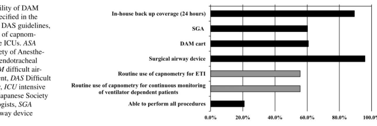
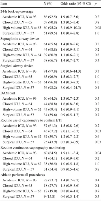
Figure 1 shows the availability of the DAM resources specified in the JSA, ASA, and DAS guidelines [10–12], and routine use of capnometry in Japanese ICUs. Accord- ing to our feasibility definitions, back-up staff was always deemed available in 89.3 % of ICUs, and surgical airway devices in 95.9 %. The feasibility of the remaining out- comes were all approximately 60 % of ICUs. There were 41 (20.9 %) facilities in which all steps were deemed achiev- able in the ICUs. Table 7 shows the associations between the feasibility of outcomes of interest and ICU type. There was a general trend that academic, closed, and high-volume ICUs were well-resourced. The rate of routine use of cap- nometry to confirm ETI was significantly higher in closed ICU [odds ratio 2.0, 95 % confidence interval (CI) 1.1–3.7, p = 0.03], but significantly lower in surgical ICUs (odds ratio 0.5, 95 % CI 0.3–0.9, p = 0.03). The percentage of routine continuous capnography monitoring of ventilated
Table 1 Demographic data of 196 Japanese intensive care units (ICUs)
Based on the replies of 196 of the 289 ICUs queried
Basic information N (inter-quartile range)
Hospital beds 613 (500–832)
ICU beds 10 (6–12)
Annual ICU admissions 688 (530–1000)
ICU type N (%)
By funding institute (N = 196)
Academic 93 (47.4)
Community 103 (52.6)
By management (N = 192)
Closed 65 (33.9)
Non-closed 127 (66.1)
By patient characteristic (N = 193)
Surgical 57 (29.5)
Emergency 67 (34.7)
Other 69 (35.8)
Table 2 Intubation equipment, alternate intubation equipment, and alternate ventilation equipment in 196 Japanese intensive care units (ICUs)
Based on the replies of 196 of the 289 ICUs queried SGA supraglottic airway device
a
ICUs may have more than one of the specified equipment items
Equipment item N (%)
1. Direct laryngoscope and adjunct equipment
aA. Curved laryngoscope blade (Macintosh type) 196 (100)
Assorted sizes 192 (98.0)
B. Straight laryngoscope blade (Miller type) 93 (47.4)
Assorted sizes 80 (40.8)
C. McCoy laryngoscope 32 (16.3)
D. Stylet 196 (100)
E. Gum elastic bougie 119 (60.7)
F. Tube exchanger catheter 154 (78.6)
G. Local anesthetic spray 156 (79.6)
2. Alternate intubation equipment
A. Rigid video laryngoscope
a165 (84.2)
a. Airway scope
®134 (68.4)
b. McGRATH MAC
®102 (52.0)
c. GlideScope
®11 (5.6)
d. C-MAC
®3 (1.5)
e. Airtraq
®2 (1.0)
f. King Vision
®1 (0.5)
g. MultiViewScope
®1 (0.5)
h. COOPDECH Video Laryngoscope
®1 (0.5)
B. Flexible fiberscope 182 (92.9)
C. Retrograde intubation kit 11 (5.6)
D. Surgical airway equipment 188 (95.9)
a. Cricothyroidotomy kit 166 (84.7)
b. Scalpel and hemostat 22 (11.2)
3. Alternate ventilation equipment
aA. SGA
a118 (60.2)
Assorted sizes 110 (56.1)
a. I-gel
®68 (34.7)
b. Air-Q
®16 (8.2)
c. LMA ProSeal
®40 (20.4)
d. LMA Classic
®28 (14.3)
e. LMA Supreme
®3 (1.5)
f. LMA Flexible
®1 (0.5)
g. Laryngeal tube
®2 (1.0)
h. Ambu AuraOnce
®2 (1.0)
i. Ambu Aura-i
®1 (0.5)
j. Combitube
®1 (0.5)
B. Oral airway 183 (93.4)
C. Nasal airway 192 (98.0)
patients was significantly higher in academic ICUs (odds ratio 1.9, 95 % CI 1.1–3.4, p = 0.04). Table 8 compares the availability of DAM resources and the use of capnometry in ICUs in Japan versus other nations.
Discussion
This national survey clarified the currently available DAM resources and extent of capnometry use in Japanese
ICUs and revealed the areas in need of improvement. To comply with current recommendations, as specified in JSA [10], ASA [11], and DAS [12] guidelines, the avail- ability of SGAs and dedicated DAM carts in Japanese
Table 3 Portable storage unit (DAM cart) and its contents in 196 Japanese intensive care units (ICUs)
Based on the replies of 196 of the 289 ICUs queried
DAM difficult airway management, SGA supraglottic airway device
Item N (%)
Portable storage unit (DAM cart) 119 (60.7)
Contents of the DAM cart N = 119
Rigid laryngoscope blades in various designs and sizes 110 (92.4)
Rigid video laryngoscope 85 (71.4)
Tracheal tubes in assorted sizes 111 (93.3)
Magill forceps 94 (79.0)
Gum elastic bougie 57 (47.9)
Tube exchanger catheter 62 (52.1)
SGA 74 (62.2)
Airway (oral/nasal) 105 (88.2)
Surgical airway device 69 (58.0)
Capnometry 36 (30.3)
Sugammadex 16 (13.4)
Bag valve mask 87 (73.1)
Yankauer suction tip 27 (22.7)
Other devices 13 (10.9)
Table 4 Frequency of using capnometry for ETI, and continuous capnography monitoring of ventilated patients in 196 Japanese inten- sive care units (ICUs)
Based on the replies of 196 of the 289 ICUs queried ETI endotracheal intubation
Item N (%)
Capnometry 182 (92.9)
Use of capnometry to confirm ETI
Routinely 109 (55.6)
Sometimes 51 (26.0)
Never 36 (18.4)
Continuous capnography monitoring of ventilated patients
Routinely 109 (55.6)
Sometimes 63 (32.1)
Never 24 (12.2)
Table 5 Neuromuscular blocking agents used to facilitate ETI, and reversal agents in 196 Japanese intensive care units (ICUs)
Based on the replies of 196 of the 289 ICUs queried
a
ICUs may have more than one drug
Item N (%)
A. Neuromuscular blocking agents
aa. Rocuronium 167 (85.2)
b. Vecuronium 68 (34.7)
c. Pancuronium 3 (1.5)
d. Succinylcholine 25 (12.8)
e. Other neuromuscular blocking agents 0 (0) B. Reversal agents
aa. Sugammadex 128 (65.3)
b. Flumazenil 124 (63.3)
c. Naloxone 94 (48.0)
d. Neostigmine 57 (29.1)
Table 6 Number of on-duty intensive care unit (ICU) physicians and their specialty
Based on the replies of 196 of the 289 ICUs queried
a
Back-up from anesthesiology or emergency department
b
Physicians may have more than one board certification
Item N (%)
Number of on-duty ICU physicians N = 196 A. Day time
a. One 58 (29.6)
b. Two or more 138 (70.4)
B. Overnight
a. One 128 (65.3)
b. Two or more 68 (34.7)
c. In-house experienced back-up coverage
aavailable 107 (54.6) Board certification of ICU physicians
bN = 2546
a. Anesthesiology 626 (24.6)
b. Emergency medicine 633 (24.9)
c. Intensive care 474 (18.6)
d. General surgery 271 (10.6)
e. Cardiovascular surgery 87 (3.4)
f. Cranial surgery 101 (4.0)
g. Orthopedics 83 (3.3)
h. Cardiovascular medicine 213 (8.4)
i. Respiratory medicine 34 (1.3)
j. Renal medicine 33 (1.3)
k. Pediatrics 101 (4.0)
l. Other board certification 158 (6.2)
649 J Anesth (2016) 30:644–652
ICUs must be improved. Capnometry is not universally used to confirm correct tube placement, nor is it being used for continuous monitoring of ventilator-dependent patients. End-tidal carbon dioxide (ETCO
2) confirmation of tube placement and the continuous monitoring of ven- tilator-dependent patients are ideal safety management practices.
Use of capnometry in Japanese intensive care units Our results showed that only 55.6 % of ICUs routinely use capnometry for ETI verification, and the same per- centage always monitor capnography in ventilator- dependent patients. However, this percentage was much higher than in previous studies conducted in other coun- tries (Table 8). Our results suggest that ETCO
2monitor- ing was successfully transferred from ORs to ICUs to a certain extent in Japan, but that there is still room for improvement.
The increased use of capnography in the ICU is the single change with the greatest potential to prevent deaths from airway complications in ICUs and elsewhere, outside the OR [14]. A national audit in the UK [8] found that fail- ure to use capnometry in treating a difficult airway contrib- uted to at least some of the fatal outcomes in ICUs. Jaber et al. [1] recently reported that after the introduction of an
“intubation bundle” including the routine use of capnome- try, ETI-related complications in critically ill patients were significantly reduced.
Displaced tracheostomy and tracheal tubes were the greatest cause of major morbidity and mortality in ICUs [8, 14]. In fact, failure to use capnography in ven- tilated patients likely contributed to more than 70 % of ICU deaths [8, 14]. Accordingly, further incorporation of ETCO
2confirmation and continuous monitoring in Japa- nese ICUs would improve patient management by critical care medical staff.
Neglect of the importance of a supraglottic airway device as a rescue ventilation device in Japanese intensive care units
In this study, an SGA was available in only 60.2 % of Japanese ICUs. In other nations, SGAs are available in 80–100 % of critical care departments (Table 8). Therefore, in Japan, SGAs have been undervalued as rescue ventila- tion devices in critical care settings. Our previous study found the same trend in the pre-hospital setting [16], which further supports this undervaluation. Each ICU must have back-up ventilation strategies [10–12] because: (1) the con- sequences of failed intubation, especially in the ICU, can be devastating [8, 14]; and (2) airway difficulties are far more likely in the ICU than in the OR [2–6]. Since the use of SGAs is well supported in rescue ventilation strategies [10–12], the standardization of airway equipment, includ- ing SGAs, would be beneficial for Japanese ICUs.
Presence of a dedicated difficult airway management cart in Japanese intensive care units
Our survey results revealed that a dedicated DAM cart was present in 60.7 % of Japanese ICUs, and that the con- tents varied considerably. This percentage is smaller than previous reports (Table 8). In an ICU, the availability of a DAM cart may have an even greater impact than in the OR, because areas outside the OR, including critical care departments, may not otherwise have immediate access to equipment for airway management [24]. Generally, time is very limited in airway management of a critically ill patient, because these patients have very little physiologi- cal reserve. Therefore, every ICU should have immediate access to at least one DAM cart [8, 10–12], which should have the same contents and layout as those used in that hos- pital’s OR [8]. Suggestions for the contents of DAM carts are in the JSA [10], ASA [11], and DAS [12] guidelines.
Fig. 1 Availability of DAM resources as specified in the JSA, ASA, and DAS guidelines, and routine use of capnom- etry in Japanese ICUs. ASA American Society of Anesthe- siologists, ETI endotracheal intubation, DAM difficult air- way management, DAS Difficult Airway Society, ICU intensive care unit, JSA Japanese Society of Anesthesiologists, SGA supraglottic airway device
0.0% 20.0% 40.0% 60.0% 80.0% 100.0%
In-house back up coverage (24 hours) SGA DAM cart Surgical airway device Routine use of capnometry for ETI Routine use of capnometry for continuous monitoring
of ventilator dependent patients Able to perform all procedures
The association between the type of intensive care unit, and the availability of difficult airway management resources and the use of capnometry
This study found a general trend showing that high-vol- ume, closed, and academic ICUs had well-prepared DAM equipment. We also noted that capnometry was more likely to be used for ETI verification in closed ICUs, and continuous capnography monitoring was more likely in
academic ICUs. It is well known that these types of ICUs have improved patient outcomes compared with other ICU types [38–40, 42, 43]. Therefore, it is possible, at least in part, that having well-prepared DAM resources is respon- sible for improved outcomes. This fact, and our data, fur- ther support current recommendations that every ICU have DAM resources at the same level as that of hospital ORs [8, 13–15]. We also observed that surgical ICUs were less likely to use capnometry for ETI verification, but we could not explain the reason for this finding, based on our survey results.
Study limitations and advantages
There are four major limitations to this study. First, we did not include non-JSICM certified ICUs, which com- prise another 50 % of all critical-care beds in Japan. This is because a complete list of non-JSICM certified ICUs was not available. However, it is likely that DAM resources are less available and capnometry is used less often in non-JSICM-certified training facilities, because most such ICUs are not academic or closed units. Our recommenda- tions regarding DAM resources and the use of capnometry can also be applied to non-JSICM-certified ICUs. Second, our survey did not determine the frequencies of difficult airways situations (i.e., cannot ventilate, cannot intubate), nor did our survey obtain information on the clinical pro- tocols for DAM in Japanese ICUs. Third, because our questionnaire was self-administered, reporting bias is pos- sible. Fourth, we did not clarify why SGAs and DAM carts were less available, nor why capnometry was under-used in Japanese ICUs. These points require further investigation.
Despite these limitations, this study has several strengths.
First, the response rate was quite high (196 of 289 surveyed ICUs responded), and our survey successfully captured the findings in various types of ICU from all geographic areas in Japan including closed, surgical, emergency, and other types. Our study provides an accurate depiction of the current state of advanced airway management in Japa- nese ICUs. Second, our findings provided the associations between ICU type, and DAM resources and the use of cap- nometry. To the best of our knowledge, this relationship has not been clarified previously. We believe that this study is a meaningful first approach to improve DAM in Japanese ICUs.
In conclusion, this nationwide cross-sectional study clarified the available DAM equipment in Japanese ICUs, as well as areas that warrant improving. Available best evi- dence clearly states that DAM resources in ICUs should be consistent with those in the OR [8, 13–15], and the use of capnometry should meet the same standards that apply in the hospital’s OR [8, 13–15]. Therefore, it would
Table 7 Association between the availability of DAM resources and the use of capnometry, and ICU type
CI confidence interval, DAM difficult airway management, ETI endotracheal intubation, ICU intensive care unit
Item N (%) Odds ratio (95 % CI) p 24-h back-up coverage
Academic ICU, N = 93 86 (92.5) 1.9 (0.7–5.0) 0.2 Closed ICU, N = 65 59 (90.8) 1.3 (0.5–3.4) 0.8 High-volume ICU, N = 63 60 (95.2) 3.1 (0.9–10.3) 0.08 Surgical ICU, N = 57 51 (89.5) 1.0 (0.4–2.8) 1.0 Supraglottic airway device
Academic ICU, N = 93 61 (65.6) 1.4 (0.8–2.6) 0.2 Closed ICU, N = 64 44 (68.8) 1.6 (0.9–3.1) 0.2 High-volume ICU, N = 64 39 (60.9) 1.1 (0.6–2.1) 0.8 Surgical ICU, N = 57 38 (66.7) 1.4 (0.7–2.7) 0.3 Surgical airway device
Academic ICU, N = 93 91 (97.8) 3.0 (0.6–14.3) 0.3 Closed ICU, N = 65 63 (96.9) 1.5 (0.3–7.7) 1.0 High-volume ICU, N = 63 61 (96.8) 1.4 (0.3–7.3) 1.0 Surgical ICU, N = 57 56 (98.2) 3.0 (0.4–24.7) 0.4 DAM cart
Academic ICU, N = 93 60 (64.5) 1.3 (0.7–2.3) 0.5 Closed ICU, N = 64 44 (68.8) 1.6 (0.8–3.0) 0.2 High-volume ICU, N = 62 43 (69.4) 1.6 (0.9–3.1) 0.2 Surgical ICU, N = 57 34 (59.6) 0.9 (0.5–1.7) 0.7 Routine use of capnometry to confirm ETI
Academic ICU, N = 93 57 (61.3) 1.5 (0.8–2.6) 0.2 Closed ICU, N = 64 43 (67.2) 2.0 (1.1–3.7) 0.03 High-volume ICU, N = 62 37 (59.7) 1.2 (0.7–2.2) 0.6 Surgical ICU, N = 57 25 (43.9) 0.5 (0.3–0.9) 0.03 Routine continuous capnography monitoring
Academic ICU, N = 93 60 (64.5) 1.9 (1.1–3.4) 0.04 Closed ICU, N = 64 41 (64.1) 1.6 (0.9–3.0) 0.2 High-volume ICU, N = 62 35 (56.5) 1.0 (0.5–1.8) 1.0 Surgical ICU, N = 57 31 (54.4) 0.9 (0.5–1.6) 0.8 Able to perform all procedures
Academic ICU, N = 93 22 (23.7) 1.4 (0.7–2.7) 0.4
Closed ICU, N = 65 18 (27.7) 1.8 (0.9–3.6) 0.1
High-volume ICU, N = 63 12 (19.0) 0.8 (0.4–1.8) 0.7
Surgical ICU, N = 57 9 (15.8) 0.6 (0.3–1.4) 0.3
651 J Anesth (2016) 30:644–652
be helpful for many Japanese ICUs to standardize DAM equipment, including an SGA and a DAM cart, and to incorporate the routine use of capnometry into their clini- cal practices.
Acknowledgments We thank all of the participating ICUs for their earnest and generous cooperation in this project. We also thank the Japanese Ministry of Health, Labour and Welfare for kindly providing essential data on the trends in the number of ICU beds in Japanese ICUs. Finally, we thank Nozomi Ono, M.D. (Department of Psy- chiatry, Hoshigaoka Hospital, Koriyama, Japan), for her assistance in reviewing the manuscript.
Compliance with ethical standards
Conflict of interest This study was conducted by a divisional fund, only. The authors have no competing interests to declare.
Open Access This article is distributed under the terms of the Crea- tive Commons Attribution 4.0 International License (http://crea- tivecommons.org/licenses/by/4.0/), which permits unrestricted use, distribution, and reproduction in any medium, provided you give appropriate credit to the original author(s) and the source, provide a link to the Creative Commons license, and indicate if changes were made.
References
1. Jaber S, Jung B, Corne P, Sebbane M, Muller L, Chanques G, Verzilli D, Jonquet O, Eledjam JJ, Lefrant JY. An intervention to decrease complications related to endotracheal intubation in the
intensive care unit: a prospective, multiple-center study. Inten- sive Care Med. 2010;36:248–55.
2. Jaber S, Amraoui J, Lefrant JY, Arich C, Cohendy R, Landreau L, Calvet Y, Capdevilla X, Mahamat A, Eledjam JJ. Clinical practice and risk factors for immediate complications of endotra- cheal intubation in the intensive care unit: a prospective, multi- ple-center study. Crit Care Med. 2006;34:2355–61.
3. Heuer JF, Barwing TA, Barwing J, Russo SG, Bleckmann E, Quintel M, Moerer O. Incidence of difficult intubation in inten- sive care patients: analysis of contributing factors. Anaesth Intensive Care. 2012;40:120–7.
4. Martin LD, Mhyre JM, Shanks AM, Tremper KK, Kheter- pal S. Emergency tracheal intubations at a university hos- pital: airway outcomes and complications. Anesthesiology.
2011;114:42–8.
5. De Jong A, Molinari N, Pouzeratte Y, Verzilli D, Chanques G, Jung B, Futier E, Perrigault PF, Colson P, Capdevila X, Jaber S.
Difficult intubation in obese patients: incidence, risk factors, and complications in the operating theatre and in intensive care units.
Br J Anaesth. 2015;114:297–306.
6. De Jong A, Molinari N, Terzi N, Mongardon N, Arnal JM, Guit- ton C, Allaouchiche B, Paugam-Burtz C, Constantin JM, Lefrant JY, Leone M, Papazian L, Asehnoune K, Maziers N, Azoulay E, Pradel G, Jung B, Jaber S, AzuRéa Network for the Frida-Réa Study Group. Early identification of patients at risk for difficult intubation in the intensive care unit: development and validation of the MACOCHA score in a multicenter cohort study. Am J Respir Crit Care Med. 2013;187:832–9.
7. Mort TC. Emergency tracheal intubation: complications asso- ciated with repeated laryngoscopic attempts. Anesth Analg.
2004;99:607–13.
8. Cook TM, Woodall N, Harper J, Benger J, Fourth National Audit Project. Major complications of airway management in the UK: results of the Fourth National Audit Project of the Royal College of Anaesthetists and the Difficult Airway Society. Part Table 8 International comparison of available DAM resources and routine use of capnometry in ICUs
DAM difficult airway management, ETI endotracheal intubation, ICU intensive care unit, N/R not recorded, SGA supraglottic airway device
a
![Figure 1 shows the availability of the DAM resources specified in the JSA, ASA, and DAS guidelines [10–12], and routine use of capnometry in Japanese ICUs](https://thumb-ap.123doks.com/thumbv2/123deta/6047254.2076408/5.892.75.434.120.435/figure-availability-resources-specified-guidelines-routine-capnometry-japanese.webp)