福島県立医科大学 学術機関リポジトリ
This document is downloaded at: 2021-11-07T23:24:34Z
Title Clinical features of liver dysfunction in collagen diseases
Author(s) Takahashi, Atsushi; Abe, Kazumichi; Yokokawa, Junko;
Iwadate, Haruyo; Kobayashi, Hiroko; Watanabe, Hiroshi;
Irisawa, Atsushi; Ohira, Hiromasa
Citation Hepatology Research. 40(11): 1092-1097
Issue Date 2010-11
URL http://ir.fmu.ac.jp/dspace/handle/123456789/357
Rights
Author posting. © The Japan Society of Hepatology 2010. This is the author s version of the work. It is posted here by
permission of Hepatology Research. 40(11):1092-1097.
http://dx.doi.org/10.1111/j.1872-034X.2010.00707.x DOI 10.1111/j.1872-034X.2010.00707.x
Text Version author
Original article
Clinical features of liver dysfunction in collagen diseases
Running title: Liver dysfunction in collagen disease.
Atsushi Takahashi, Kazumichi Abe, Junko Yokokawa, Haruyo Iwadate, Hiroko Kobayashi, Hiroshi
Watanabe, Atsushi Irisawa, Hiromasa Ohira
Department of Gastroenterology and Rheumatology, Fukushima Medical University School of
Medicine, 1 Hikarigaoka, Fukushima 960-1295, Japan.
Corresponding author: Atsushi Takahashi. Department of Gastroenterology and Rheumatology,
Fukushima Medical University School of Medicine, 1 Hikarigaoka, Fukushima 960-1295, Japan.
Tel +81-24-547-1202 Fax +81-24-547-2055 E-ma
ABSTRACT
Aim: Liver dysfunction is not rare in patients with collagen disease. We sought to elucidate the
clinical features of liver dysfunction in the presence of collagen disease.
Methods: We analyzed the frequency and causes of liver dysfunction in 607 patients (rheumatoid
arthritis (RA), n=220; systemic lupus erythematosus (SLE), n=164; systemic sclerosis (SSc), n=47;
Sjögren’s syndrome (SjS), n=44; Behçet disease, n=43; polymyositis/dermatomyositis (PM/DM),
n=27; vasculitis syndrome, n=25; mixed connective tissue disease (MCTD), n=21; and adult-onset
Still’s disease (AOSD), n=16).
Results: Liver dysfunction was observed in 238 (39.2%) of 607 patients showing collagen disease.
Patients with AOSD (81.3%), PM/DM (51.9%) and vasculitis syndrome (48.0%) frequently
displayed liver dysfunction. Liver dysfunction in collagen diseases results from many causes;
drug-induced liver injury (26.1%), fatty liver (7.6%), viral hepatitis (1.3%), AIH (4.2%), PBC
(15.9%) and the collagen disease itself (15.5%). Conversely, primary biliary cirrhosis was a leading
cause in SSc (76.1%) and SjS (70.0%). Liver dysfunction in collagen disease tended to be mild. In
addition, alanine aminotransferase levels correlated positively with ferritin levels in AOSD (R=0.708,
P<0.05). Moreover, alkaline phosphatase levels correlated positively with C reactive protein levels in
vasculitis syndrome(R=0.833, P<0.05).
Conclusions: Liver dysfunction in the presence of collagen disease has various causes, and
dysfunction associated with collagen disease reflects the activity of the collagen disease itself.
Key words: collagen disease; liver dysfunction
INTRODUCTION
Collagen diseases affect multiple organs, including the liver. Liver dysfunction can arise
not only due to the collagen disease itself, but also from various other causes, such as drug toxicity,
fatty infiltration, or overlapping autoimmune liver disease1. Collagen diseases are systemic, but tend
to affect specific organs. Liver dysfunction is not rare in patients with collagen diseases1,2 and is
associated with specific problems in this situation. For example, liver dysfunction in the presence of
systemic lupus erythematosus (SLE) is difficult to distinguish from autoimmune hepatitis (AIH),
with both SLE and AIH showing similar laboratory findings. In addition, patients with collagen
diseases are often treated using corticosteroids, immunosuppressive drugs and biological agents such
as anti-tumor necrosis factor and anti-interleukin (IL)-6. As a result, these patients are at risk of de
novo hepatitis B. Despite these problems, few reports have described liver dysfunction against a
background of collagen disease. Liver dysfunction in the presence of collagen disease is typically
mild and temporary, so the causes are often overlooked. This study examined the clinical features of
liver dysfunction in patients with collagen diseases and discussed the problems in identifying causes
and in managing such dysfunction.
METHODS Patients
We analyzed 607 patients who had been clinically diagnosed with collagen diseases.
Underlying pathologies comprised rheumatoid arthritis (RA) in 220 patients, SLE in 164 patients,
systemic sclerosis (SSc) in 47 patients, Sjögren’s syndrome (SjS) in 44 patients, Behçet disease (BD)
in 43 patients, polymyositis/dermatomyositis (PM/DM) in 27 patients, vasculitis syndrome in 25
patients, mixed connective tissue disease (MCTD) in 21 patients, and adult-onset Still’s disease
(AOSD) in 16 patients. Diagnoses of collagen diseases were made on the basis of the criteria
described by the American College of Rheumatology for RA3, SLE4, and SSc5, the European
community for SjS6, Bohan and Peter7 for PM/DM, and Kasukawa et al.8 for MCTD.
Study protocols
Patients were examined for evidence of liver dysfunction, defined by elevations in serum levels of
alanine aminotransferase (ALT) (normal, <42 IU/l) or alkaline phosphatase (ALP) (normal, <359
IU/l). Moreover, gamma-glutamyl transpeptide (γGTP) (normal, <48 IU/l) was evaluated. Patients
were classified as showing liver dysfunction when the results of at least two different tests were
outside the normal range. Patients were categorized according to clinical diagnosis. The diagnosis of
drug-induced liver injury was based on the Japanese diagnostic scoring system9. We diagnosed as
drug-induced liver injury when the score was above 5, indicating a high possibility that the case was
a drug-induced liver injury. Structural abnormality and diagnosis of fatty liver were evaluated by
ultrasonography and computed tomography. As virus markers, hepatitis B surface antigen and
hepatitis C antibody were screened for. Diagnoses of AIH and primary biliary cirrhosis (PBC) were
based on the criteria provided by Jonson and McFarlane10 and Sasaki et al.11, respectively. Liver
dysfunction without any cause other than the collagen disease itself or that improved in parallel with
recovery of collagen diseases was classified as liver disease associated with collagen diseases.
Moreover, the degree of liver dysfunction was compared with the activity of each category. Liver
histology was evaluated for diagnosis in some patients (RA in 4 patients, SLE in 10 patients, SSc in
16 patients, SjS in 16 patients, PM/DM in 3 patients). Patients for whom causes of liver dysfunction
were not examined were classified as “unknown”. Continuous variables are expressed as mean
±standard deviation. Non-parametric values are denoted by median values. Values of p<0.05 were
considered statistically significant.
RESULTS
Incidence of liver dysfunction in collagen diseases (Table 1)
Liver dysfunction was observed in 238 (39.2%) of 607 patientswith collagen disease.
Frequency of liver dysfunction was highest in AOSD (81.3%), high in PM/DM (51.9%) and
vasculitis syndrome (48.0%), and low in BD (27.9%).
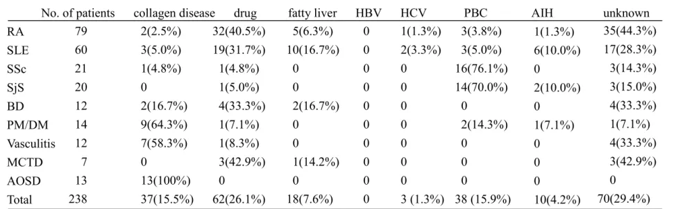
Causes of liver dysfunction (Table 2)
Liver dysfunction in patients with collagen disease can result from many causes:
drug-induced liver injury (26.1%); fatty liver (7.6%); viral hepatitis (1.3%); AIH (4.2%); PBC
(15.9%); and the collagen disease itself (15.5%). The causes of liver dysfunction differ with each
disease. The collagen disease itself was the main cause in patients with AOSD (100%), PM/DM
(64.3%), and vasculitis syndrome (58.3%). Drug-induced liver injury was a common cause of liver
dysfunction and was particularly prevalent in MCTD (42.9%), RA (40.5%), BD (33.3%) and SLE
(31.7%), respectively. The major causative agent for drug-induced liver dysfunction was
methotrexate in RA and antibiotics in SLE. PBC was a leading cause of liver dysfunction in SSc
(76.1%) and SjS (70.0%). Moreover, the cause was often unknown in patients other than those with
AOSD. Few patients showed liver dysfunction due to hepatitis B or C.
Degree of liver dysfunction in collagen disease (Figure 1)
Degree of liver dysfunction in patients with collagen disease was, on the whole, mild. ALT
levels (a) are higher in patients with AOSD than those with other collagen diseases .Conversely, ALP
levels (b) are higher in patients with SSc, vasculitis syndrome and SjS than those with other collagen
diseases. Moreover, γGTP levels (c) are higher in patients with vasculitis syndrome than those with
other collagen diseases.
Relationship between degree of liver dysfunction and activity of each collagen disease
In patients with AOSD, ALT levels correlated positively with ferritin (r=0.708; p<0.05). In
patients with vasculitis syndrome, ALP levels correlated positively with levels of C-reactive protein
(CRP) (r=0.833; p<0.05). In patients with other collagen diseases, neither ALT nor ALP levels
showed correlations with markers of disease activity.
DISCUSSION
In the present study, liver dysfunction was seen in 39.2% of collagen disease patients. The
frequency of liver dysfunction appears to depend on the original collagen disease, with dysfunction
seen more frequently in patients with AOSD, PM/DM, and vasculitis syndrome. Previous papers
have reported similar tendencies. Rates of liver dysfunction in AOSD, PM/DM and vasculitis
syndrome have been reported as about 35-85%, 30% and 54%, respectively1,12-14. Interestingly, the
main cause of liver dysfunction in these diseases is associated with the original collagen disease. The
degree of liver dysfunction thus reflects the activity of the original disease and liver enzymes
improve in parallel with recovery of the collagen disease following corticosteroids treatment. In
fact, ALT or ALP levels correlated with the disease activity in patients with AOSD and vasculitis syndrome. In contrast to them, in patients with PM/DM, ALT levels didn’t correlate
with creatine phosphokinase (CK). CK levels generally reflect the activity of myositis15, however,
dermatomyositis without CK elevation are rarely observed16. In this study, one DM patient with liver
dysfunction showed normal CK levels. Consequently, ALT levels didn’t correlate with CK levels in
patients with PM/DM. Although autoimmune mechanism is concerned with the liver dysfunction in
collagen diseases, detailed pathogenesis is unknown. It is reported that their main histological
findings of the liver are arteritis, and some patient shows chronic active hepatitis or nonspecific
reactive hepatitis17. In this study, one SLE patient with liver dysfunction showed nonspecific reactive
hepatitis.
The causes of liver dysfunction showed some tendency toward associations with the
original collagen disease. In the present study, drug-related effects were the most frequent causes of
liver dysfunction in patients with RA and SLE. Conversely, the leading cause of liver dysfunction in
SLE has been reported as SLE itself, with the degree of liver dysfunction correlating with disease
activity1. These differences may depend on elapsed time from onset of the disease. In this study, liver
dysfunction appeared long after onset of SLE, so activity of SLE was relatively mild. Moreover,
many cases were not examined for causes of liver dysfunction because the dysfunction was mild or
temporary, although histological examination of the liver is useful in making a differential diagnosis
between AIH and SLE-associated hepatitis18. The frequency of SLE-associated liver dysfunction
might thus be lower than previously reported.
In the present study, no patients showed liver dysfunction due to hepatitis B. However, de
novo hepatitis B has recently become a serious problem in patients undergoing chemotherapy or
steroid therapy19. Fulminant hepatic failure due to de novo hepatitis B is lethal20. The possibility of
hepatitis B should thus be investigated in patients receiving steroid therapy to prevent fulminant
hepatic failure. In particular, checking the HB core and HB surface antibodies is important in HBs
antigen negative patients. Moreover, anti-viral drugs should be started according to the guidelines if
the existence of HBV is confirmed21.
Liver dysfunction is generally classifiable into liver cell damage and biliary tract damage.
The pattern of liver dysfunction depends on the cause and underlying collagen disease. In this study,
ALP levels were elevated in patients with SSc and SjS, in whom liver dysfunction was caused by
PBC. Moreover, ALT and ALP levels were elevated in patients with AOSD and vasculitis syndrome,
respectively. Liver dysfunction is a major feature in patients with AOSD, and is thus included among
the criteria for AOSD12. We supposed that the pattern of liver dysfunction in AOSD was liver cell
damage on the basis of elevated ALT levels and histological findings, although the precise
mechanisms remain unclear. In this study, ALT levels correlated positively with ferritin, reflecting
the activity of AOSD22. Previous studies have reported elevated ALP levels as characteristic in
patients with vasculitis, reflecting the involvement of small intrahepatic vessels causing ischemic
cholangitis23,24. In the present study, ALP levels correlated positively with CRP, reflecting the
activity of vasculitis syndrome 25
Autoimmune liver diseases such as AIH and PBC sometimes complicate collagen disease.
In particular, the present study revealed PBC as a leading cause of liver dysfunction in SSc and SjS.
PBC should be considered when liver dysfunction is identified in patients with SSc or SjS.
No patients in this study exhibited severe liver failure. Liver dysfunction in patients with
collagen disease is generally mild and the prognosis is good irrespective of the cause1,26,27. The use
of steroid administration is one probably reason for such positive outcomes. On the other hand, some
investigations have reported severe hepatic failure in AOSD28-30. Such liver failure developed during
steroid therapy long after onset of AOSD. Moreover, Ott et al. reported a patient with AOSD in
whom hepatic failure developed when other symptoms were well controlled by corticosteroid
treatment30. Clinicians thus need to remain vigilant for liver dysfunction in AOSD.
In conclusion, liver dysfunction in patients with collagen diseases shows specific
tendencies for each collagen disease. Understanding the features of liver dysfunction is necessary
when treating collagen disease.
REFERENCES
1. Kojima H, Uemura M, Sakurai S, et al. Clinical features of liver disturbance in rheumatoid
disease: clinicopathological study with special reference to the cause of liver disturbance. J
Gastroenterol 2002; 37:617-25
2. Abraham S, Begum S, Isenberg D. Hepatic manifestations of autoimmune rheumatic diseases.
Ann Rheum Dis 2004; 63:123-29
3. Arnett FC, Edworthy SM, Bloch DA, et al. The American Rheumatism Association 1987 revised
criteria for the classification of rheumatoid arthritis. Arthritis Rheum 1988; 31: 315-24
4. Tan EM, Cohen AS, Fries JF, et al. The 1982 revised criteria for the classification of systemic
lupus erythematosus. Arthritis Rheum 1982; 25: 1271-7
5. Subcommittee for Scleroderma Criteria of the American Rheumatism Association Diagnostic
and Therapeutic Criteria Committee. Preliminary criteria for the classification of systemic
sclerosis (scleroderma). Arthritis Rheum 1980; 23: 581-90
6. Vitali C, Bombardieri S, Moutsopoulos HM, et al. Preliminary criteria for the classification of
Sjögren’s syndrome. Results of a prospective concerted action supported by the European
Community. Arthritis Rheum 1993; 36: 340-7
7. Bohan A, Peter JB. Polymyositis and dermatomyositis. N Engl J Med 1975 292: 344-7
8. Kasukawa R, Tojo T, Miyawaki S, et al. Mixed connective tissue disease – a preliminary
diagnostic criteria. Jpn J Rheumatol 1988; 1: 263-70
9. Takikawa H, Takamori Y, Kumagi T, et al. Assessment of 287 Japanese cases of drug induced
liver injury by the diagnostic scale of the International Consensus Meeting. Hepatol Res 2003;
27: 192-5
10. Jonson PJ, McFarlane IG. Meeting report: International Autoimmune Hepatitis Group.
Hepatology 1993; 18: 998-1005
11. Sasaki H, Inoue K, Higuchi K, et al. Primary biliary cirrhosis in Japan: national survey by the
Subcommittee on Autoimmune Hepatitis. Gastoenterol Jpn 1985; 20: 476-85
12. Yamaguchi M, Ohta A, Tsunematsu T, et al. Preliminary criteria for classification of adult Still’s
disease. J Rheumatol 1992; 19: 424-30
13. Reginato AJ, Schumacher HR, Baker DG, et al. Adult onset Still’s disease: Experience in 23
patients and literature review with emphasis on organ failure. Semin Arthritis Rheum 1987; 17:
39
14. Pouchot J, Sampalis JS, Beaudet F, et al. Adult Still’s disease: manifestation, disease course, and
outcome in 62 patients. Medicine 1991; 70: 118
15. Kroll M, Otis J, Kagen L. Serum enzyme, myoglobin and muscle strength relationships in
polymyositis and dermatomyositis. J Rheumatol 1986; 13: 349-55
16. Fudman EJ, Schnitzer TJ. Dermatomyositis without creatine kinase elevation. A poor prognostic
sign. Am J Med 1986; 80: 329-32
17. Matsumoto T, Kobayashi S, Shimizu H et al. The liver in collagen diseases: pathologic study of
160 cases with particular reference to hepatic arteritis, primary biliary cirrhosis, autoimmune
hepatitis and nodular regenerative hyperplasia of the liver. Liver 2000; 20: 366-73
18. Iwai M, Harada Y, Ishii M, et al. Autoimmune hepatitis in a patient with systemic lupus
erythematosus. Clin Rheumatol 2003; 22: 234-6
19. Hoofnagle JH. Reactivation of hepatitis B. Hepatology 2009; 49: S156-65
20. Umemura T, Tanaka E, Kiyosawa K, Umemura T, Kumada H; Japan de novo Hepatitis B
Research Group. Mortality secondary to fulminant hepatic failure in patients with prior
resolution of hepatitis B virus infection in Japan. Clin Infect Dis 2008; 47: e52-6
21. Tsubouchi H, Kumada H, Kiyosawa K, et al. Prevention of immunosuppressive therapy or
chemotherapy-induced reactivation of hepatitis B virus infection – Joint report of the intractable
liver disease study group of Japan and the Japanese study group of the standard antiviral therapy
for viral hepatitis– Kanzo 2009; 50: 38-42
22. Ohta A, Yamaguchi M, Kaneoka H, et al. Adult Still’s disease: Review of 228 cases from the
literature. J Rheumatol 1987; 14: 1139
23. Takebayashi K, Aso Y, Kitamura H, Sakurai Y, Wakabayashi S, Inukai T. Microscopic
polyangiitis presenting with liver dysfunction preceding rapidly progressive necrotizing
glomerulonephritis. South Med J 2004; 97: 911-4
24. Nakamoto T, Yoshikawa M, Nakatani T, et al. Microscopic polyangiitis that presented liver
dysfunction prior to noted renal manifestations. Intern Med 2000; 39: 517-21
25. Weyand CM, Fulbright JW, Hunder GG, Evans JM, Goronzy JJ. Treatment of giant cell
arteritis: Interleukin-6 as a biologic marker of disease activity. Arthritis Rheum 43:1041-8
26. Chowdhary VR, Crowson CS, Poterucha JJ, Moder KG. Liver involvement systemic lupus
erythematosus: case review of 40 patients. J Rheumatol 2008; 35: 2159-64
27. Zhu G, Liu G, Liu Y, Xie Q, Shi G. Liver abnormalities in adult onset Still’s disease: a
retrospective study of 77 Chinese patients. J Clin Rheumatol 2009; 15: 284-8
28. Dino O, Provenzano G, Giannuoli G, Sciarrino E, Pouyet M, Pagliaro L. Fulminant hepatic
failure in adult onset Still’s disease. J Rheumatol 1996; 23: 784-5
29. Nagashima T, Aoki Y, Onishi S, Iwamoto M, Okazaki H, Minota S. Steroid-refractory severe
hepatic failure in adult onset Still’s disease responding to cyclosporine. Clin Rheumatol 2008;
27: 1451-3
30. Ott Sj, Baron A, Berghaus T, Lamerz R, Beuers U. Liver failure in adult onset Still’s disease
during corticosteroid treatment. Eur J Gastoroenterol Hepatol 2003; 15: 80-90
Figure legends.
Degree of liver dysfunction in collagen disease. ALT, ALP, and γGTP levels were
evaluated. ALT levels (a) are higher in patients with AOSD than those with other collagen diseases .
ALP levels (b) are higher in patients with SSc, vasculitis syndrome and SjS than those with other
collagen diseases. γGTP levels (c) are higher in patients with vasculitis syndrome than those with
other collagen diseases. The data represent mean±SD. *Statistically significant differences between
the indicated diseases (P<0.05). RA, rheumatoid arthritis; SLE, systemic lupus erythematosus; SSc,
systemic sclerosis;
SjS, Sjögren’s syndrome;
BD, Behçet disease; PM, polymyositis; DM,dermatomyositis; MCTD, mixed connective tissue disease; AOSD, adult onset Still’s disease