2019
4.2.11. His-tag ArcA ... 48
4.2.12. Cy3 ... 48
4.2.13. Electrophoresis mobility shift assay (EMSA) ... 49
5.2.11. Electrophoresis mobility shift assay (EMSA) ... 79
5.3.
... 79
5.3.1. NADH dehydrogenase ... 79 5.3.2. NADH dehydrogenase ... 80 5.3.3. Nqr ... 82 5.3.4. Nqr ... 825.4.
... 83
... 87
6.
... 97
6.1.
... 97
6.2.
... 97
6.2.1. ... 97 6.2.2. ... 97 6.2.3. × ... 97 6.2.4. × ... 98 6.2.5. ... 98 6.2.6. ... 98 6.2.7. His-tag ArcA ... 99 6.2.8. Cy3 ... 996.2.9. Electrophoresis mobility shift assay (EMSA) ... 99
1.
1.1.
2 2 (ATP) 3 2 × ATP 2 3 (NADH NADPH) 2 2 3 2 3 2 3 2 3 4 4 3 2 NAD+/NADH 2 2 3 2 31.2.
2 3(electrochemically active bacteria; EAB) 2
(Rabaey and Rozendal, 2010)3 2
2 EAB (Kracke et al., 2015)3
electrotroph 2 chemotroph phototroph 3 Acidithiobacillus Desulfovibrio 2 (Deng et al., 2018)3 (Nakamura et al., 2010)2 3 2 (Reysenbach and Shock, 2002)2 3 2 2 “ 3
1.3.
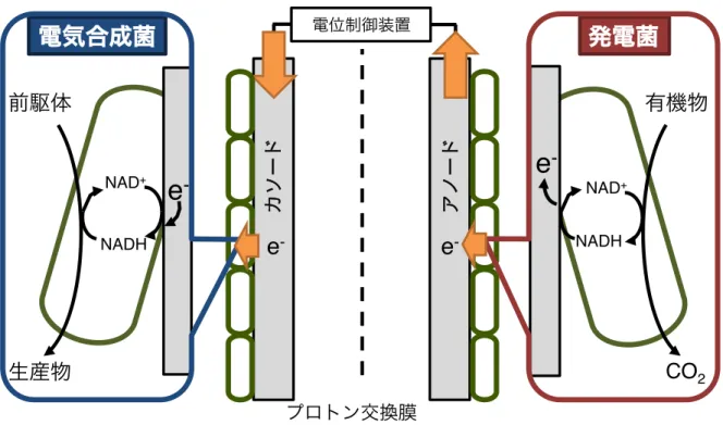
2EAB 3 EAB(bioelectrochemical systems; BES) 3 BES Fig. 1-1
3BES ( )
( ) 3 2
2
(EAB ) 3
2
(Rabaey and Rozendal, 2010)3BES 2EAB 3
2 3 2 2 3EAB 2 BES 3
1.4.
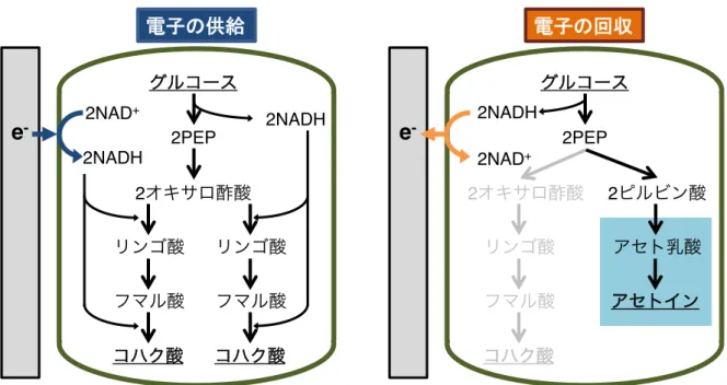
BES(Moscoviz et al., 2016)3 Fig. 1-2 3
2 2
(Förster et al., 2017)3 2 2
4 3
3 2BES
2
3 2
(Förster et al., 2017; Wu et al., 2019a, 2019b) (Fig. 1-2)3Förster
(2017) 2 2 3 Wu (2019a, 2019b) 2 2 1.7 3 2 3 2 2 EAB 2
Δ 10 µA/cm2 (Förster et al., 2017)3
(Sturm-richter et al., 2015)2 3 2 EAB 2EAB ′ 3 2 EAB 2 3 2 2 2 ′ EAB 3
1.5. Shewanella oneidensis MR-1
Shewanella oneidensis MR-1 γ- 2 2 (Venkateswaran et al., 1999)3 2 2(Myers and Nealson, 1988)3MR-1 “
2
(Rodionov et al., 2010)3 2 2 2
2 2 2 (DMSO 2
(Nealson and Saffarini, 1994)3Shewanella
3 2 3 MR-1 2 (Heidelberg et al., 2002)2 2 2 3 2 2 (Tefft and Teravest, 2019)3 2MR-1 3
1.6. S. oneidensis MR-1
MR-1 Fig. 1-3 (Hirose et al.,
2019a)3MR-1 glucose uniporter (Glf) glucokinase (Glk)
2 3 2 N- 2 6-3MR-1 Entner-Doudoroff (ED) 2 Embden-Meyerhof-Parnas (EMP) 1 ATP 1 3 3
Δ NADH 2acetyl-CoA pyruvate dehydrogenase
(PDH) 2 Δ 2acetyl-CoA pyruvate-formate lyase (PFL) 3PFL 2 formate dehydrogenase (FDH) CO2 3MR-1 3 FDH (fdnGHI2fdhABC-12fdhABC-2) 2 3 2 Δ 2 Δ 3 2 2NADH lactate
dehydrogenase (LdhA) D- (Kasai et al.
2019)3 2
3
MR-1 N- 2 (D- L- )
3D- L
2 D- 3 2L- 2LldEFG
3 2 L
-3
Acetyl-CoA Δ Δ 3 Δ
acetyl-CoA TCA 2NADH
3 2 Δ ( Δ ) acetyl-CoA
2 MR-1 × ATP 3
Δ ATP ( )
(Hunt et al., 2010)3 2 Δ 2TCA
NADH ′ isocitrate dehydrogenase (Icd) 2-oxoglutarate
dehydrogenase (Suc) 2 TCA 3 2
MR-1 NADH NADH (NAD+ ) 2 3 3MR-1 8 (UQ8) 7 (MK7) 2 Δ UQ8 (+0.11 V vs. ; 2 )2 Δ MK7 (-0.074 V) 3 Δ UQ8 PetA 2 c 2 3 PetA
” 2ATP synthase ATP 3 2 Δ
2CymA MK7 2
3 FccA 2
3 2NapB
2 NapA 3
NapB NrfA 3DMSO
DmsE DmsABF 2DMSO
3 / FccA/CctA 2
MtrCAB/OmcA 3 (TMAO)
2CymA (TorC) 2TMAO
TorA 3
MR-1 Δ ( Δ )
-2 Δ
(Kasai et al., 2019)3 2MR-1 NADH dehydrogenase FDH
Δ 2
2
3 2MR-1 2 2
hydrogenase
(Meshulam-simon et al., 2007)3MR-1 hydrogenase Hya Hyd
2 3 2 Δ 3 2 hydrogenase 3
1.7. MR-1
MR-1 (Fig. 1-4) (Hirose et al., 2019b)3 2 c 2 3 2 (IV) 2 (III)2 3MR-1 CymA2 FccA/CctA2MtrCAB2OmcA 3CymA 2 2 2FccA/CctA 2 (Myers and Myers,
2000)3MtrCAB OmcA 2MtrA
(Hartshorne et al., 2009)3MtrB β × 2
(Beliaev and Saffarini, 1998)3MtrC OmcA
2MtrC OmcA = 2:1 (Shi et al., 2006)3FccA
CctA 2CymA Mtr
(Sturm et al., 2015)3 2FccA 2
CymA (Myers and Myers, 2000)3
( ) 2 “
(Firer-Sherwood et al., 2008)3 2MR-1 3
protein (CRP) (Kasai et al., 2015)3CRP
× × 2MR-1
(Saffarini et al., 2003)3
2 Δ MetR
× × CRP (Mogi et al.,
unpublished data)3 DNA
2MR-1
3 2MR-1 2
(Gorby et al., 2006; Pirbadian et al., 2014)3 MtrCAB/OmcA
2
(Pirbadian et al., 2014)3
1.8.
“ 2
3 “
sensor kinase 2DNA 2
response regulator 3 sensor kinase (Hpt)
3 2
4 3 “
2 3
2Arc system 3
γ-2S. oneidensis2Escherichia coli2Serratia marcescens2Klebsiella pneumoniae2Vibrio choleae2Salmonella Typhimurium Haemophilus
influenzae (Hirose et al., 2019b)3Arc system E. coli ( )
2sensor kinase PAS
(Bekker et al., 2010)3
Arc system ArcB/ArcA 3 2
Δ sensor kinase ArcB 2
ArcB 3 response regulator
ArcA 2 ArcA (ArcA-P)
3 2 Δ 2ArcB
2ArcA-P 2ArcA DNA 3DNA ArcA
2 ArcA “ ′
(Park et al., 2013)3 Arc system
2 (Park
et al., 2013)3
2MR-1 Arc system sensor kinase ArcS2 HptA2
response regulator ArcA (Lassak et al., 2010)3
2HptA ArcS ArcA 2
3MR-1 ArcA ArcA 2 DNA
” (Gao et al., 2008)3 2 sensor kinase 2
MR-1 ArcS PAS 2 Chache sensor
2 ArcB (Lassak et al., 2013)3
2ArcS ArcB 3
2 2 Fnr (fumarate nitrate
reductase regulator) (Gunsalus and Park, 1994)3
“ DNA 2 3
2DNA 3 2
3 2Fnr Δ 2
′ (Myers et al., 2013)3MR-1 Fnr
EtrA (electron transport regulator A) 2 ArcA ′
(Cruz-García et al., 2011)3
1.9.
2S. oneidensis MR-1 4 2
2
(Grobbler et al., 2018, 2014; Kitayama et al., 2017; Nakagawa et al., 2015)3 2
EAB 2MR-1 Shewanella decolorationis S12 2
Desulfovibrio ferrophilus IS5 Geobacter sulfurreducens 2
(Deng and Okamoto, 2018; Li et al., 2019; Lian
et al., 2016)3 2EAB 2
× 3
Fig. 1-3 MR-1 (Hirose et al., 2019a)3GlcNAc: N-acetylglucosamine; GlcNAc-6P: N-acetylglucosamine-6-phosphate; KDG-6P: 2-keto-3-deoxygluconate 6-phosphate; PEP: phosphoenolpyruvate; Q: quinone; QH2: quinol; NagP: N-acetylglucosamine transporter; NagK: N-acetylglucosamine kinase; NagA: N-acetylglucosamine-6-phosphate deacetylase; NagB: glucosamine/fructose-6-phosphate aminotransferase; Fbp: fructose-1,6-bisphosphatase; Fba: fructose-bisphosphate aldolase; TpiA: triosephosphate isomerase; Pgi: glucose-6-phosphate isomerase; Zwf: glucose-6-phosphate 1-dehydrogenase; Edd: phosphogluconate dehydratase; Eda: 2-dehydro-3-deoxyphosphogluconate aldolase/ (4S)-4-hydroxy-2-oxoglutarate aldolase; PpsA: pyruvate, water dikinase; PckA: phosphoenolpyruvate carboxykinase; Pdh: pyruvate dehydrogenase; GltA: citrate synthase; Acn: aconitate hydratase; Icd: isocitrate dehydrogenase; SucAB: 2-oxoglutarate dehydrogenase; SucCD: succinyl-CoA synthetase; Sdh: succinate dehydrogenase/fumarate reductase; Fum: fumarate reductase; Mdh: malate dehydrogenase; AceB: malate synthase; AceA: isocitrate lyase; Pta: phosphate acetyltransferase; AckA: acetate kinase; Acs: acetyl-CoA synthetase; Fdh: formate dehydrogenase; Dld: d-lactate dehydrogenase, quinone dependent; Lld: l-lactate dehydrogenase; LdhA: d-lactate dehydrogenase, NAD dependent; Glf: glucose uniporter; GalP: galactose:H+ symporter; Glk: glucokinase; Xks1: xylulokinase; Xyl2: xylitol dehydrogenase; Xyl1: NAD(P)H-dependent xylose reductase repressor.
Glucose external Glucose-6P Gluconate-6P Pyruvate Acetyl-CoA Lactate Formate CO2 Acetyl-P Acetate Xylulose-5P Xylose Xylitol Xylulose GlcNAc external GlcNAc-6P GlcN-6P Fructose-6P Fructose-1,6P Glyceraldehyde-3P GlcNAc internal KDG-6P PEP Glucose internal Xyl1 Xks1 NagP NagK NagA Eda Pgl, Zwf Glk Acs AckA Pta GltA Oxaloacetate Malate Fumarate Succinate Succinyl-CoA Citrate Isocitrate 2-oxogulutarate Glyoxylate Acn AceA Icd SucAB SucCD Sdh Fum Mdh PckA PykA PpsA Edd Fba, TpiA Pgi Chitin Pfl*† Pdh Fdh Dld Lld NagB Chitinase AceB LdhA ATP
ADP ADPATP
Fig. 1-4 MR-1 (Hirose et al., 2019b)3LDH, lactate dehydrogenase; FDH, formate dehydrogenase; NDH, NADH dehydrogenase; Q, quinone; QH2, quinole; OM, outer
membrane; IM, inner membrane.
4.
4.1.
2EAB 4 2
2 2
(Bosch et al., 2014; Grobbler et al., 2014; Li et al., 2019; Lian et al., 2016)3 EAB
2 × 3 4 2EAB 3 MR-1 3
4.2.
4.2.1.
Appendix 1 3E. coli S. oneidensis MR-1
LB 3 Δ 5 ml LB 2 100 ml 50 ml LB 2 40 mM 3 10 mM (PMM; 9 mM (NH4)2SO4, 5.7 mM K2HPO4, 3.3 mM KH2PO4, 30 mM HEPES-NaOH (pH7.4)) 2 5 ml PMM 30 ml 2S. oneidensis MR-1 OD600 0.05 2 3 30°C2180 rpm 2 3 8 ml PMM 13 ml 2 20 mM 2.5 mM 2S. oneidensis MR-1 OD600 = 0.01 2 3 5 min 2 “ Δ 230°C 3OD600
mini photo 518R photometer ( ) 21 h 3
(MnO2) 2100 ml PMM 80 ml 2
20 mM MnO2 2MR-1 OD600 = 0.01 2
3 10 min “ Δ 224 h 1 ml
2MnO2 3MnO2 Kouzuma et al. (2012) 2leucoberblin
blue 3
4.2.2.
Appendix 2 3
DISMIC-25HP; Advantec) 1.5 ml 2 3
B-PER bacterial protein extraction reagent (Thermo Fisher Scientific)
3 1 ml 3 ml 2 15min 3
— 2 1.5ml 2 3
Micro BCA protein assay kit (Thermo Fisher Scientific) 2 3
4.2.5.
2 2 (HPLC) (Agilent 1100
series) 3 1 ml 2
(0.2 µm pore size, DISMIC-25HP; Advantec) 3HPLC
Zorbax column (SB-Aq, 4.6 by 150 mm; Agilent)2 10%
20 mM potassium phosphate buffer (pH 2) 3 , 1 ml min-1; , 5µl;
, 25°C; , UV 210 nm 3 2
3
4.2.6. RNA
Total RNA Trizol (Invitrogen) 2RNeasy Mini kit RNase-free
DNase Set (QIAGEN) 3 RNA Agilent 2100 Bioanalyzer RNA
pico Chips (Agilent) 2 3
4.2.7. qRT-PCR
PCR_(qRT-PCR) LightCycler 1.5 instrument LightCycler SYBR Green I (Roche) 3PCR 15 ng total RNA21.3 µl 50 mM Mn(OAc)22
7.5 µl RNA Master SYBR Green I20.15 µM 3
Appendix File 2 3nuoI (SO_1014)2SO_09392SO_45092atpG (SO_4748)2
dld (SO_1521)2SO_15382SO_4360216S rRNA PCR
3 16S rRNA 3
4.2.8. DNA
×
S. oneidensis MR-1 ×
FairPlay III Labeling kit (Agilent) 3 cDNA
× 2WT n = 52∆arcS n = 6 ( ×
; n = 32 × ; n = 2) 3
GeneSpring GX version 11.5 (Agilent Technologies) 3 Student’s T-test Benjamini-Hochberg false discovery rate (BH-FDR) 3 Δ fold change (FC) 1 2.0 0.5 (|log2FC| 1 1.0)2P < 0.05
3DNA × 6 (SO_09392
dld2SO_15382SO_43602SO_45092atpG ) qRT-PCR ′ 36
qRT-PCR 3 Cluster 3.0 Treeview software
3
4.2.9. NADH/NAD
+NADH/NAD+ NAD+/NADH assay kit (Colorimetric; Abcam)
3 × 600 µl Extraction buffer 2 Lysing
Matrix E tube (MPB) 3Fast prep FP120 (MPB) Speed: 4 40 sec
3— (15000 rpm, 4°C, 10 min) 2 10 kD spin column (Abcam) 3 3
4.2.10.
WT 150 ml OD600 1.0 2
+0.5 V –0.1 V 2 3
Bekker et al. (2007)2Grammel et al. (2008) 3
Δ 2 3 3 ml (60%) : buffer (10 mM) 3 ml 3 2 3 ml 3 — ( ) 3 2 3 Δ 2 × 2- (4:1, v/v) 120 µl 2 3
MR-1 UQ8 MK7 (Venkateswaran et al. 1999)2
3 2Coenzyme Q8 (AVT) -7
(WAKO) 3 HPLC (Prominence series, shimadzu) 2 L-
3 ° × UV (2 2MK7; 248 nm UQ8; 290 nm) 2 (λmax) (RT) 3
4.2.11. His-tag
ArcA
Appendix 2 3 2pET 3 (His-tag) ArcA 2MR-1total DNA PCR 2arcA DNA 3PCR QIA
quick PCR purification kit (QIAGEN) 3 2BamHI NdeI
2 DNA mini Elute reaction clean up kit (Qiagen)
3 DNA BamHI NdeI pET-28a (+) (Novagen)
3 (pET-28_arcA) BL21(DE3) (Novagen)
3 BL21(DE3)(pET-28_arcA) Km 2×YT
237°C2180 rpm OD600 0.7 3300 ml
Km 2×YT 100 ml 2OD600 0.05 3
30 C2180 rpm OD600 0.7 2Isopropyl-β-D-galactopyranoside
(IPTG) 500 µM 225°C2180 rpm 3 50 ml
2— × 3 × IMAC wash buffer
(Bio-Nobile) 300 µl 2 3 buffer 10 2
Misonix 2— 3
His-tag ArcA Quickpick IMAC metal affinity kit
(Bio-Nobile) 2 3 His-tag ArcA
SDS-PAGE 2 3 micro BCA
protein assay kit (Thermo Fisher Scientific) 2 3
4.2.12. Cy3
Appendix 2 3SO_00112sucA (SO_1930)2nuoA
(SO_1021)2atpI (SO_4754) DNA 3Cy3
DNA PCR 3PCR DNA QIAXII
gel extraction kit (QIAGEN) 3Nano drop (ND-10002 )
4.2.13. Electrophoresis mobility shift assay (EMSA)
EMSA Gao et al. (2008) 3 His-tag
ArcA 2× (200 mM Tris/HCl (pH 7)220 mM MgCl22250 mM KCl2
100 mM (Sigma)) 1:1 1 h
His-tag ArcA 3DNA 100 mM Tris/HCl (pH7.4)210 mM MgCl22
20 mM KCl22 mM DTT20.2 µg/ml poly (dI-dC)210% 22 nM Cy3 DNA
2 His-tag ArcA (0, 100, 200, 300 ng) 12 µl 2
(Takara) 15°C21 h 3 × 1 h
12.5 % 3
0.5×Tris-borate-EDTA (TBE) buffer 2200 V 3 Typhoon
FLA 9000 (FUJIFILM2GE healthcare) 2 3
4.2.14. ×
nuoA lacZ 3nuoA MR-1
total DNA PCR 3 nuoA 5
100 bp 3 Appendix 2
3PCR QIA quick PCR purification kit (Qiagen) 2
3 PCR × pMElacZ (Endoh et al., 2003)
EcoRI BamHI 3 pMElacZ Antactic phosphatase (New
England Biolabs) 3pMElacZ PCR
2pMElacZ 3
(pMElacZ-R) 2 ”
3
4.2.15. ×
S. oneidensis MR-1 × Kasai et al. (2015)
3× 3
MR-1 LB 5 ml 22 ml — (16000 g21 min24°C) 3 ×
300 mM 2 2 100 µl 300 mM 3
50 µl 1-3 µg
Gene Pulser cuvette, gap: 0.1 cm (Bio Rad)
2
20 min
3
Micropulser (Bio Rad)
180 kV
4 sec
3
2
500 µl LB
µg/ml) × × 3 ×
30
°C 2 34.2.16.
β- ° 3 Kasai et al. (2015) 310 mM 40 mM MM × OD600 = 0.01 2 3 500 µl 2 ml 2Z buffer (60mM Na2HPO4240 mM NaH2PO4 2H2O210 mM KCl2MgSO4 7H2O250 mM
2-) 500 µl2 15 µl 0.1% SDS 10 µl 215 sec
35 min 2200 µl 2- -β- (ONPG)
3 2stop solution (1 M Na2CO3)
β 3OPNG stop solution 3
— (12,000 × g22 min24°C) 2 300 µl × 2
× ° (SH-12002 ) 420 nm (Abs420)
3β- ° Miller unit = (1000×Abs420) / ( (min) ×
(ml) × OD600) 3 3 3
4.3.
4.3.1.
MR-1
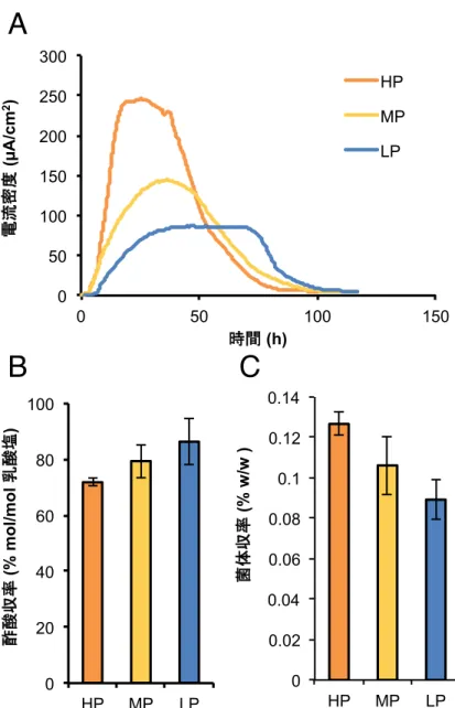
S. oneidensis MR-1 ( ; WT)
218 ml 2
+0.5 V (high potential; HP)2+0.2 V (middle potential; MP) 0 V (low potential;
LP) 3 2
(Fig. 4-1A)3 2 2
(data not shown)3 2 (Fig. 4-1B)3 2 (
) 2 (Fig.
4-1C)3 MR-1
4.3.2.
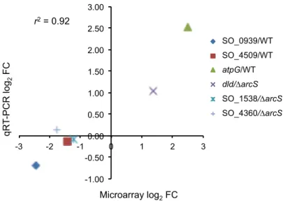
MR-1 2DNA × 3MR-1 +0.2 V (MP) 2 +0.5 V (HP) –0.1V (LP) 3MP Δ +0.2 V 32 h 2 2DNA × 3 Fig. 4-2 3HP2MP2LP Δ 2 MP Δ HP Δ LP Δ 3 2HP Δ LP Δ 2HP Δ LP Δ 322 2274 248 (Appendix 3)3 × 2 6 2qRT-PCR × (Fig. 4-3)3 2 ′ (r2 = 0.92) 2 × 3 HP Δ LP Δ 2 ′(Fig. 4-4)3 Table 4-1 3 D-lactate
dehydrogenase (dld)2NADH dehydrogenase (nuo) F0F1-ATP synthase (atp) 2
HP Δ LP Δ
3 2
2 3
MR-1 Δ ( Δ ) ×
ATP 2 (NADH dehydrogenase F0F1-ATP synthase)
(Hunt et al., 2010)3 NADH
(pyruvate dehydrogenase (PDH)2NADH dehydrogenase succinate dehydrogenase)
2 pyruvate-formate lyase (PFL) formate
dehydrogenase (FDH) CO2 (Pinchuk et al., 2011)3
2 × 2HP Δ pyruvate dehydrogenase (aceF)2succinate
dehydrogenase (sucAB)2nuo2 atp 2
MR-1 NADH 2 ATP
3 2FDH 3 2fdnGHI (SO_0101-SO_0103)
fdhABC2 (SO_4513-SO_4515) HP Δ 2fdhABC1 (SO_4509-SO_4511) LP Δ
3 2MR-1 FDH 3 2 FDH – 3
4.3.3.
2MR-1 2 PFL FDH ( ) 2 PDH Nuo NADH 3 2MR-1 PDH (∆PDH) PFL (∆PFL) 2HP2MP2LP Δ 2 Δ 3 2HP Δ ∆PDH WT 20%2∆PFL 80% (Fig. 4-5)3 PDH HP Δ 2 PFL 3 LP Δ ∆PFL (Fig. 4-5)2 Δ PFL 3 2∆PDH MP Δ LP Δ WT (Fig. 4-5)2PDH PFL 3 2PFL ′ 2 Δ PDH “ 3 MR-1 ( ) 2 (NADH ) CymA ” 2(Fig. 1-4) (Simon et al., 2008)3 2Geobacter CymA 2
(Levar et al., 2017)3 cymA ′
dehydrogenase 3 Δ
NADH dehydrogenase 3MR-1 Nuo2Ndh2Nqr1
Nqr2 4 NADH dehydrogenase 2 NADH dehydrogenase
(∆NDH) 2HP2MP LP Δ (Fig. 4-5)3 2∆NDH HP Δ 2MP LP Δ 3 HP Δ NADH dehydrogenase ′ 2 NADH 3 2 ∆PDH ∆PFL (Fig. 4-5) HP Δ 80% NADH ( ) 2HP Δ ∆NDH 3 2HP Δ ∆NDH 2 3 2∆NDH NADH 2HP Δ NADH 2 3 2HP2MP LP Δ WT ∆NDH NADH/NAD+ (Fig. 4-6)3 2∆NDH WT HP Δ NADH/NAD+ 2∆NDH NADH 3∆NDH NADH MP LP Δ 2NADH/NAD+ (Fig. 4-6)3 2MP Δ ∆NDH WT (Fig. 4-5)2HP Δ MP Δ NADH/NAD+ 3 2 2MR-1 2 NADH 3
4.3.4. NADH dehydrogenase
′
2 MR-1 3 2 2MR-1 3WT ∆NDH (E0, pH 7.0) : (+0.82 V)2MnO2 (+0.53 V)2 (+0.43 V)2 (-0.03 V) 2 Δ ∆NDH WT(Fig. 4-7A)3 2 Δ 0 V (LP) Δ 2NADH
3 2∆NDH (Fig. 4-7B) MnO2 (Fig.
2 NADH 3 2MR-1 2 3
4.3.5. Arc system
2MR-1 3 2MR-1 3 Arc system 3MR-1 2 ( MnO2) (Fig. 1-4)3 2 2Arc system 2 × 3 2 3 MR-1 UQ8 MK7 2UQ8 +0.11 V 2 3 22HP Δ NADH:ubiquinone oxidoreductase (nuo)
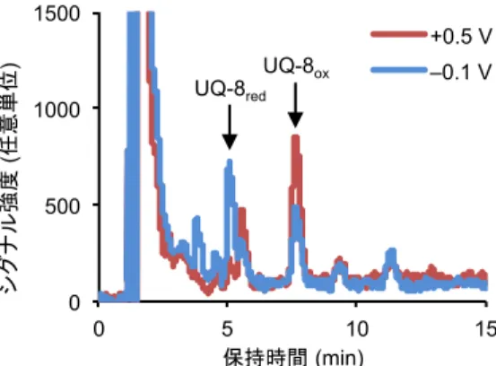
2HP Δ UQ8 3 2 2 UQ 3 UQ8 2HP Δ LP Δ UQ8 UQH2 UQ 3 2HP Δ LP Δ UQ8 2 (Fig. 4-8)3 2 3 2 2 HP Δ UQ 3 2Arc system ′ 3MR-1
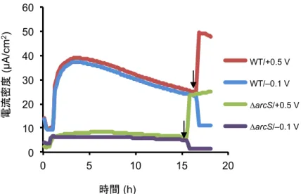
Arc system sensor kinase (ArcS)2 (HptA)2response regulator (ArcA)
3 ArcS ∆arcS
2 (+0.5 V vs. –0.1 V)
2Arc system ′ 3 ∆arcS
Fig. 4-2 Appendix 4
′ (Fig. 4-2)3
2WT 322 2305 ∆arcS
(Fig. 4-9A)3 2105 ∆arcS
2
3 2WT ∆arcS mean-average (MA) plot
2∆arcS (fold change) (Fig. 4-9B)3
2Arc system 3
Arc system 2qRT-PCR
WT ∆arcS nuoI (Nuo ) HP LP
Δ 3 2WT HP Δ nuoI
2∆arcS HP2LP Δ WT nuoI 2
Δ (Fig. 4-10)3 2Arc system nuoI
′ 3
2
nuoI (Fig. 4-11)3 2arcS
3 2MR-1
Arc system 2
3 2 2WT
∆arcS 17 (Fig. 4-9A)3
Arc system 2 D-lactate dehydrogenase
dld 3 cyclic-AMP receptor protein (CRP)
(Kasai et al., 2017)3 2CRP (mtrCAB ) 2dld Arc system CRP ′ 3 qRT-PCR 2 nuo Arc system ′ 3 2 Arc system 3
nuoA DNA ArcA electrophoresis mobility shift
assay (EMSA) 3 2 ArcA nuoA
DNA (Fig. 4-12)2nuo (nuoA–N )
ArcA 3ArcA
DNA 2 DNA ArcA
2 “ ArcA
3 2 Arc system
atp suc ArcA (Fig. 4-13)3
Arc system 3
ArcA nuo 2nuoA
lacZ × 3EMSA nuoA 5
100 bp DNA ” 2× (pMElacZ) )
- ° (lacZ) 3 WT ∆arcA
2β- ° (LacZ) (Fig. 4-14A B)3 2
(+1) –112 (Fig. 4-14A) 2WT ∆arcA
LacZ (Fig. 4-14B)3 2–112 –63 2
∆arcA LacZ 2WT 3
2ArcA ′ nuoA –63 2–63 –112
2 (Fig. 4-14A)3EMSA (Fig. 4-12)
nuoA ArcA 2 3 (Table 4-1) (Fig. 4-5)2∆NDH (Fig. 4-7) 2ArcA (LP Δ ) 2nuoA (PnuoA) 3 2–273 2 2–112
ArcA LacZ 2∆arcA
(HP) Δ Δ 2 Δ PDH Nuo NADH (Fig. 4-15)3Nuo
2 2 H+/e
-2 (Efremov et al., 2010)3Nuo CymA
2 NADH 3 3 (Fig. 4-1B) (Table 4-1) 2HP Δ “ acetyl-CoA 2TCA 3 2MR-1 2 (Fig. 4-15)3 2 3 2 2Arc system 3 2MnO2 NADH (Fig. 4-7C)3 2MnO2 2 ( 2 ) 3MR-1 2
(Nealson and Saffarini, 1994)3MR-1
Arc system 2 2
3 2MR-1 Shewanella Nuo
2 Nqr 3
MR-2 Shewanella ′
2Nuo Arc system MR-1
3 2Shewanella NADH dehydrogenase
– 3
(Table 4-1) 2 2 Arc
system 3
cyclic AMP (cAMP) receptor protein (CRP)
2Arc system ′ (Kasai et al., 2015)3
2MnO2 4
3
MR-1 Arc system
(Gao et al., 2008)3 TCA ′
Arc system 2MR-1
Arc system (Gao et al., 2008)3 2
× (Table 4-1) 2TCA 2-oxogulutarate dehydrogenase
suc Arc system Δ 3 2MR-1
Δ suc 2 TCA
(Tang et al., 2007)3 2EMSA (Fig. 4-13) suc Arc system
2MR-1 TCA 2
3 TCA
3
Arc system TCA ′ 2nuo
(Bongaerts et al., 1995; Lin, 1996; Park et al., 2013)3 2
3 2MR-1 nuo Arc
system (Fig. 4-12)3 2
Arc system
3 (Table 4-1) qRT-PCR (Fig. 4-10)2
∆NDH (Fig. 4-5) (Fig. 4-7) 2 Nuo
2
3 2lacZ × (Fig. 4-14B) 2ArcA
nuoA 2PnuoA “
3 2ArcS Δ
HptA ArcA 2 ArcA
“ nuoA 3 2
∆arcS nuo (Fig. 4-10 4-11)2 ArcA
nuo 3
2ArcA ( )
3ArcS
(Lassak et al., 2013)2∆arcS ArcA 2 nuo
3
3 2 Δ
UQ (Fig. 4-8)3
2 - 3 2UQ
MK 3
2 2 Mtr
CymA MK (McMillan et al., 2012)3CymA UQ
Table 4-1 ( )
Process Locus tag Gene Annotation Log2 FC*
Lactate and pyruvate oxidation
SO_1521 dld Respiratory FAD-dependent D-lactate dehydrogenase
2.32
SO_0425 aceF Dihydrolipoamide acetyltransferase 1.52
Formate oxidation SO_0101 fdnG Nitrate-inducible formate dehydrogenase molybdopterin-binding subunit
2.97 SO_0102 fdnH Nitrate-inducible formate dehydrogenase
iron-sulfur subunit
3.33 SO_0103 fdnI Nitrate-inducible formate dehydrogenase
cytochrome b subunit
2.89 SO_4509 fdhA Formate dehydrogenase molybdopterin-binding
subunit
–1.43
SO_4510 fdhB Formate dehydrogenase fes subunit –1.08
SO_4511 fdhC Formate dehydrogenase cytochrome b subunit –1.00 SO_4513 fdhA Fnr-inducilble formate dehydrogenase
molybdopterin-binding subunit
1.96 SO_4515 fdhC Fnr-inducible formate dehydrogenase
cytochrome b subunit
1.94
TCA cycle SO_1930 sucA 2-Oxoglutarate dehydrogenase complex
dehydrogenase E1 component
1.75 SO_1931 sucB 2-Oxoglutarate dehydrogenase complex
succinyl-CoA:dihydrolipoate S-succinyltransferase E2 component
1.77
SO_1933 sucD Succinyl-CoA synthase alpha subunit 1.66 NADH oxidation SO_1010 nuoM NADH-ubiquinone oxidoreductase subunit M 2.85 SO_1012 nuoK NADH-ubiquinone oxidoreductase subunit K 2.68 SO_1013 nuoJ NADH-ubiquinone oxidoreductase subunit J 2.58 SO_1014 nuoI NADH-ubiquinone oxidoreductase subunit I 2.46 SO_1015 nuoH NADH-ubiquinone oxidoreductase subunit H 2.89 SO_1016 nuoG NADH-ubiquinone oxidoreductase subunit G 2.61 SO_1017 nuoF NADH-ubiquinone oxidoreductase subunit F 2.46 SO_1018 nuoE NADH-ubiquinone oxidoreductase subunit E 1.78 SO_1019 nuoCD NADH-ubiquinone oxidoreductase subunit CD 1.90
ATP synthesis SO_4746 atpC ATP synthase F1 epsilon subunit 2.19
SO_4747 atpD ATP synthase F1 beta subunit 2.22
SO_4748 atpG ATP synthase F1 gamma subunit 2.50
SO_4749 atpA ATP synthase F1 alpha subunit 2.43
SO_4750 atpH ATP synthase F1 delta subunit 1.94
SO_4751 atpF ATP synthase F0 B subunit 1.85
SO_4752 atpE ATP synthase F0 C subunit 1.71
SO_4753 atpB ATP synthase F0 A subunit 1.37
*Log
Fig. 4-4 (COG) 3
0 1 2 3 4 5 6 7
Energy production and conversion [C] Cell cycle control, cell division, chromosome partitioning [D] Amino acid transport and metabolism [E] Nucleotide transport and metabolism [F] Carbohydrate transport and metabolism [G] Coenzyme transport and metabolism [H] Lipid transport and metabolism [I] Translation, ribosomal structure and biogenesis [J] Transcription [K] Replication, recombination and repair [L] Cell wall/membrane/envelope biogenesis [M] Cell motility [N] Posttranslational modification, protein turnover, chaperones [O] Inorganic ion transport and metabolism [P] Secondary metabolites biosynthesis, transport and catabolism [Q] General function prediction only [R] Function unknown [S] Signal transduction mechanisms [T] Intracellular trafficking, secretion, and vesicular transport [U] Defense mechanisms [V]
Fig. 4-8 3WT +0.5 V –0.1 V
2 (UQ-8red) (UQ-8ox) 3
Fig. 4-9 Arc system ′ 3(A) WT ∆arcS 3(B) × mean-average 3 305 17 105 ΔarcS WT
A
B
-6 -3 0 3 6 9 -6 -3 0 3 6 M: lo g2 fo ld ch an geA: log2 signal intensity
WT
∆arcS ΔarcS
Fig. 4-10 nuoI 3WT ∆arcS +0.5 V –0.1 V 2qRT-PCR nuoI 3 3 3 (P < 0.05) 3 0 0.5 1 1.5 2
WT_high WT_low ΔarcS_high ΔarcS_low
Fig. 4-11 3
WT ∆arcS
HP LP HP LP
Normalized signal intensity
Fig. 4-12 EMSA nuoA ArcA (ArcA-P) 3
Competitor DNA 2DNA ” 3
ArcA-P (ng)
nuoA
Fig. 4-13 EMSA sucA atpI ArcA-P 3
SO_00112 nuoA ” 3
ArcA-P – + – + – + – +
Fig. 4-15 Δ Δ MR-1 3Fdh: formate
dehydrogenase2Nuo: NADH dehydrogenase2 c (FccA
Abe, H., Doi, Y., Fukushima, T., Eya, H., 1994. Biosynthesis from gluconate of a random copolyester consisting of 3-hydroxybutyrate and medium-chain-length 3-hydroxyalkanoates by
Pseudomonas sp. 61-3. Int. J. Biol. Macromol. 16, 115–119.
https://doi.org/10.1016/0141-8130(94)90036-1
Bekker, M., Alexeeva, S., Laan, W., Sawers, G., De Mattos, J.T., Hellingwerf, K., 2010. The ArcBA two-component system of Escherichia coli is regulated by the redox state of both the ubiquinone and the menaquinone pool. J. Bacteriol. 192, 746–754. https://doi.org/10.1128/JB.01156-09 Beliaev, A.S., Saffarini, D.A., 1998. Shewanella putrefaciens mtrB encodes an outer membrane
protein required for Fe(III) and Mn(IV) reduction. J. Bacteriol. 180, 6292–6297.
Bongaerts, J., Zoske, S., Weidner, U., Linden, G., 1995. Transcriptional regulation of the proton translocating NADH dehydrogenase (nuoA‐N) of Escherichia coli by electron acceptors, electron donors and gene regulators. Mol. Microbiol. 16, 521–534.
https://doi.org/10.1111/j.1365-2958.1995.tb02416.x
Bosch, J., Lee, K.Y., Hong, S.F., Harnisch, F., Schröder, U., Meckenstock, R.U., 2014. Metabolic efficiency of Geobacter sulfurreducens growing on anodes with different redox potentials. Curr.
Microbiol. 68, 763–768. https://doi.org/10.1007/s00284-014-0539-2
Bretschger, O., Obraztsova, A., Sturm, C.A., In, S.C., Gorby, Y.A., Reed, S.B., Culley, D.E., Reardon, C.L., Barua, S., Romine, M.F., Zhou, J., Beliaev, A.S., Bouhenni, R., Saffarini, D., Mansfeld, F., Kim, B.H., Fredrickson, J.K., Nealson, K.H., 2007. Current production and metal oxide
reduction by Shewanella oneidensis MR-1 wild type and mutants. Appl. Environ. Microbiol. 73, 7003–7012. https://doi.org/10.1128/AEM.01087-07
Call, D.F., Wagner, R.C., Logan, B.E., 2009. Hydrogen production by Geobacter species and a mixed consortium in a microbial electrolysis cell. Appl. Environ. Microbiol. 75, 7579–7587.
https://doi.org/10.1128/AEM.01760-09
Claassens, N.J., Sousa, D.Z., Dos Santos, V.A.P.M., De Vos, W.M., Van Der Oost, J., 2016. Harnessing the power of microbial autotrophy. Nat. Rev. Microbiol. 14, 692–706. https://doi.org/10.1038/nrmicro.2016.130
Cruz-García, C., Murray, A.E., Rodrigues, J.L.M., Gralnick, J. a, McCue, L.A., Romine, M.F., Löffler, F.E., Tiedje, J.M., 2011. Fnr (EtrA) acts as a fine-tuning regulator of anaerobic metabolism in
Shewanella oneidensis MR-1. BMC Microbiol. 11, 64. https://doi.org/10.1186/1471-2180-11-64
https://doi.org/10.1126/sciadv.aao5682
Deng, X., Okamoto, A., 2018. Electrode potential dependency of single-cell activity identifies the energetics of slow microbial electron uptake process. Front. Microbiol. 9, 1–8.
https://doi.org/10.3389/fmicb.2018.02744
Dibrov, P., Dibrov, E., Pierce, G.N., 2018. Na + -NQR ( Na + -translocating NADH(: ubiquinone oxidoreductase ) as a novel target for antibiotic. FEMS Microbiol. Rev. 41.5,653–671. https://doi.org/10.1093/femsre/fux032
Duhl, K. L.; Tefft, N. M.; TerAvest, M.A., 2018. Shewanella oneidensis MR-1 utilizes both sodium- and proton-pumping NADH dehydrogenases during aerobic growth. Appl. Environ. Microbiol. https://doi.org/10.1128/AEM.00415-18
Efremov, R.G., Baradaran, R., Sazanov, L. a, 2010. The architecture of respiratory complex I. Nature 465, 441–445. https://doi.org/10.1038/nature09066
Endoh, T., Habe, H., Yoshida, T., Nojiri, H., Omori, T., 2003. A CysB-regulated and σ54-dependent regulator, SfnR, is essential for dimethyl sulfone metabolism of Pseudomonas putida strain DS1.
Microbiology 149, 991–1000. https://doi.org/10.1099/mic.0.26031-0
Firer-Sherwood, M., Pulcu, G.S., Elliott, S.J., 2008. Electrochemical interrogations of the Mtr
cytochromes from Shewanella: Opening a potential window. J. Biol. Inorg. Chem. 13, 849–854. https://doi.org/10.1007/s00775-008-0398-z
Förster, A.H., Beblawy, S., Golitsch, F., Gescher, J., 2017. Biotechnology for Biofuels Electrode ‑ assisted acetoin production in a metabolically engineered Escherichia coli strain. Biotechnol.
Biofuels 1–11. https://doi.org/10.1186/s13068-017-0745-9
Gao, H., Wang, X., Yang, Z.K., Chen, J., Liang, Y., Chen, H., Palzkill, T., Zhou, J., 2010.
Physiological roles of ArcA, Crp, and EtrA and their interactive control on aerobic and anaerobic respiration in Shewanella oneidensis. PLoS One 5, e15295.
https://doi.org/10.1371/journal.pone.0015295
Gao, H., Wang, X., Yang, Z.K., Palzkill, T., Zhou, J., 2008. Probing regulon of ArcA in Shewanella
oneidensis MR-1 by integrated genomic analyses. BMC Genomics 9, 42.
https://doi.org/10.1186/1471-2164-9-42
Gorby, Y.A., Yanina, S., Mclean, J.S., Rosso, K.M., Moyles, D., Dohnalkova, A., Beveridge, T.J., Chang, I.S., Kim, B.H., Kim, K.S., Culley, D.E., Reed, S.B., Romine, M.F., Saffarini, D.A., Hill, E.A., Shi, L., Elias, D.A., Kennedy, D.W., Pinchuk, G., Watanabe, K., Ishii, S., Logan, B., Nealson, K.H., Fredrickson, J.K., 2006. Electrically conductive bacterial nanowires produced by
11358-11363. https://doi.org/10.1073/pnas.0604517103
Grobbler, C., Virdis, B., Nouwens, A., Harnisch, F., Rabaey, K., Bond, P.L., 2018. Effect of the anode potential on the physiology and proteome of Shewanella oneidensis MR-1. Bioelectrochemistry 119, 172–179. https://doi.org/10.1016/j.bioelechem.2017.10.001
Grobbler, C., Virdis, B., Nouwens, A., Harnisch, F., Rabaey, K., Bond, P.L., 2014. Use of SWATH mass spectrometry for quantitative proteomic investigation of Shewanella oneidensis MR-1 biofilms grown on graphite cloth electrodes. Syst. Appl. Microbiol. 1–5.
https://doi.org/10.1016/j.syapm.2014.11.007
Gunsalus, R.P., Park, S.J., 1994. Aerobic-anaerobic gene regulation in Escherichia coli: control by the ArcAB and Fnr regulons. Res. Microbiol. 145, 437–450.
https://doi.org/10.1016/0923-2508(94)90092-2
Hartshorne, R.S., Reardon, C.L., Ross, D., Nuester, J., Clarke, T.A., Gates, A.J., Mills, P.C., Fredrickson, J.K., Zachara, J.M., Shi, L., Beliaev, A.S., Marshall, M.J., Tien, M., Brantley, S., Butt, J.N., Richardson, D.J., 2009. Characterization of an electron conduit between bacteria and the extracellular environment. Proc. Natl. Acad. Sci. 106, 22169–22174.
https://doi.org/10.1073/pnas.0900086106
Heidelberg, J.F., Paulsen, I.T., Nelson, K.E., Gaidos, E.J., Nelson, W.C., Read, T.D., Eisen, J.A., Seshadri, R., Ward, N., Methe, B., Clayton, R.A., Meyer, T., Tsapin, A., Scott, J., Beanan, M., Brinkac, L., Daugherty, S., DeBoy, R.T., Dodson, R.J., Durkin, A.S., Haft, D.H., Kolonay, J.F., Madupu, R., Peterson, J.D., Umayam, L.A., White, O., Wolf, A.M., Vamathevan, J., Weidman, J., Impraim, M., Lee, K., Berry, K., Lee, C., Mueller, J., Khouri, H., Gill, J., Utterback, T.R., McDonald, L.A., Feldblyum, T. V., Smith, H.O., Venter, J.C., Nealson, K.H., Fraser, C.M., 2002. Genome sequence of the dissimilatory metal ion-reducing bacterium Shewanella oneidensis. Nat.
Biotechnol. 20, 1118–1123. https://doi.org/10.1038/nbt749
Hirose, A., Kasai, T., Koga, R., Suzuki, Y., Kouzuma, A., Watanabe, K., 2019a. Understanding and engineering electrochemically active bacteria for sustainable biotechnology. Bioresour.
Bioprocess. 1–15. https://doi.org/10.1186/s40643-019-0245-9
Hirose, A., Kouzuma, A., Watanabe, K., 2019b. Towards development of electrogenetics using electrochemically active bacteria. Biotechnol. Adv.
https://doi.org/10.1016/j.biotechadv.2019.02.007
Hunt, K.A., Flynn, J.M., Naranjo, B., Shikhare, I.D., Gralnick, J.A., 2010. Substrate-level
phosphorylation is the primary source of energy conservation during anaerobic respiration of
https://doi.org/10.1128/JB.00090-10
Ishii, M., Takishita, S., Iwasaki, T., Peerapornpisal, Y., Yoshino, J., Kodama, T., Igarashi, Y., 2000. Purification and Characterization of Membrane-bound Hydrogenase from Hydrogenobacter
thermophilus Strain TK-6, an Obligately Autotrophic, Thermophilic, Hydrogen-oxidizing
Bacterium. Biosci. Biotechnol. Biochem. 64, 492–502. https://doi.org/10.1271/bbb.64.492 Ishii, S., Kosaka, T., Hori, K., Hotta, Y., Watanabe, K., 2005. Coaggregation facilitates interspecies
hydrogen transfer between Pelotomaculum thermopropionicum and Methanothermobacter
thermautotrophicus. Appl. Environ. Microbiol. 71, 7838–7845.
https://doi.org/10.1128/AEM.71.12.7838-7845.2005
Ishii, T., Kawaichi, S., Nakagawa, H., Hashimoto, K., Nakamura, R., 2015. From
chemolithoautotrophs to electrolithoautotrophs: CO2 fixation by Fe(II)-oxidizing bacteria coupled with direct uptake of electrons from solid electron sources. Front. Microbiol. 6, 1–9. https://doi.org/10.3389/fmicb.2015.00994
Jones, S.W., Fast, A.G., Carlson, E.D., Wiedel, C.A., Au, J., Antoniewicz, M.R., Papoutsakis, E.T., Tracy, B.P., 2016. CO2 fixation by anaerobic non-photosynthetic mixotrophy for improved carbon conversion. Nat. Commun. 7, 12800. https://doi.org/10.1038/ncomms12800
Kane, A.L., Brutinel, E.D., Joo, H., Maysonet, R., VanDrisse, C.M., Kotloski, N.J., Gralnick, J.A., 2016. Formate metabolism in Shewanella oneidensis generates proton motive force and prevents growth without an electron acceptor. J. Bacteriol. 198, 1337–1346.
https://doi.org/10.1128/JB.00927-15
Karthikeyan, R., Singh, R., Bose, A., 2019. Microbial electron uptake in microbial electrosynthesis(: a mini ‑ review. J. Ind. Microbiol. Biotechnol. https://doi.org/10.1007/s10295-019-02166-6 Kasai, T., Kouzuma, A., Nojiri, H., Watanabe, K., 2015. Transcriptional mechanisms for differential
expression of outer membrane cytochrome genes omcA and mtrC in Shewanella oneidensis MR-1. BMC Microbiol. 15, 68. https://doi.org/10.1186/s12866-015-0406-8
Kasai, T., Kouzuma, A., Watanabe, K., 2017. CRP regulates D-lactate oxidation in Shewanella
oneidensis MR-1. Front. Microbiol. 8, 1–11. https://doi.org/10.3389/fmicb.2017.00869
Kasai, T., Suzuki, Y., Kouzuma, A., Watanabe, K., 2019. Roles of D -Lactate Dehydrogenases in the Anaerobic Growth of Shewanella oneidensis MR-1 on Sugars. Appl. Environ. Microbiol. 85, 1– 11. https://doi.org/10.1128/AEM.02668-18
Kimura, Z.I., Okabe, S., 2013. Acetate oxidation by syntrophic association between Geobacter
sulfurreducens and a hydrogen-utilizing exoelectrogen. ISME J. 7, 1472–1482.
Kitayama, M., Koga, R., Kasai, T., Kouzuma, A., Watanabe, K., 2017. Structures, compositions, and activities of live Shewanella biofilms formed on graphite electrodes in electrochemical flow cells.
Appl. Environ. Microbiol. 83, 1–11. https://doi.org/10.1128/AEM.00903-17
Kouzuma, A., Hashimoto, K., Watanabe, K., 2012. Roles of siderophore in manganese-oxide reduction by Shewanella oneidensis MR-1. FEMS Microbiol. Lett. 326, 91–98.
https://doi.org/10.1111/j.1574-6968.2011.02444.x
Kracke, F., Vassilev, I., Kr??mer, J.O., 2015. Microbial electron transport and energy conservation - The foundation for optimizing bioelectrochemical systems. Front. Microbiol. 6, 1–18.
https://doi.org/10.3389/fmicb.2015.00575
Lassak, J., Bubendorfer, S., Thormann, K.M., 2013. Domain analysis of ArcS, the hybrid sensor kinase of the Shewanella oneidensis MR-1 Arc two-component system, reveals functional differentiation of its two receiver domains. J. Bacteriol. 195, 482–92.
https://doi.org/10.1128/JB.01715-12
Lassak, J., Henche, A.-L., Binnenkade, L., Thormann, K.M., 2010. ArcS, the cognate sensor kinase in an atypical Arc system of Shewanella oneidensis MR-1. Appl. Environ. Microbiol. 76, 3263–74. https://doi.org/10.1128/AEM.00512-10
Le Laz, S., Kpebe, A., Lorquin, J., Brugna, M., Rousset, M., 2014. H2-dependent azoreduction by
Shewanella oneidensis MR-1: Involvement of secreted flavins and both [Ni-Fe] and [Fe-Fe]
hydrogenases. Appl. Microbiol. Biotechnol. 98, 2699–2707. https://doi.org/10.1007/s00253-013-5208-z
Levar, C.E., Hoffman, C.L., Dunshee, A.J., Toner, B.M., Bond, D.R., 2017. Redox potential as a master variable controlling pathways of metal reduction by Geobacter sulfurreducens. ISME J. 11, 741–752. https://doi.org/10.1038/ismej.2016.146
Li, D.B., Li, J., Liu, D.F., Ma, X., Cheng, L., Li, W.W., Qian, C., Mu, Y., Yu, H.Q., 2019. Potential regulates metabolism and extracellular respiration of electroactive Geobacter biofilm. Biotechnol.
Bioeng. 116, 961–971. https://doi.org/10.1002/bit.26928
Lian, Y., Yang, Y., Guo, J., Wang, Y., Li, X., Fang, Y., Gan, L., Xu, M., 2016. Electron acceptor redox potential globally regulates transcriptomic profiling in Shewanella decolorationis S12. Sci.
Rep. 6, 1–9. https://doi.org/10.1038/srep31143
Light, S.H., Su, L., Rivera-lugo, R., Cornejo, J.A., Louie, A., Iavarone, A.T., 2018. A flavin-based extracellular electron transfer mechanism in diverse Gram-positive bacteria. Nature.
https://doi.org/10.1038/s41586-018-0498-z
protein of Escherichia coli(: characterization of DNA binding at target promoters. J. Bacteriol. 178, 6238–6249.https://doi.org/10.1128/jb.178.21.6238-6249.1996
Liu, C., Gorby, Y.A., Zachara, J.M., Fredrickson, J.K., Brown, C.F., 2002. Reduction kinetics of Fe(III), Co(III), U(VI), Cr(VI), and Tc(VII) in cultures of dissimilatory metal-reducing bacteria. Biotechnol. Bioeng. 80, 637–649. https://doi.org/10.1002/bit.10430
Logan, B.E., 2009. Exoelectrogenic bacteria that power microbial fuel cells. Nat. Rev. Microbiol. 7, 375–381. https://doi.org/10.1038/nrmicro2113
Madsen, C.S., TerAvest, M.A., 2019. NADH dehydrogenases contribute to extracellular electron transfer by Shewanella oneidensis MR-1 in bioelectrochemical systems. bioRxiv 1–15. https://doi.org/http://dx.doi.org/10.1101/657668
Marshall, M.J., Plymale, A.E., Kennedy, D.W., Shi, L., Wang, Z., Reed, S.B., Dohnalkova, A.C., Simonson, C.J., Liu, C., Saffarini, D.A., Romine, M.F., Zachara, J.M., Beliaev, A.S., Fredrickson, J.K., 2008. Hydrogenase- and outer membrane c-type cytochrome-facilitated reduction of technetium(VII) by Shewanella oneidensis MR-1. Environ. Microbiol. 10, 125–136. https://doi.org/10.1111/j.1462-2920.2007.01438.x
McMillan, D.G.G., Marritt, S.J., Butt, J.N., Jeuken, L.J.C., 2012. Menaquinone-7 is specific cofactor in tetraheme quinol dehydrogenase CymA. J. Biol. Chem. 287, 14215–14225.
https://doi.org/10.1074/jbc.M112.348813
Melo, A.M.P., Bandeiras, T.M., Teixeira, M., 2004. New Insights into Type II NAD ( P ) H(: Quinone Oxidoreductases. Microbiol. Mol. Biol. Rev. 68, 603–616.
https://doi.org/10.1128/MMBR.68.4.603
Meshulam-simon, G., Behrens, S., Choo, A.D., Spormann, A.M., 2007. Hydrogen Metabolism in
Shewanella oneidensis MR-1. Appl. Environ. Microbiol. 73, 1153–1165.
https://doi.org/10.1128/AEM.01588-06
Moscoviz, R., Toledo-Alarcón, J., Trably, E., Bernet, N., 2016. Electro-Fermentation: How To Drive Fermentation Using Electrochemical Systems. Trends Biotechnol. 34, 856–865.
https://doi.org/10.1016/j.tibtech.2016.04.009
Myers, C.R., Nealson, K.H., 1988. Bacterial manganese reduction and growth with manganese oxide as the sole electron acceptor. Science. 240, 1319–1321.
https://doi.org/10.1126/science.240.4857.1319
Myers, J.M., Myers, C.R., 2000. Role of the tetraheme cytochrome CymA in anaerobic electron transport in cells of Shewanella putrefaciens MR-1 with normal levels of menaquinone. J.
Myers, K.S., Yan, H., Ong, I.M., Chung, D., Liang, K., Tran, F., Keleş, S., Landick, R., Kiley, P.J., 2013. Genome-scale Analysis of Escherichia coli FNR Reveals Complex Features of
Transcription Factor Binding. PLoS Genet. 9, 11–13. https://doi.org/10.1371/journal.pgen.1003565
Nakagawa, G., Kouzuma, A., Hirose, A., Kasai, T., Yoshida, G., Watanabe, K., 2015. Metabolic Characteristics of a Glucose-Utilizing Shewanella oneidensis Strain Grown under
Electrode-Respiring Conditions. PLoS One 10, e0138813. https://doi.org/10.1371/journal.pone.0138813
Nakamura, R., Takashima, T., Kato, S., Takai, K., Yamamoto, M., Hashimoto, K., 2010. Electrical current generation across a black smoker chimney. Angew. Chemie - Int. Ed. 49, 7692–7694. https://doi.org/10.1002/anie.201003311
Nealson, K.H., Saffarini, D., 1994. Iron and manganese in anaerobic respiration: environmental significance, physiology, and regulation. Annu. Rev. Microbiol. 48, 311–343.
https://doi.org/10.1146/annurev.micro.48.1.311
Pankratova, G., Szypulska, E., Pankratov, D., Leech, D., Gorton, L., 2019. Electron Transfer between the Gram-Positive Enterococcus faecalis Bacterium and Electrode Surface through Osmium Redox Polymers. ChemElectroChem 6, 110–113. https://doi.org/10.1002/celc.201800683 Park, D.M., Akhtar, M.S., Ansari, A.Z., Landick, R., Kiley, P.J., 2013. The bacterial response
regulator ArcA uses a diverse binding site architecture to regulate carbon oxidation globally.
PLoS Genet. 9, e1003839. https://doi.org/10.1371/journal.pgen.1003839
Pinchuk, G.E., Geydebrekht, O. V, Hill, E. a, Reed, J.L., Konopka, A.E., Beliaev, A.S., Fredrickson, J.K., 2011. Pyruvate and lactate metabolism by Shewanella oneidensis MR-1 under fermentation, oxygen limitation, and fumarate respiration conditions. Appl. Environ. Microbiol. 77, 8234–40. https://doi.org/10.1128/AEM.05382-11
Pinchuk, G.E., Hill, E. a., Geydebrekht, O. V., de Ingeniis, J., Zhang, X., Osterman, A., Scott, J.H., Reed, S.B., Romine, M.F., Konopka, A.E., Beliaev, A.S., Fredrickson, J.K., Reed, J.L., 2010. Constraint-based model of Shewanella oneidensis MR-1 metabolism: A tool for data analysis and hypothesis generation. PLoS Comput. Biol. 6, 1–8.
https://doi.org/10.1371/journal.pcbi.1000822
Pirbadian, S., Barchinger, S.E., Man, K., Suk, H., Jangir, Y., Bouhenni, R.A., 2014. Shewanella
oneidensis MR-1 nanowires are outer membrane and periplasmic extensions of the extracellular
Pradella, S., Hippe, H., Stackebrandt, E., 1998. Macrorestriction analysis of Desulfurella acetivorans and Desulfurella multipotens. FEMS Microbiol. Lett. 159, 137–144.
https://doi.org/10.1016/S0378-1097(97)00561-2
Rabaey, K., Rozendal, R.A., 2010. Microbial electrosynthesis - Revisiting the electrical route for microbial production. Nat. Rev. Microbiol. 8, 706–716. https://doi.org/10.1038/nrmicro2422 Reysenbach, A.L., Shock, E., 2002. Merging genomes with geochemistry in hydrothermal ecosystems.
Science. 296, 1077–1082. https://doi.org/10.1126/science.1072483
Rodionov, D.A., Yang, C., Li, X., Rodionova, I.A., Wang, Y., Obraztsova, A.Y., Zagnitko, O.P., Overbeek, R., Romine, M.F., Reed, S., Fredrickson, J.K., Nealson, K.H., Osterman, A.L., 2010. Genomic encyclopedia of sugar utilization pathways in the Shewanella genus. BMC Genomics 11. https://doi.org/10.1186/1471-2164-11-494
Ross, D.E., Flynn, J.M., Baron, D.B., Gralnick, J.A., Bond, D.R., 2011. Towards Electrosynthesis in
Shewanella(: Energetics of Reversing the Mtr Pathway for Reductive Metabolism. PLoS One 6.
https://doi.org/10.1371/journal.pone.0016649
Rowe, A.R., Rajeev, P., Jain, A., Pirbadian, S., Okamoto, A., Gralnick, J.A., El-Naggar, M.Y., Nealson, K.H., 2018. Tracking electron uptake from a cathode into Shewanella cells: Implications for energy acquisition from solid-substrate electron donors. MBio 9, 1–19. https://doi.org/10.1128/mBio.02203-17
Saffarini, D. a, Schultz, R., Beliaev, A., 2003. Involvement of Cyclic AMP ( cAMP ) and cAMP Receptor Protein in Anaerobic Respiration of Shewanella oneidensis. J. Bacteriol. 185, 3668– 3671. https://doi.org/10.1128/JB.185.12.3668
Sharma, P., Stagge, S., Bekker, M., Bettenbrock, K., Hellingwerf, K.J., 2013. Kinase Activity of ArcB from Escherichia coli Is Subject to Regulation by Both Ubiquinone and Demethylmenaquinone.
PLoS One 8. https://doi.org/10.1371/journal.pone.0075412
Shi, L., Chen, B., Wang, Z., Elias, D.A., Mayer, M.U., Gorby, Y.A., Ni, S., Lower, B.H., Kennedy, D.W., Wunschel, D.S., Mottaz, H.M., Marshall, M.J., Hill, E.A., Beliaev, A.S., Zachara, J.M., Fredrickson, J.K., Squier, T.C., 2006. Isolation of a high-affinity functional protein complex between OmcA and MtrC: Two outer membrane decaheme c-type cytochromes of Shewanella
oneidensis MR-1. J. Bacteriol. 188, 4705–4714. https://doi.org/10.1128/JB.01966-05
Simon, J., van Spanning, R.J.M., Richardson, D.J., 2008. The organisation of proton motive and non-proton motive redox loops in prokaryotic respiratory systems. Biochim. Biophys. Acta -
Bioenerg. 1777, 1480–1490. https://doi.org/10.1016/j.bbabio.2008.09.008
Physiological Role of the Proton-Translocating NADH:Quinone Oxidoreductase (Complex I) Across Bacteria. MBio 6, e00389-15-. https://doi.org/10.1128/mBio.00389-15
Sturm-richter, K., Golitsch, F., Sturm, G., Kipf, E., Dittrich, A., Beblawy, S., Kerzenmacher, S., Gescher, J., 2015. Unbalanced fermentation of glycerol in Escherichia coli via heterologous production of an electron transport chain and electrode interaction in microbial electrochemical cells. Bioresour. Technol. 186, 89–96. https://doi.org/10.1016/j.biortech.2015.02.116
Sturm, G., Richter, K., Doetsch, A., Heide, H., Louro, R.O., Gescher, J., 2015. A dynamic periplasmic electron transfer network enables respiratory flexibility beyond a thermodynamic regulatory regime. ISME J. 9, 1802–1811. https://doi.org/10.1038/ismej.2014.264
Tang, Y.J., Hwang, J.S., Wemmer, D.E., Keasling, J.D., 2007. Shewanella oneidensis MR-1 fluxome under various oxygen conditions. Appl. Environ. Microbiol. 73, 718–29.
https://doi.org/10.1128/AEM.01532-06
Tefft, N.M., Teravest, M.A., 2019. Reversing an Extracellular Electron Transfer Pathway for Electrode-Driven Acetoin Reduction. ACS Synth. Biol.
https://doi.org/10.1021/acssynbio.8b00498
Teran-Melo, J.L., Peña-Sandoval, G.R., Silva-Jimenez, H., Rodriguez, C., Alvarez, A.F., Georgellis, D., 2018. Routes of phosphoryl-group transfer during signal transmission and signal decay in the dimeric sensor histidine kinase ArcB. J. Biol. Chem. 293, jbc.RA118.003910.
https://doi.org/10.1074/jbc.RA118.003910
Tran, Q.H., Bongaerts, J., Vlad, D., Unden, G., 1997. Requirement for the proton-pumping NADH dehydrogenase I of Escherichia coli in respiration of NADH to fumarate and its bioenergetic implications. Eur. J. Biochem. 244, 155–160.
https://doi.org/10.1111/j.1432-1033.1997.00155.x
Venkateswaran, K., Moser, D.P., Dollhopf, M.E., Lies, D.P., Saffarini, D. a, MacGregor, B.J.,
Ringelberg, D.B., White, D.C., Nishijima, M., Sano, H., Burghardt, J., Stackebrandt, E., Nealson, K.H., 1999. Polyphasic taxonomy of the genus Shewanella and description of Shewanella
oneidensis sp. nov. Int. J. Syst. Bacteriol. 49, 705–724.
https://doi.org/10.1099/00207713-49-2-705
Wu, Z., Wang, J., Liu, J., Wang, Y., Bi, C., Zhang, X., 2019a. Engineering an electroactive
Escherichia coli for the microbial electrosynthesis of succinate from glucose and CO2. Microb. Cell Fact. 18, 1–14. https://doi.org/10.1186/s12934-019-1067-3
(1) Nakagawa, G., Kouzuma, A., Hirose, A., Kasai, T., Yoshida, G., & Watanabe, K. (2015). Metabolic characteristics of a glucose-utilizing Shewanella oneidensis strain grown under electrode-respiring conditions. PloS one, 10(9), e0138813.
(2) Hirose, A., Kasai, T., Aoki, M., Umemura, T., Watanabe, K., & Kouzuma, A. (2018). Electrochemically active bacteria sense electrode potentials for regulating catabolic pathways. Nature communications, 9(1), 1-10.
2
(1) Kouzuma A, Kasai T, Hirose A, & Watanabe K (2015). Catabolic and regulatory systems in Shewanella oneidensis MR-1 involved in electricity generation in microbial fuel cells.
Frontiers in microbiology, 6, 609. (2) 2 2 . . 47:74-79. (3) 2 2 . . 58:33-38 (4) 2 2 . 12 . . CMC . (5) 2 2 2 . Shewanella . .
(6) Hirose, A., Kouzuma, A., & Watanabe, K. (2019). Towards development of
electrogenetics using electrochemically active bacteria. Biotechnology advances, 37(6), 107351.
(7) Hirose, A., Kasai, T., Koga, R., Suzuki, Y., Kouzuma, A., & Watanabe, K. (2019). Understanding and engineering electrochemically active bacteria for sustainable biotechnology. Bioresources and Bioprocessing, 6(1), 10.
(8) Inohana Y, Matsumoto A, Nagoya M, Hirose A, Kouzuma A, Watanabe K. Rice paddy-field microbial fuel cells: fundamental and recent progress. Bio-electrochemical systems: A sustainable platform for fuels and chemicals. Springer/nature. In press.
(1) ○Hirose A., Kouzuma A., Watanabe K. Molecular mechanisms for sensing and responding to electrode potentials in Shewanella oneidensis MR-1. Bio micro world 2015, Barcelona, Spain, October, 2015.
(2) ○Hirose A., Kouzuma A., Aoki M., Umemura T., Watanabe K. Electrode
(3) Hirose A., Kasai T., Kouzuma A. and ○Watanabe K. Shewanella senses electrode potential for catabolic regulation. The 3rd European Meeting of the International Society for Microbial Electrochemistry and Technology, Rome, Italy, September, 2016.
(4) Hirose A., Kasai T., Kouzuma A. and ○Watanabe K. Shewanella senses electrode potential for catabolic regulation. The 3rd Asian-Pacific Meeting of the International Society for Microbial Electrochemistry and Technology, Busan, South Korea, August,
2016. .
(5) ○Kouzuma A., Hirose A., Mogi H., Kasai T., and Watanabe K. A novel mode of regulation for electrochemical activities of Shewanella oneidensis MR-1. International Society for Microbial Electrochemistry and Technology, Okinawa, Japan, October, 2019.
(1) ○ . Shewanella oneidensis
. 2015 , , 2015 3 .
(2) ○Hirose A., Kouzuma A. and Watanabe K. Molecular mechanisms for sensing and responding to electrode potentials in Shewanella oneidensis MR-1.
2015 , , 2015 10 . (3) ○ . Shewanella oneidensis MR-1 . 2016 , , 2016 3 . (4) ○ . Shewanella oneidensis MR-1 . 2016 , , 2016 6 . (5) ○ . . 15 , , 2016 11 . (6) ○ . Shewanella oneidensis MR-1 . 2017 2017 3 . (7) ○ . Shewanella oneidensis MR-1 . 2017, , 2017 8 .
MR-1 . 2018 2018 3 .
(10) ○ . Shewanella oneidensis MR-1 NADH
Appendix
Appendix 1
Strain or plasmid Relevant characteristic Source of reference
Escherichia coli
DH5α F-, φ 80dlacZ∆M15, ∆(lacZYA-argF)U169, deoR, recA1,
endA1, hsdR17(rK-, mK+), phoA, supE44, λ-, thi-1,
gyrA96, relA1
Takara JM109 recA1. endAl, gyrA96, thi. hsdR17, supE44, relA1, λ-,
∆(lac-proAB), [F’, traD36, proAB, lacIq Z∆M15]
Takara
JM109λpir JM109 lysogenized with λpir Penfold and
Pemberton, 1992 WM6026 lacIq, rrnB3, DElacZ4787, hsdR514, DE(araBAD)567,
E(rhaBAD)568, rph-1, att-lambda::pAE12-del(oriR6K-cat::frt5), DE(endA)::frt, uidA(delMluI)::pir(wt), attHK::pJK1006-del1/2 (deloriR6K-cat::frt5, deltrfA::frt) William Metcalf, University of Illinois
BL21 DE3 F– ompT hsdR17 (rB– mB+) gal dcm(DE3) F–, ompT,
hsdSB (rB– mB–), gal(λcI 857, ind1, sam7, nin5,
lacUV5-T7gene1), dcm(DE3)
Novagen
S. oneidensis strain
MR-1 Wild type ATCC
ΔPFL SO_2912 pflB disruption This study
ΔPDH SO_0424 aceE disruption This study
ΔNDH SO_1017 nuoF , SO_3517 ndh , SO_0907 nqrF-1 ,
SO_1108 nqrF-2 disruption
This study
ΔarcS SO_0577 arcS disruption This study
ΔarcA SO_3988 arcA disruption This study
ΔhyaB SO_2098 hyaB disruption This study
ΔhydA SO_3920 hydA disruption This study
ΔhydAΔhyaB SO_2098 hyaB , SO_3920 hydA disruption This study
Δatp SO_4746 atpC to SO_4754 atpI disruption This study
ΔubiA SO_0468 (ubiA) disruption This study
ΔmenA SO_1910 (menA) disruption This study
ΔnuoF SO_1017 nuoF This study
Δndh SO_3517 ndh This study
ΔnqrF-1 SO_0907 nqrF-1 This study
ΔnqrF-2 SO_1108 nqrF-2 This study
ΔnuoFΔndh SO_1017 nuoF , SO_3517 ndh disruption This study
ΔnuoFΔNDHΔubiA SO_1017 (nuoF), SO_3517 (ndh), SO_0468 (ubiA) disruption
This study
ΔnuoFΔNDHΔmenA SO_1017 (nuoF), SO_3517 (ndh), SO_1910 menA
disruption
This study
NUO SO_3517 ndh , SO_0907 nqrF-1 , SO_1108 nqrF-2
disruption
This study
NDH SO_1017 nuoF , SO_0907 nqrF-1 , SO_1108
nqrF-2 disruption
This study
NQR1 SO_1017 nuoF , SO_3517 ndh , SO_1108 nqrF-2
disruption
This study
NQR2 SO_1017 nuoF , SO_3517 ndh , SO_0907 nqrF-1
disruption
This study
ΔcymA SO_4591 (cymA) disruption Bretschger et al.
Appendix 1
Strain or plasmid Relevant characteristic Source of reference
(omcA(mtrC SO_1779 (omcA), SO_1778 (mtrC) disruptionn Bretschger et al. 2007
ΔetrA SO_2356 (etrA) disruption This study
Plasmid
pET-28 a Expression vector, T7 promoter Novagen
pET-arcA pET-28(a) containing N terminal His-tag-arcA This study
pSMV-10 9.1 kb mobilizable suicide vector; oriR6K, mobRP4,
sacB, Kmr, Gmr
Chad Saltikov, California Inst. of
Tech. pSMV-pfl 1.6 kb fusion PCR fragment containing ∆pfl cloned into
the SpeI site of pSMV-10
This study pSMV-aceE 1.6 kb fusion PCR fragment containing ∆aceE cloned
into the SpeI site of pSMV-10
This study pSMV-nuoF 1.6 kb fusion PCR fragment containing ∆nuoF cloned
into the SpeI site of pSMV-10
This study pSMV-ndh 1.6 kb fusion PCR fragment containing ∆ndh cloned into
the SpeI site of pSMV-10
This study pSMV-nqrF-1 1.6 kb fusion PCR fragment containing ∆nqrF-1 cloned
into the SpeI site of pSMV-10
This study pSMV-nqrF-2 1.6 kb fusion PCR fragment containing ∆nqrF-2 cloned
into the SpeI site of pSMV-10
This study pSMV-arcS 1.6 kb fusion PCR fragment containing ∆arcS cloned
into the SpeI site of pSMV-10
This study pSMV-arcA 1.6 kb fusion PCR fragment containing ∆arcA cloned
into the SpeI site of pSMV-10
This study pSMV-atp 1.6 kb fusion PCR fragment containing ∆atp cloned into
the SpeI site of pSMV-10
This study pSMV-hyaB 1.6 kb fusion PCR fragment containing ∆hyaB cloned
into the SpeI site of pSMV-10
This study pSMV-hydA 1.6 kb fusion PCR fragment containing ∆hydA cloned
into the SpeI site of pSMV-10
This study pSMV-ubiA 1.6 kb fusion PCR fragment containing ∆ubiA cloned
into the SpeI site of pSMV-10
This study pSMV-menA 1.6 kb fusion PCR fragment containing ∆menA cloned
into the SpeI site of pSMV-10
This study pSMV-etrA 1.6 kb fusion PCR fragment containing ∆etrA cloned into
the SpeI site of pSMV-10
This study
pMElacZ pME4510 derivative, lacZ Gmr Endoh et al. 2003
pME-PnuoA pMElacZ containing nuoA upstream region This study
pME-PnuoA_TSS-7 pMElacZ containing region from -7 to +163 relative to
TSSnuoA
This study pME-PnuoA_TSS-63 pMElacZ containing region from -63 to +163 relative to
TSSnuoA
This study pME-PnuoA_TSS-112 pMElacZ containing region from -112 to +163 relative
to TSSnuoA
This study pME-PnuoA_TSS-273 pMElacZ containing region from -273 to +163 relative
to TSSnuoA
This study pME-PnuoA_TSS-361 pMElacZ containing region from -361 to +163 relative
to TSSnuoA
This study pME-PnuoA_TSS-407 pMElacZ containing region from -407 to +163relative
to TSSnuoA
This study
pME-Pndh pMElacZ containing ndh upstream region This study
pME-Pnqr1 pMElacZ containing nqr1 upstream region This study
Appendix 1
Strain or plasmid Relevant characteristic Source of reference
pME-PyedY pMElacZ containing yedY SO_2042 upstream
region
This study
pME-PSO_0939 pMElacZ containing SO0939 upstream region This study
pME-PfeoA pMElacZ containing feoA b3408 upstream region This study
pME-PhyaA pMElacZ containing E. coli JM109 hyaA b0972
upstream region
This study
pME-PgadE pMElacZ containing E. coli JM109 gadE b3512
upstream region
This study pME-Pnqr1-ABS pMElacZ containing nqr1 upstream region deleted
5'-UTR
This study pME_Plac pMElacZ containing E. coli JM109 lacZ upstream
region
Appendix 2
Primer Sequence 5' to 3' Modification, for use
qRT-nuoI-F TTTCGAGATGGGCGAGTATC qRT-PCR for nuoI SO_1014 qRT-nuoI-R CGCTCATGCGATAGAAGTTG qRT-PCR for nuoI SO_1014
qRT-dld-F CATCGGCACTCAACTTCTCA qRT-PCR for dld SO_1521
qRT-dld-R CGCAGGTATCAATCACATCG qRT-PCR for dld SO_1521
qRT-SO_4509-F CAAGCCGTTTTGATCAAGGT qRT-PCR for SO_4509
qRT-SO_4509-R ATCCTTCTGTGCGATCTTGG qRT-PCR for SO_4509
qRT-SO_0939-F AATCGACGCCAAGCATTAAC qRT-PCR for SO_0939
qRT-SO_0939-R TTTTCCCCGAGTGCTAATTG qRT-PCR for SO_0939
qRT-atpG-F CGTGAAAAGCTGGAAAGAGC qRT-PCR for atpG SO_4748 qRT-atpG-R TGCTTGTGCCGATACTTGTC qRT-PCR for atpG SO_4748
qRT-SO_1538-F ACCTCAGTGATCTTGGCCTC qRT-PCR for SO_1538
qRT-SO_1538-R CGCCTTCTTCAATCACAGCA qRT-PCR for SO_1538
qRT-SO_4360-F CGTTCATCACACCCGCTAAC qRT-PCR for SO_4360
qRT-SO_4360-R CGCATGGCAGGTATAACAGG qRT-PCR for SO_4360
qRT-nuoF-F ATTTAGCAACGCTCGAGCAG qRT-PCR for nuoF
qRT-nuoF-R ATTTAATGGCGCTGGCAAGC qRT-PCR for nuoF
qRT-ndh-F TGCTTGCTTTAGGTGGTGTC qRT-PCR for ndh qRT-ndh-R AGGGCATCCAAGAGCTTTTG qRT-PCR for ndh qRT-nqrF-1-F AGCCGCTGAGAATGACAACT qRT-PCR for nqrF-1 qRT-nqrF-1-R ACCGAGGAGTTCATGATTGG qRT-PCR for nqrF-1 qRT-nqrF-2-F GCGCTTACTCGATGGCTAAC qRT-PCR for nqrF-2 qRT-nqrF-2-R ACCGAATGGACCAGAAATTG qRT-PCR for nqrF-2 Overexpression arcA_NdeI_F CGCCATATGCAAAATCCGCACATTCTGATCG ArcA SO_3988 overexpression, NdeI arcA_BamHI_R CGCGGATCCTTAGTCTTCTAAGTTACCGCAG AAACG ArcA SO_3988 overexpression, BamHI EMSA nuoA-gelshift-F GCAATACATTGGCAACCA
EMSA for nuoA SO_1021 upstream region
Cy3-nuoA-gelshift-R
GGTCACGATTAAGTTTCATCC
EMSA for nuoA SO_1021 upstream region, 5'-Cy3 SO_0011-gelshift-F
GGTATAATCGGGGAGTTTTTA
EMSA for SO_0011 upstream region
Cy3-SO_0011-gelshift-R TTCTCTGACATATTATTCTCTC
EMSA for SO_0011 upstream region, 5'-Cy3
sucA-gelshift-F
GCGAGCTGTGTAATGCAAGAA
EMSA for sucA (SO_1930) upstream region
Cy3-SO_
sucA-gelshift-R TGATGCCTTGGTGCATTTCTA
EMSA for sucA (SO_1930) upstream region, 5'-Cy3
atpI-gelshift-F
TGGCTGAATTAGACGGAATTC
EMSA for atpI SO_4754 upstream region
Cy3-atpI-gelshift-R TACTCAACTCATCTTCTCCGC
EMSA for atpI SO_4754 upstream region, 5'-Cy3
fdnG-gelshift-F GTGAGTTCGGCCCAAATAGAG
EMSA for fdnG SO_0101 upstream region
Appendix 2
Primer Sequence 5' to 3' Modification, for use
feoA_gelshift-R CGCGGATCCAGGTGCCTACTTGTTTCT
EMSA for feoA (b3408) upsteam region, 5'-Cy3
hyaA_gelshift-R CGCGGATCCATCGCACGTCTCTCCTCC
EMSA for hyaA (b0972) upstream region, 5'-Cy3
gadE_gelshift-R CGCGGATCCAACTTGCTCCTTAGCCGT
EMSA for gadE (b3512) upstream region, 5'-Cy3 Gene disruption
arcS_FO AAAGATGATGCCTTGGCTGG arcS SO_0577 disruption arcS_5O-SpeI GATGACTAGTGCTACACAAGAACGATGTG
SpeI, arcS SO_0577 disruption
arcS_5I
CTGATCGGTGCAAAAGTTGTCGATCTGCAAT
ACGTG arcS SO_0577 disruption
arcS_3I
AACTTTTGCACCGATCAGCACCGTTAATTGTT
CGCC arcS SO_0577 disruption
arcS_3O-SpeI TAGCACTAGTCGACATGAATGTACCGTCAG
SpeI, arcS SO_0577 disruption
arcS_RO TTGATACCCATCCTCTGGCA arcS SO_0577 disruption arcS_5-linker CTGATCGGTGCAAAAGTT arcS SO_0577 disruption arcS_3-linker AACTTTTGCACCGATCAG arcS SO_0577 disruption pflB_5O AGCGTGATGCTATTCACAGGG pflB SO_2912 disruption pflB_5-linker GGTCACACCAGCACCTGA pflB SO_2912 disruption pflB_3O GGCGTCTTAGTACACCGC pflB SO_2912 disruption pflB_3-linker TCAGGTGCTGGTGTGACC pflB SO_2912 disruption pflB_FO GCTAGCTGTGATGCAGAG pflB SO_2912 disruption pflB_RO CCATGGGCATACATTGCC pflB SO_2912 disruption pflB_5O-SpeI-IN GAAGGTAGACTAGTATCGTCGTTCCGTGCCT G SpeI, pflB SO_2912 disruption pflB_3O-SpeI-IN GAAGGTAGACTAGTCACCGTCGAGTTGATAG C SpeI, pflB SO_2912 disruption pflB_5I GGTCACACCAGCACCTGAAGATTTCCAATCA
CCAGG pflB SO_2912 disruption
pflBB_3I
TCAGGTGCTGGTGTGACCCAGCAAGATGTGA
TCACG pflB SO_2912 disruption
aceE_5O CTGATGTGATCATGGCGC aceE SO_0424 disruption aceE_5-linker GGTGTTAGCCACTGAAGC aceE SO_0424 disruption aceE_3O GCACTGATCATATCGCCC aceE SO_0424 disruption aceE_3-linker GCTTCAGTGGCTAACACC aceE SO_0424 disruption aceE_FO CGCCAAATGGCCTATAGC aceE SO_0424 disruption aceE_RO CAACGACAGCAGTTTGCC aceE SO_0424 disruption aceE_5-O-SpeI-IN
GAAGGTAGACTAGTAGCAAATGATCCTCGAG G
SpeI, aceE SO_0424 disruption
aceE_3-O-SpeI-IN
GAAGGTAGACTAGTCGATCACATCGACATTG C
SpeI, aceE SO_0424 disruption
aceE_5I GGTGTTAGCCACTGAAGCGGATCTACGTCTTGTAGC aceE SO_0424 disruption aceE_3I
GCTTCAGTGGCTAACACCGATCAATCCACAG
TACGC aceE SO_0424 disruption
nuoF_3O-SpeI-IN GAAGGTAGACTAGTCAAACTGCATGTCCCAC
T
SpeI, nuoF SO_1017 disruption
Appendix 2
Primer Sequence 5' to 3' Modification, for use
nuoF_5O CTTTGCTCACCTGCAACA nuoF SO_1017 disruption nuoF_FO GTTACTACCTGACCAGCG nuoF SO_1017 disruption nuoF_RO GCCGTGGAAACGGTTTTC nuoF SO_1017 disruption nuoF_3I GGTTCAGTGCAGGCTGGTGATCCAGCCCAAT
CTGCT nuoF SO_1017 disruption
nuoF_5I ACCAGCCTGCACTGAACCGAGAACTTTGCTC
TGTGG nuoF SO_1017 disruption
nuoF_5-linker ACCAGCCTGCACTGAACC nuoF SO_1017 disruption nuoF_3-linker GGTTCAGTGCAGGCTGGT nuoF SO_1017 disruption ndh_3-linker GCTGGTTCAGTGACCGCT ndh SO_3517 disruption ndh_5-linker AGCGGTCACTGAACCAGC ndh SO_3517 disruption ndh_FO TAGCGCCTTCATTTTCGG ndh SO_3517 disruption ndh_RO TGAGCTCGTTATTCACCC ndh SO_3517 disruption ndh_3-O ACCGCCACTTTCAGAAGATCC ndh SO_3517 disruption ndh_5-O GCGCTAACAATGTGTAAACGG ndh SO_3517 disruption ndh_3I GCTGGTTCAGTGACCGCTGGCCAAAGCTGAA
ATTAC ndh SO_3517 disruption
ndh_5I AGCGGTCACTGAACCAGCCACTATTCGCTTA
GTAGC ndh SO_3517 disruption
ndh_3O-SpeI GAAGGTAGACTAGTGCGAGGGGATTATAGAGC SpeI, ndh SO_3517
disruption
ndh_5-O-SpeI GAAGGTAGACTAGTATCGCATCAAGCCAAAC
G
SpeI, ndh SO_3517 disruption
nqrF-1_3-linker GGTGCTGTGTCAACCGCT nqrF-1 SO_0907 disruption
nqrF-1_5-linker AGCGGTTGACACAGCACC nqrF-1 SO_0907 disruption
nqrF-1_FO TGTGCGTGAGTTGTTAGG nqrF-1 SO_0907 disruption
nqrF-1_RO AAGAATGCCACGGTTAGC nqrF-1 SO_0907 disruption
nqrF-1_3O GTAGTGAGATCAATGCGGCAC nqrF-1 SO_0907 disruption
nqrF-1_5O GGCTGGTACTTACCTAACGAG nqrF-1 SO_0907 disruption nqrF-1_3I GGTGCTGTGTCAACCGCTCGGTGATTAAGAT GCTCG nqrF-1 SO_0907 disruption nqrF-1_5I AGCGGTTGACACAGCACCATGCCTATGCCTA TTGCC nqrF-1 SO_0907 disruption nqrF-1_3O-SpeI GAAGGTAGACTAGTCCTAACTACAACACAGG G SpeI, nqrF-1 SO_0907 disruption nqrF-1_5O-SpeI GAAGGTAGACTAGTGCATTAACGTGATCCAG C SpeI, nqrF-1 SO_0907 disruption
nqrF-2_3-linker AACTCAATCGTGGCTCAG nqrF-2 SO_1108 disruption
nqrF-2_5-linker CTGAGCCACGATTGAGTT nqrF-2 SO_1108 disruption
nqrF-2_FO GGATGGTATTGGTAACGG nqrF-2 SO_1108 disruption