Genomic insights on secondary metabolism in symbiotic dinoflagellates
Author Girish Beedessee Degree Conferral
Date
2019‑04‑30
Degree Doctor of Philosophy Degree Referral
Number
38005甲第33号 Copyright
Information
(C) 2019 The Author.
URL http://doi.org/10.15102/1394.00000779
Okinawa Institute of Science and Technology Graduate University
Thesis submitted for the degree Doctor of Philosophy
Genomic insights on secondary metabolism in symbiotic dinoflagellates
by
Girish Beedessee
Noriyuki Satoh
April 2019
Declaration of Original and Sole Authorship
I, Girish Beedessee, declare that this thesis entitled “Genomic insights on secondary metabolism in symbiotic dinoflagellates” and the data presented in it are original and my own work.
I confirm that:
● This work was done solely while a candidate for the research degree at the Okinawa Institute of Science and Technology Graduate University, Japan.
● No part of this work has previously been submitted for a degree at this or any other university.
● References to the work of others have been clearly attributed. Quotations from the work of others have been clearly indicated, and attributed to them.
● In cases where others have contributed to part of this work, such contribution has been clearly acknowledged and distinguished from my own work.
● None of this work has been previously published elsewhere, with the exception of the following:
1.Beedessee G, Hisata K, Roy MC, van Dolah F, Satoh N, Shoguchi E. (2019) Diversified secondary metabolite biosynthesis gene repertoire revealed in symbiotic dinoflagellates. Sci Reports 9:1204
2.Beedessee G, Hisata K, Roy MC, Satoh N, Shoguchi E. (2015) Multifunctional polyketide synthase genes identified by genomic survey of the symbiotic dinoflagellate, Symbiodinium minutum. BMC Genomics 16:941
Signature
Date: 04/19/2019
ABSTRACT
Dinoflagellates (division Pyrrhophyta, class Dinophyceae) are an important group of phytoplankton found in a wide range of environment reflecting a remarkable diversity in form and nutrition styles. They are typically unicellular, photosynthetic, free-swimming and form part of freshwater, brackish and marine phytoplankton communities. Dinoflagellates also produce a wide variety of secondary metabolites including toxins that are dangerous to man, marine animals, fish and other member of food chains. At present, the only available genomes of dinoflagellates are that of the family Symbiodiniaceae. Decoding higher order dinoflagellates remains a challenge because of their large nuclear genomes (up to 250 Gbp).
Dinoflagellates highlight the extent of divergence that has taken place in the evolution of
eukaryotic life. Taking together the economical, ecological and evolutionary importance of
dinoflagellates, undertaking their genome sequencing is a valuable venture. For these reasons,
this dissertation aims at understanding how the chemical diversity arises in the family
Symbiodiniaceae and explain what evolutionary drivers contribute to this diversity. Next, I
decode the genome of a basal dinoflagellate, Amphidinium gibossum, known to produce
interesting small molecules of biological importance. The purpose of this new genome was to
investigate if A. gibossum secondary metabolism differs from that of the family
Symbiodiniaceae. I found that the underlying chemistry is similar, and I attempt to explain how
specialized enzymes generate unique chemical diversity in them. Lastly, I focus on how
nutrient starvation affect secondary metabolism in A. gibossum. In several dinoflagellates,
phosphate and nitrate stress are known to increase or decrease toxin production, but the
underlying transcriptomic mechanism remains limited. During such stress conditions,
expression of membrane transporters for import of specific ions is upregulated and expression
of secondary metabolism is correlated with nutrient availability, involving the action of
miRNAs.
Acknowledgments
I wish to thank my supervisor, Prof. Noriyuki Satoh. He gave me the freedom to drive this project the way I wanted and provided all the resources that I could imagine for completing this thesis work. His fresh eyes helped a lot during proofreading of this thesis. I am grateful to Dr. Eiichi Shoguchi, who introduced the amazing world of dinoflagellates to me. He has been very patient throughout this work and has polished my scientific skills.
I also thank all the members of the Marine Genomics Unit, who provide important advice on technical and analysis aspects of this project. Special thanks to Dr. Asuka Arimoto and Dr.
Koki Nishitsuji for their help in troubleshooting during experimental and computational analysis.
I would like to acknowledge assistance received from the Dr. Miyuki Kanda (DNA Sequencing Section), Dr. Koji Koizumi (OIST Imaging), Dr. Micheal Roy (Instrumental Analysis Section) and Scientific Computing Section for technical support.
I greatly appreciate the constant support for the OIST graduate school; all the members of this
team have taken care of my student life at OIST, allowing me to focus exclusively on my
research work. Finally, I would like to thank my wife, Ashmika, who has been very supportive
over the past four years, allowing me to lead my work style.
ABBREVIATIONS
HAB harmful algal bloom
PCP peridinin-chlorophyll a-protein
AZP azaspiracid poisoning
ASP amnesic shellfish poisoning
CFP ciguatera fish poisoning
DSP diarrheic shellfish poisoning
NSP neurotoxic shellfish poisoning
PSP paralytic shellfish poisoning
DNA deoxyribonucleic acid
EST expressed sequenced tags
PKS polyketide synthase
ACP acyl carrier protein
KS ketosynthase
AT acyl transferase
KR ketoreductase
DH dehydratase
ER enoylreductase
TBP TATA binding protein
NRPS non-ribosomal peptide synthetase
ORF Open reading frame
DMF N, N-dimethylformamide
Table of contents
1 Introduction ………
1.1 General features of dinoflagellates ………...
1.2 Dinoflagellate genome organization ……….
1.3 Transcription in dinoflagellates ………
1.4 Mitochondrial and Chloroplast genomes ……….…….
1.5 Toxin biosynthesis in dinoflagellates ……….…. ….
1.6 Biotechnological applications of dinoflagellates ……….
1.7 Aims of this thesis ……….…….………….
2 Secondary metabolite genes in Symbiodiniaceae ……….…….
2.1 Introduction ……….…….…….…….…. ….
2.2 Materials and methods ……….…….………
2.2.1 Symbiodiniaceae cultures ……….
2.2.2 Data retrieval ……….…….………
2.2.3 Phylogenetic analysis ……….
2.2.4 Genomics locations and in silico analysis of PKS and NRPS Genes ………
2.2.5 Polyol extraction and mass spectrometry analysis of Symbiodiniaceae cultures.
2.2.6 KS protein localization ……….……….…….…………
2.3 Results ………...
2.3.1 Phylogenetic analyses of ketosynthase and acyltransferase domains………...
2.3.2 Phylogenetic analysis of adenylation and condensation domain in NRPS ……….
2.3.3 Identification of biosynthetic gene clusters from Symbiodiniaceae ………
2.4 Discussion ……….
2.4.1 Evolution of modularity within Symbiodiniaceae genomes ………...
1
1
3
4
5
5
9
9
10
10
12
12
12
13
14
15
15
16
16
22
24
25
25
2.4.2 Evolution of polyketide biosynthesis ……….
2.4.3 Evolution of non-ribosomal peptide biosynthesis ………...
2.4.4 Secondary metabolic pathways are conserved in the family Symbiodiniaceae ……
3 Genome of Amphidinium gibossum ………...
3.1 Introduction ……….……… …..
3.2 Materials and methods ……….………….
3.2.1 Biological sample and genome size estimation ……….
3.2.2 Genome size estimation ………...
3.2.3 DNA sample preparation and sequencing ………...
3.2.4 Evaluation of genome completeness and removal of bacterial/viral sequences ….
3.2.5 Transcriptome assembly for generating gene models ……….
3.2.6 cDNA construction, Iso-seq sequencing and data processing ………
3.2.7 Annotation of repetitive elements and gene models generation ……….
3.2.8 Pfam and KEGG pathway analysis ……….…….…….…….…….
3.2.9 Phylogenetic analysis of PKS and NRPS proteins ……….
3.2.10 PKS protein immunolocalization ……….…….………...
3.3 Results ……….…….…….…….…….…...
3.3.1 Genomic features of A. gibossum ……….…….…….…….
3.3.2 Evidence of multifunctional PKS transcripts in A. gibossum ……….
3.3.3 Features of abundant domains, pathway and repetitive elements analysis ……….
3.3.4 Analyses of ketosynthase, acyltransferase, adenylation and condensation domains 3.4 Discussion ……….…….…….…….…….
3.4.1 The advances of genomic findings of A. gibossum ……….
3.4.2 Biochemistry of secondary metabolism in dinoflagellates ……….
27 28 29
31
31
33
33
34
34
35
35
36
36
37
37
38
39
39
42
43
44
49
49
49
46
3.4.3 Secondary metabolism machinery is conserved in dinoflagellates ……….
4 Transcriptome of Amphidinium gibossum ………
4.1 Introduction ……….…….…….…….…...
4.2 Materials and methods ……….…….……
4.2.1 Biological sample ……….…….
4.2.2 Culture and nutrient treatment ………
4.2.3 Transcriptome analysis, annotation and differential gene expression ………
4.2.4 Bioinformatic analysis of small RNA ……….…….……….
4.2.5 Identification of key proteins in microRNA biogenesis pathways ……….
4.2.6 Mass spectrometry ……….….…
4.2.7 NanoLC-MS analysis of the Amphidinium extract ……….
4.3 Results ……….…….…….…….………...
4.3.1 Transcriptome assembly and functional annotation ………...
4.3.2 Differential expression analysis under nitrogen starvation ………
4.3.3 Differential expression analysis under phosphate starvation ……….
4.3.4 Identification of miRNAs, differential expression and target prediction ………...
4.3.5 Metabolomics analysis ……….…….
4.4 Discussion ……….…….…….…….….…
4.4.1 Nitrogen metabolism ……….…….
4.4.2 Phosphate metabolism ……….…...
4.4.3 Secondary metabolism during nutrient starvation ……….…….
4.4.4 Amphidinium gibossum RNAi pathway and its role in nutrient starvation ………
50
53 53 54 54 54 55 56 57 58 58 59 59 60 61 65 66 70 70 70 71 72
69
5 Final Conclusion ……….
5.1 Symbiodiniaceae genomes generate chemical diversity by expanding its secondary metabolism genes ……….…….…….………….
5.2 A. gibossum genome illuminates a conserved secondary metabolism in
dinoflagellates ……….…….…….………….
5.3 Transcriptome approaches to understand A. gibossum secondary metabolism ……
5.5 Concluding remarks ……….……….
6 References ……….
Appendices
74
74
74
75
75
76
List of Figures
Figure 1.1 | Diagrammatic cross-section of a dinoflagellate and phylogenetic relationship of dinoflagellates and acquisition of special characters………...
Figure 1.2 | Simplified scheme of PKS and NRPS subtypes………...
Figure 2.1 | Phylogenetic analysis of ketosynthase (KS) domains of eukaryotic and
prokaryotic polyketide and fatty acid synthases……….
Figure 2.2 | Phylogenetic analysis of acyltransferase (AT) domain of eukaryotic and
prokaryotic polyketide and fatty acid synthases……….
Figure 2.3 | Pathway duplication and conservation within and across Symbiodiniaceae……...
Figure 2.4 | Phylogenetic comparison of adenylation (A) and condensation (C) domains of prokaryotic and eukaryotic NRPS……….
Figure 2.5 | Multifunctional PKS genes in Symbiodiniaceae .………
Figure 3.1 | General features of Amphidinium gibossum ………
Figure 3.2 | PKS transcripts recovered from Iso-Seq ……….
Figure 3.3 | KEGG pathway analysis in A. gibossum ……….
Figure 3.4 | Phylogenetic analysis of ketosynthase (KS) and acyltransferase (AT) domains ....
Figure 3.5 | Phylogenetic comparison of adenylation (A) and condensation (C) domains ……
Figure 3.6 | Immunofluorescent staining of Amphidinium cells with anti-KS and anti-KR antibodies ………
Figure 3.7 | Biosynthesis of specialized metabolites from Symbiodiniaceae and A. gibossum dinoflagellates ……….
Figure 4.1 | Gene annotation of Amphidinium gibossum unigenes using gene ontology (GO)...
Figure 4.2 | Global expression profile of differentially expressed genes under nitrogen
starvation ………
2 8
18
20 21
23 25 40 42 44 46 47
48
52 60
62
59
Figure 4.3 | Global expression profile of differentially expressed genes under phosphate starvation ………
Figure 4.4 | NanoLC-MS profile of the methanol extract of Amphidinium gibossum at three time points ………
Figure 4.5 | Summary of cellular overview of the main differential expressed genes during nitrogen and phosphate starvation ……….
Figure 4.6 | Alignment of functional domains of the A. gibossum homolog……….
64
67
68
69
List of Tables
Table 3.1 | Genome statistics of Amphidinium gibossum and other Symbiodiniaceae…………
Table 3.2 | Top 30 abundant domains in A. gibossum ……….
Table 4.1 | Significantly enriched KEGG pathways upregulated under N starvation ………….
Table 4.2 | Significantly enriched KEGG pathways downregulated under N starvation ………
Table 4.3 | Significantly enriched KEGG pathways upregulated under P starvation ………….
41
41
63
63
63
Appendix
Appendix A | Figure showing GC plots of 4 scaffolds associated with dinoflagellate PKS-I Appendix B | Table showing features of LTR-retrotransposons identified from PKS and NRPS-associated scaffolds
Appendix C | Figure showing nanoLC-MS profile and mass spectrum of methanol fraction of clade A3, B1 and C.
Appendix C | Figure showing similarity profile of methanol extract of clade B1 and C
Appendix E | Figure showing immunofluorescent staining of Cladocopium sp. (clade C) cells Appendix F | Phylogenetic analysis of alignment of Amphidinium partial LSU rDNA
Appendix G | Figure showing comparison with FACS of A. gibossum
Appendix H | Table showing details of genome assembly and annotation statistics Appendix I | Figure showing recovery of BUSCO and CEGMA genes in A. gibossum Appendix J | Table showing A. gibossum repeat content
Appendix K | Table showing examples of some potent amphidinolides
Appendix L | Figure showing NMR profile of methanol extract of A. gibossum
Appendix M | Figure showing physiological parameters of A. gibossum under N-P depletion Appendix N | Figure showing top 10 represented KEGG pathways
Appendix O | Figure showing length and distribution of microRNAs detected
Appendix P | Figure showing gene ontology of predicted target unigenes of 1 differentially expressed miRNA under nitrate stress
Appendix Q | Figure showing gene ontology of predicted target unigenes of 4 differentially
expressed miRNA under phosphate stress
Chapter 1| General features of dinoflagellates 1 1 General features of dinoflagellates
1.1 Introduction
Dinoflagellates are a phylum of unicellular eukaryotes, mostly 10-100 µm in size, living in diverse ecosystems. They are characterized by two flagella and a unique cell-covering called the theca (Lin, 2011). Dinoflagellates belong to the group Alveolata, which also contains two other phyla, Ciliata and Apicomplexa. The ciliates are mostly unicellular heterotrophs or parasitic while apicomplexans are mostly animal parasites and contain a nonphotosynthetic plastid (apicoplast) (Wisecaver & Hackett, 2011). Dinoflagellates are important eukaryotic producers in the ocean and play important roles as symbionts in reef-forming corals (Coffroth
& Santos, 2005). They also produce a wide range of secondary metabolites that have significant impact on the fisheries and marine ecosystems (Wang, 2008). Based on theca, two different cell types can be seen: (1) fragile and naked unarmored cells that have an outer plasmalemma surrounding a single layer of flattened vesicles and (2) rigid armored dinoflagellates that have cellulose or other polysaccharides within vesicles (Hackett et al., 2004).
The two flagella facilitate motility; one is rooted in the sulcus (longitudinal groove) and directs
the cell while the second is found in the cingulum (transverse groove) and is involved on
propelling (Figure 1.1a). In alveolates, dinoflagellates form a monophyletic group and are
closely related to apicomplexans, having diverged 800-900 million years ago (Hackett et al.,
2007; Bhattacharya et al., 2007). Dinoflagellates consist of eight major classes, namely
Gonyaulacales, Prorocentrales, Gymnodiniales, Peridiniales, Suessiales, Noctilucales,
Syndiniales and Blastodiniales. The basal lineages and evolutionary relationships among the
classes still remain detabable (Hoppenrath & Leander, 2010; Janouskovec et al., 2017) (Figure
1.1b)
a b
Figure 1.1 | (a) Diagrammatic cross-section of a dinoflagellate. (Redrawn from Taylor,
1980) (b) Phylogenetic relationship of dinoflagellates and acquisition of special characters
during evolution. The shaded box represents the core dinoflagellates. (Modified from
Wisecaver & Hackett (2011)).
Chapter 1| General features of dinoflagellates 3 1.2 Dinoflagellates genome organization
Dinoflagellates have a number of unique features that distinguish them from other eukaryotes, namely large amount of DNA (LaJeunesse, 2005), unusual bases (Rae, 1976), and absence of nucleosomes (Rizzo, 1972; Haapala,1973). The occurrence of these characteristics justifies the need to elucidate structure and composition of dinoflagellate genomes. A 616-Mbp gene-rich nuclear DNA assembly from an estimated 1.5-Gbp of the coral symbiont, Symbiodinium minutum was the first dinoflagellate genome decoded (Shoguchi et al., 2013). In the past few years, several other Symbiodinium genomes have been decoded (Lin et al., 2015; Aranda et al., 2016; Shoguchi et al., 2018; Liu et al., 2018). These reports showed the uniqueness and divergent characteristics of dinoflagellates genomes when compared to other eukaryotes.
Symbiodinum spp. are reported to possess the smallest genomes in dinoflagellates, ranging from 1.5-4.8 pg DNA per haploid genome (LaJeunesse, 2005) while the largest genome is found in Prorocentrum micans (250 pg DNA per haploid genome) (Veldhuis, 1997).
Genes usually occur in multiple copies in tandem arrays, with the number of copies varying between 20-10,000 (e.g. protein kinases in L. polyedrum, actin in A. carterae and rDNA in Alexandrium spp., respectively) (Salois & Morse, 1997; Bachvaroff & Place, 2008; Galluzzi et al., 2009). Using a regression model (Hou and Lin, 2009), a recent estimate of 34,156 and 75,461 genes was proposed for small and large dinoflagellates, respectively (Murray, 2016).
To accommodate such large amount of genetic material, dinoflagellate nuclei contain large numbers of chromosomes, up to 270 (Rizzo, 2003).
Nuclear DNA in dinoflagellates occurs in liquid crystalline form (Bouligand, 2001;
Chow et al., 2010) and chromosomes are permanently condensed and appear as “bands” under
the electron microscope (Rizzo, 2003). Dinoflagellate nuclear DNA is found to be extensively
methylated; up to 70% of the thymine is replaced by 5-hydroxymethyuracil (Rae, 1978). A
potential gene involved in methylation regulation, S-adenosylmethionine (SAM) has been
associated with saxitoxin synthesis (Harlow, 2007). Dinoflagellate introns are also unusual and have been found not to obey any known splice site consensus sequence MAG ¦ GTRAGT at the 5′ splice site and CAG ¦ G at the 3′ splice-site (Mount, 1992; Zhang, 1998). The Symbiodinium genomes have been shown that GC and GA are also present 5′ splice site, in addition to GT. Additional features include the unusual arrangement of genes, namely a unidirectionally aligned gene and a cluster-like gene organization (Shoguchi et al., 2013; Lin et al., 2015; Aranda et al., 2016; Shoguchi et al., 2018; Liu et al., 2018).
1.3 Transcription in dinoflagellates
One major feature of dinoflagellate transcription is the addition of conserved sequence, spliced leader (SL) at the 5’ end of mRNA molecules. The presence of the this 22-nt leader sequence on the end of 5’ end of transcripts was revealed in expressed sequenced tags (ESTs) from several dinoflagellates (Zhang et al., 2007b; Lidie & Van Dolah, 2007). The role of SL trans- splicing is to convert polycistronic mRNA to monocistronic mRNA, and this might regulate gene expression (Zhang et al., 2007b). cis-regulatory elements such as TATA box appear to be absent in dinoflagellate genomes; however, a new class of transcription initiation factor with strong homology to TATA box-binding proteins (TBP) has been found in dinoflagellates (Guillebault et al., 2002). Recent data identified TTTT and TTTG as the most represented and conserved motifs in S. kawagutii, suggesting the possibility of replacement of TATA box conserved position with TTTT in dinoflagellates (Lin et al., 2015).
Transcriptional regulation in dinoflagellates is a feature that differs from other
eukaryotes; lesser genes (~5-30%) appear to be regulated at the transcription level compared
to post-translational stage (Johnson et al., 2012). MicroRNAs (miRNAs) are likely involved in
controlling gene expression post-transcriptionally. In recent years, relatively few studies have
reported the presence of miRNAs in dinoflagellates (Baumgarten et al., 2013; Gao et al., 2013;
Chapter 1| General features of dinoflagellates 5 Lin et al., 2015). In S. kawagutti, miRNAs are believed to control 6026 genes, mostly linked with metabolic processes, and interestingly, some target genes in the coral host Acropora digitifera (Lin et al., 2015). During phosphorus limitation in Prorocentrum donghaiense, miRNA sequencing revealed 17 miRNAs, possibily regulating 3268 protein-coding genes (Shi et al., 2017).
1.4 Mitochondrial and Chloroplast genomes
In comparison to their nuclear genomes, organelle genomes of dinoflagellates are smaller in terms of number of genes. Dinoflagellate mitochondrial genomes are highly reduced with only three protein-coding genes (cob1, cox1 and cox3) and two highly fragmented rRNAs (Jackson et al., 2007; Kamikawa et al., 2009; Nash et al., 2007). No tRNAs have been found in the mitochondrial genomes, suggesting the total dependence on imported tRNAs for protein translation (Waller & Jackson, 2009). Dinoflagellate mitochondrial and chloroplast mRNAs undergo extensive and diverse editing compared to the largely limited A ® G and C ® U changes that occur in other eukaryotes. Nine types of editing have been reported in dinoflagellates (Lin, 2008). RNA editing is absent from ciliates and apicomplexans and has evolved independently in dinoflagellates, acting mainly at protein-coding and rRNA gene level.
Many chloroplast and mitochondrial genes have been transferred to the nucleus (Zhang, 1999;
Hackett et al., 2004; Howe et al., 2008). Once these transferred genes are transcribed and translated, their protein products are imported into their respective organelles (Jackson et al., 2007; Nash et al., 2008; Slamovits et al., 2007).
1.5 Toxin biosynthesis in dinoflagellates
Marine algal toxins have been grouped in relation to six human illnesses: azaspiracid poisoning
(AZP), amnesic shellfish poisoning (ASP), ciguatera fish poisoning (CFP), diarrheic shellfish
poisoning (DSP), neurotoxic shellfish poisoning (NSP), and paralytic shellfish poisoning (PSP), respectively. Four of these are caused by dinoflagellate-derived polyketide toxins (Rein
& Snyder, 2006). Some toxins are small heterocyclic guanidinium alkaloids while others are derivatives of polyketides. Polyketides are biosynthesized by specific enzymes called polyketide synthases (PKSs) via the sequential Claisen condensations of small carboxylic acid subunits in a fashion similar to fatty acid biosynthesis. Traditionally, polyketide synthases have been classified into three types (Type I, II and III); however, there have been suggestions to re- consider this classification scheme (Shen, 2003). Dinoflagellate-derived polyketides are grouped based on their structural type; (i) polyether ladders, (ii) macrocycles (including macrolides and non-macrolides), and (iii) linear polyethers (Rein, 1999). Polyketide synthase (PKS) and non-ribosomal peptide synthase (NPRS) are two important classes of modular enzymes involved in secondary metabolite biosynthesis, where modules integrate building blocks into a growing chain like an assembly line. As shown in Figure 1.2a, the core enzymes of PKSs include ketosynthase (KS), acyl transferase (AT), and acyl carrier protein (ACP) (PP- binding) domains. In addition, polyketide synthesis may involve three optional domains:
ketoreductase (KR), dehydratase (DH), and enoylreductase (ER) (Figure 1.2a). Type I PKSs are large multifunctional enzymes in which several domains are found in a single protein (Figure 1.2c). Type II PKSs are multiprotein complexes of several individual enzymes. Type III PKSs are mainly involved in flavonoid biosynthesis in plants.
On the other hand, NRPSs are modular multi-enzyme complexes that synthesize a diverse array of biologically active peptides or lipopeptides (Schwarzer et al., 2003).
Biosynthesis of non-ribosomal peptides occurs via the action of catalytic modules within
NRPS, that are composed of three compulsory domains; adenylation (A), thiolation (T) and
condensation (C). The process involves recognition of amino acid (or hydroxyl acid) by the A-
domain, covalent attachment of the adenylated amino acid to a phosphopantetheine carrier of
Chapter 1| General features of dinoflagellates 7 the T-domain, and finally peptide bond formation between two consecutively bound amino acids to a growing peptide chain by the C-domain. These core domains are often supported by domains such as an epimerization (E) domain, a dual/epimerization (E/C) domain, a reductase (R) domain, a methylation (MT) domain, and a cyclization (C) domain or an oxidation (Ox) domain, respectively (Marahiel et al.,1997). Finally, PKSs and NRPSs have a fourth common domain, the thioesterase (TE) domain, that releases the assembled polypeptide and polyketide chains from the enzyme complex (Figure 1.2b). PKS and NRPS pathways often cross-talk such that a polyketide product is elongated by NRPS or vice versa to produce hybrid natural products. The role of several transcriptionally regulated genes during the subphase stage of cell cycle has even been linked to toxin biosynthesis in the dinoflagellate Alexandirum fundyense (Taroncher-Oldenburg, G & Anderson, 2000).
Type I PKS genes were first identified using a PCR approach in several dinoflagellates and several experiments supported a dinoflagellate origin for most of the PKS genes (Snyder et al., 2003). Over the years there have been reports of monofunctional PKS genes being characterized from several dinoflagellates (Monroe & Van Dolah, 2008; Eichholz et al., 2012;
Salcedo et al., 2012; Pawlowiez et al., 2014; Meyer et al., 2015; Kohli et al., 2015). However,
recent surveys have started to reveal the presence of multifunctional PKS domains within
dinoflagellates along with the commonly found monofunctional domains (Beedessee et al.,
2015; Kohli et al., 2017; Van Dolah et al., 2017).
Figure 1.2 | Simplified scheme of PKS (a) and NRPS subtypes (b). Blue shapes are
compulsory domains while red shapes are optional domains. (c) An example of a modular
polyketide synthase for pikromycin, consisting of 6 modules made of PIKAI-IV polypeptides
for polyketide biosynthesis (Modified from Dutta et al., 2014).
Chapter 1| General features of dinoflagellates 9 1.6 Biotechnological applications of dinoflagellates
Dinoflagellate toxins have gained increasing interest for biotechnology and potential medical applications. Okadaic acid, causative agent for DSP, was linked to several health risks and been useful for understanding cellular role of phosphatases (Tunez, 2003). It is also a model potent neurotoxin for studying changes in schizophrenia and other neurodegenerative diseases (He et al., 2005). Okadaic acid can behave as an inhibitor of protein phosphatase 2A and thus has been used to investigate mechanisms of anti-tumor agents on breast cancer (Liu & Sidell, 2005).
Compounds known as zooxanthellatoxins (ZTs) and zooxanthellamides (ZADs) with potent vasoconstrictive and cytotoxic activity have been isolated from several strains of cultured dinoflagellate Symbiodinium sp. (Nakamura et al., 1995a; Nakamura et al., 1995b;
Onodera, 2005; Fukatsu, 2007). Symbioimine, obtained from the same dinoflagellate is a potential drug for prevention and treatment of osteoporosis in postmenopausal women and maybe useful in development of anti-inflammatory drugs against cyclooxygenase-2-associated diseases (Kita et al., 2005). Antifungal agents, gambieric acids A-D, have been isolated from the marine dinoflagellate Gambierdiscus toxicus (GIII strain) and have been found to display significant activity against filamentous fungi, in some cases 2000-fold more active than amphotericin B (Nagai, 1992; Nagai, 1993).
1.7 Aims of this thesis
Based on the background mentioned above, this thesis aims to address three questions, namely (1) how chemical diversity arises in the late-branching dinoflagellate family Symbiodiniaceae;
(2) whether the genome of the early-branching dinoflagellate, Amphidinium gibossum, follows
the same metabolic code as Symbiodiniaceae; and (3) does nutrient stress affect secondary
metabolism in Amphidinium gibossum
2 Secondary metabolite genes in Symbiodiniaceae 2.1 Introduction
Dinoflagellates of the family Symbiodiniaceae (LaJeunesse et al., 2018) have symbiotic associations with many invertebrates, such as corals and clams. This invertebrate- Symbiodiniaceae relationship appears to provide a competitive advantage (Trench, 1979), causing the production and exchange of metabolites by members of this mutualism (Lewis &
Smith, 1971). This genus is known to be sources of unusual, large, polyhydroxyl and polyether compounds or “super-carbon-chain compounds (SCC),” made of long-chain scaffolds functionalized by oxygen (Uemura, 1971). Molecular phylogenetic analysis has also classified diverse members of this family into nine clades (A to I) by molecular phylogenetic analysis (Pochon & Gates, 2010). Zooxanthellatoxins (ZTs) and zooxanthellamides (ZADs) are some of these compounds that have been isolated from numerous clades and a clade-to-metabolite connection has been suggested and experimentally supported, in which specific Symbiodiniaceae can produce particular metabolites (Fukatsu et al., 2007). Nakamura et al.
(1998) proposed the existence of common biogenetic processes, such as the polyketide pathway, that generates products similar to palytoxins and zooxanthellatoxins. Several other secondary metabolites have been characterized from these clades, but their ecological functions and biosynthetic pathways are yet to be identified (Gordon & Leggat, 2010).
A genomic survey revealing how secondary metabolite genes are organized in Breviolum minutum, added much information to prior transcriptomic analyses (Beedessee et al., 2015). New Symbiodinaceae genomes are now available that permit us to survey and compare genes involved with metabolite biosynthesis (Shoguchi et al., 2013; Lin et al., 2015;
Aranda et al., 2016; Shoguchi et al., 2018). However, the question of how chemical diversity
arises in Symbiodiniaceae remains unanswered. The evolution of novel chemistry is depended
on diversity-generating metabolism, which encompasses broad-substrate enzymes (Williams
Chapter 2| Secondary metabolite genes in Symbiodiniaceae 11
et al., 1989). Metabolic pathways can accept several different substrates, producing diverse chemical products and this offers organisms a unique chemistry to face environmental challenges (Murray et al., 2016). There are two main classes of modular enzymes that are involved in secondary metabolite biosynthesis, namely polyketide synthase (PKS) and non- ribosomal peptide synthase (NRPS), that function like an assembly line where modules incorporate building blocks into a growing chain (Wang et al., 2014). PKS and NRPS pathways often cross-talk where a polyketide product can be elongated by NRPS or vice versa to make hybrid natural products, thereby increasing structural diversity (Du et al., 2001).
Pathways that play a role in secondary metabolite biosynthesis are among the most fast evolving genetic elements (Fischbach et al., 2008). Many processes such as gene loss, duplication, and horizontal gene transfer (HGT) have played important roles in spreading of PKSs in fungi and bacteria (Kroken et al., 2003; Jenke-Kodama et al., 2005). Within PKS and NRPS genes, mutations, domain rearrangements, and module duplications are known to generate novel, diverse small-molecules (Fischbach et al., 2008). Several entry points exist where combinatorial potential arises. The AT domain in PKS shows specificity for malonyl- CoA, methylmalonyl-CoA, or other malonyl-CoAs, while the KR domain can produce two stereoisomers (Caffrey, 2003). On the contrary, NRPS can accept 500 different monomers such as nonproteinogenic amino acids, fatty acids and α-hydroxyl acids (Caboche et al., 2008;
Strieker et al., 2010). Different tailoring enzymes such as glycosyltransferases, halogenases, methyltransferases, and oxidoreductases can additionally modify the chemical structure of secondary metabolites by adding various functional groups (Rix et al., 2002).
To probe the existence of shared biosynthetic pathways, three Symbiodiniaceae (clades
A3, B1, and C) were investigated, these being known to synthesize different metabolites, and
I surveyed their genomes for genes implicated in polyketide and non-ribosomal peptide
biosynthesis. I further examined how these genomes are armed to enlarge their gene catalogue
for biosynthesis of complex secondary metabolites and propose possible diversification strategies that have contributed to such chemical diversity.
2.2 Material and methods 2.2.1 Symbiodiniaceae cultures
Symbiodinium tridacnidorum (Clade A3) and Cladocopium sp. (clade C) were collected from the clam Tridacna crocea and bivalve Fragum sp., respectively, by late Dr. Terufumi Yamasu (University of the Ryukyus, Okinawa, Japan). Breviolum minutum (Clade B1) was collected from the stony coral, Montastraea faveolata by Dr. Mary Alice Coffroth (University of New York, Buffalo, USA). The cultures were grown in autoclaved, artificial seawater containing 1X Guillard’s (F/2) marine-water enrichment solution (Sigma-Aldrich: G0154), complemented with antibiotics (ampicillin (100 μg/mL), kanamycin (50 μg/mL), and streptomycin (50 μg/mL).
The protocol of Shoguchi et al. (2013) was followed for culturing and sampling of the dinoflagellates.
2.2.2 Data retrieval
PKS (KS & AT), FAS (FabB-KASI, FabF-KASII & FabD) and NRPS (A & C) sequences for
the clades A3, B1, C, and Fugacium kawagutii were accessed from two genome browser
(http://marinegenomics.oist.jp/genomes/gallery/ , http://web.malab.cn/symka_new/genome.js)
(Koyanagi et al., 2013; Lin et al., 2015). Additionally, transcriptome data for several
dinoflagellates, apicomplexans, stramenopiles, and haptophytes were retrieved from the
Marine Microbial Eukaryote Transcriptome Sequencing Project (MMETSP)
(http://datacommons.cyverse.org/browse/iplant/home/shared/imicrobe/camera) and reviewed
for comparative analysis (Keeling et al., 2014). Amino acid sequences of PKS and NRPS
domains of other animals, prokaryotes, fungi, and chlorophytes were obtained from Genbank
Chapter 2| Secondary metabolite genes in Symbiodiniaceae 13 with bonus sequences from dinoflagellates (Eichholz et al., 2012; Kohli et al., 2017).
Supplementary NRPS sequences from Proteobacteria, Firmicutes, and Cyanobacteria were retrieved from Wang et al. (2014). Conserved active-site residues and functional prediction in sequences were identified using Pfam (Punta et al., 2012). PKS, FAS, and NRPS sequences with full domains and conserved active sites were used. Throughout this chapter, gene models from the three Symbiodiniaceae genomes (A3, B1 and C) are tagged with the letters A, B, and C to improve the readability and interpretation.
2.2.3 Phylogenetic analysis
For Bayesian inference and maximum likelihood analysis, Type I and II PKS/FAS and condensation (C) and adenylation (A) domain sequences representing different taxa were used.
Domain sequence datasets were aligned separately using the MUSCLE algorithm, which
consisted of 233 KS sequences (226 aa), 96 AT sequences (208 aa), 117 A-sequences (400 aa),
and 110 C-sequences (260 aa) (Edgar et al., 2004). Unaligned regions (e.g. large insertions and
deletions) were removed before phylogenetic analyses. Maximum likelihood phylogenetic
analysis was conducted using RaxML with 1000 bootstraps using the GAMMA and Le-
Gasquel amino acid replacement matrix (Stamatakis et al., 2014). Bayesian inference was
implemented with MrBayes v.3.2 using the same replacement model (maximum of six million
generations and four chains or until the posterior probability approached 0.01) (Ronquist et al.,
2012). Trees and statistics were summarized using a 25% burn-in of the data. The two methods
estimate phylogeny based on different assumptions and algorithms. Figtree
(http://tree.bio.ed.ac.uk/software/figtree/) was used to edit trees.
2.2.4 Genomic locations and in silico analysis of PKS and NRPS genes
The Latent Semantic Indexing of the LSI-based A-domain predictor was used to determine the specificity of the A-domain (Baranašić et al., 2014). In order to determine C-domain types, NaPDos was used (Ziemert et al., 2012). Symbiodiniaceae AT sequences were compared to the Hidden Markov Model-based ensemble (HMM) of Khayatt et al. (2013). Additional information on possible substrate specificity was predicted using I-TASSER (Zhang, 2008).
To identify NRPS and PKS gene clusters within given scaffold regions, AntiSMASH (Antibiotics & Secondary Metabolite Analysis SHell) version 4.1.0 was used with default settings using nucleotides sequences as queries (Blin et al., 2017). The subcellular localization of PKS proteins (e.g. chloroplast and mitochondria) and the presence of signal peptide or membrane anchor were predicted using ChloroP 1.1 and TargetP 1.1 (cut-off score of ≥ 0.50) and the subcellular localization predictor, DeepLoc, respectively (Emanuelsson et al., 1997;
Emanuelsson et al., 2007; Armenteros et al., 2017). To align and visualize syntenic
relationships between the three genomes, NUCmer operation of SyMap v4.2 (Synteny
Mapping and Analysis Program) was used (Soderlund et al., 2011). GFFs (General Feature
Files) containing scaffold information and descriptions of these genomes were imported into
SyMap. An all-against-all BLAST search of PKS-coding scaffolds of one genome against itself
was conducted at a BLAST bit score cutoff of ≥ 100 and e-value ≤ e
-20, so as to determine
orthologs. Outputs were parsed, and orthologous pair detection was completed using custom
perl scripts. All possible segmental duplications were visualized using Circos (Krzywinski et
al., 2009). GC-profile was used to analyse GC content variations in PKS-coding scaffolds using
a halting parameter of 100 (Gao & Zhang, 2006). Long terminal repeat (LTR) retrotransposon-
specific features were detected using LTR Finder 1.05 with defaults parameters (Xu & Wang,
2007).
Chapter 2| Secondary metabolite genes in Symbiodiniaceae 15 2.2.5 Polyol extraction and mass spectrometry analysis of Symbiodiniaceae cultures Cultured cells were collected by centrifugation (9000 xg,14,000 g, 10 min, 10°C) and extracted with methanol (3 times at RT). Subsequent extraction was conducted following Beedessee et al. (2015). All crude extracts were lyophilized and stored at -30 °C. MS data was acquired using A Thermo Scientific hybrid (LTQ Orbitrap) mass spectrometer, and high-resolution MS spectrum was collected at 60,000 resolution in FTMS mode (Orbitrap), at full mass range (m/z 400-2,000 Da) with spray voltage (1.9 kV), capillary temperature (200 ºC), and both negative and positive ion modes. Crude extract was diluted (1:50) and separated on a capillary ODS column. A 20-min gradient was used for polyol separation.
2.2.6 Immunofluorescence
KS proteins were visualized using a modified protocol of Berdieva et al. (2018). Cells were prefixed in methanol: F/2 medium (1:1) at RT for 15 min. After overnight fixation in methanol at -20 ºC, cells were washed in PBS, followed by permeabilization (1% Triton X-100 for 15 min except for 5 min for clade B1). Cells were then washed with PBS and blocked with 5%
normal goat serum-PBST (1h). After overnight incubation at 4ºC with primary anti-KS antibodies (provided by Dr. Frances Van Dolah, College of Charleston, USA) (1:100 dilution in blocking solution), primary antibody solution was removed, followed by 3 x 5-min PBS washes. Cells were then incubated with Alexa Fluor 488 (Abcam Cat #ab150077) secondary antibody (1h at RT in a 1:100 dilution with blocking solution) ending with several PBS washes.
Cells were visualized using a Zeiss Axio-Observer Z1 LSM780 confocal microscope under a
Plan-APOCHROMAT 63X/1.4 oil DIC objective lens. Primary antibodies were omitted for
negative controls. ImageJ was used to analyzed Z-stacks profiles (Schindelin et al., 2012).
2.3 Results
2.3.1 Syntenic and phylogenetic analyses of ketosynthase and acyltransferase domains In order to understand molecular evolution and diversification of PKS and FAS, an extensive search for PKS (KS and AT) and FAS (FabB-KASI, FabF-KASII and FabD) genes within three Symbiodiniaceae genomes was conducted since these domains are conserved (Kroken et al., 2003). The sequences were integrated into a dataset of well-characterized sequences from multiple taxa and subjected to phylogenetic analysis. Majority KS domains clustered according to their domain organization types under a reliable node (Bayesian Inference posterior probability, 0.79 and maximum likelihood bootstrap support, 99%) (Figure 2.1).
Recently, contigs encoding multiple PKS domains were reported in the dinoflagellates, Gambierdiscus excentricus and Gambierdiscus polynesiensis (Kohli et al., 2017). The dataset also included those sequences and they clustered into three dinoflagellate groups (Dinoflagellate PKS I, II and III clades; blue highlighted inset of Figure 2.1). 25 KS sequences each from clades A3, B1 and C were confirmed. The present analysis showed only one gene model, B1030341.t1, to be associated with Type II fatty acid synthesis (FabF-KASII) and one gene model, B1027279.t1, in the FabB-KASI group. There is a clear separation between Type I PKS / FAS and Type II FAS, an observation in agreement to that reported by Kohli et al.
(2016).
Additionally, the present analysis exposes the expanded nature of KS genes into nine
PKS groups (Dinoflagellate PKS I-III and Symbiodininaceae PKS I-VI) associated with either
multi- or monofunctional domains (Figure 2.1). Interestingly, one clade (Dinoflagellate PKS-
I) was found to be closely related to cyanobacterial KS sequences. The GC profile of PKS-I
clade scaffolds from clade C showed some regions of higher GC content (45-46.5%), in
comparison to the average genomic GC content of 43.0%, suggestive of gene transfer event
Chapter 2| Secondary metabolite genes in Symbiodiniaceae 17
(Appendix Figure A). cTP (chloroplast transit peptide) signal was detected in ~3% (3/83) of
the sequences while 12% (10/83) of sequences contained mitochondrial targeting peptide
(mTP) or secretory signal each (Figure 2.1).
Figure 2.1 | Phylogenetic analysis of ketosynthase (KS) domains of eukaryotic and prokaryotic polyketide and fatty acid synthases. Analysis of ketosynthase, FabB-KASI, and FabF-KASII domains displays extensive diversification of these domains into nine groups.
Posterior probabilities generated by Bayesian inference are indicated by dots (0.70-0.89) and
squares (0.9-1.0). M, S, and C denote mitochondria, secretory, and chloroplasts signal peptide,
respectively
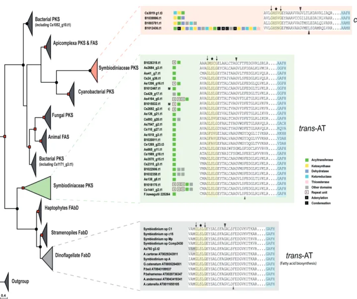
Chapter 2| Secondary metabolite genes in Symbiodiniaceae 19 An unusual feature among the three genomes is the high number (26) of trans-AT genes in contrast to cis-AT (4). A phylogenetic tree of the AT domain consisted of two main nodes, cis-AT and trans-AT (Bayesian Inference posterior probability, 1.00 and maximum likelihood probability, 81%) (Figure 2.2), deviating from the classical substrate-based clustering (Khayatt et al., 2013). Alignment of the trans-AT motif revealed a deviation from the usual GHSxG conserved motif to GLSxG where x can be any residue; thus, a change from a basic amino acid (histidine) to an aliphatic one (leucine) while cis-AT maintained their GHSxG motif. The implication of His à Leu remains to be investigated (Figure 2.2). Use of the HMMs by Khayatt et al. (2013) did not suggest any clear distinction regarding which substrates are being incorporated into biosynthetic pathways. However, I-TASSER predicted that most Symbiodinium AT sequences pertain to the family of malonyl-CoA ACP transferase.
Downstream of the active site serine, a motif (YASH or HAFH) is involved in the choice of either methylmalonyl-CoA or malonyl-CoA, respectively (Tang et al., 2006). The motif, GAFH, present in most Symbiodinium sequences reflects the prediction of I-TASSER. ~9 % (3/33) of AT gene models contained the cTP or mTP signals (Figure 2.2).
Comparative visualization of PKS-containing scaffolds from the three genomes showed
extensive duplication events in the three clades between genes associated with polyketide
biosynthetic clusters (Figure 2.3a). Genomic synteny was observed between clades B1 and A3
(8 syntenic blocks), clades B1 and C (10 syntenic blocks), and clades A3 and C (7 syntenic
blocks) (Figure 2.3b-d), respectively while only four PKS-containing gene clusters were found
to be shared among all the three clades (green boxes in Figure 2.3b-d). The observed
rearrangements within the syntenic scaffolds included mainly deletions. Transposons were
found on scaffolds carrying PKS- and NRPS-encoding genes, suggesting that these genes can
be influenced by transposable elements. 47% (52/110) of PKS- and 34% (14/41) NRPS-
containing scaffolds possessed LTR signatures (Appendix Table B). Taken together, these
results indicate that PKS genes have diversified in each Symbiodinium clade by several evolutionary processes.
Figure 2.2 | Phylogenetic analysis of acyltransferase (AT) domain of eukaryotic and
prokaryotic polyketide and fatty acid synthases. A clear demarcation between cis- and
trans-AT is detectable. Bayesian inference posterior probability are shown by dots (0.70-0.89)
and square (0.9-1.0). Black triangles show conserved residues characteristic to specific
substrate groups, asterisk indicates active site residue, and black arrows indicate conserved
residues used by HMM (Khayatt et al.,2013). C, M and S depict chloroplast, mitochondria, and
secretory signal peptide, respectively.
Chapter 2| Secondary metabolite genes in Symbiodiniaceae 21
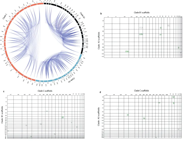
Figure 2.3 | Pathway duplication and conservation within and across Symbiodiniaceae. (a)
Plot showing duplicate gene distribution within PKS-containing scaffolds of three
Symbiodiniaceae genomes. Colored sections (black = clade B1, orange = clade C, blue = clade
A3) represent scaffolds studied in Fig. 1. A link represents a possible duplication event between
two domains. (b) Synteny plot of clade A3 and B1 PKS-containing scaffolds. (c) Synteny plot
of clade B1 and C PKS-containing scaffolds. (d) Synteny plot of clade A3 and C PKS-
containing scaffolds. Dotted boxes highlight regions of significant homology between
genomes. Green colored dotted boxes show common regions shared among the three genomes.
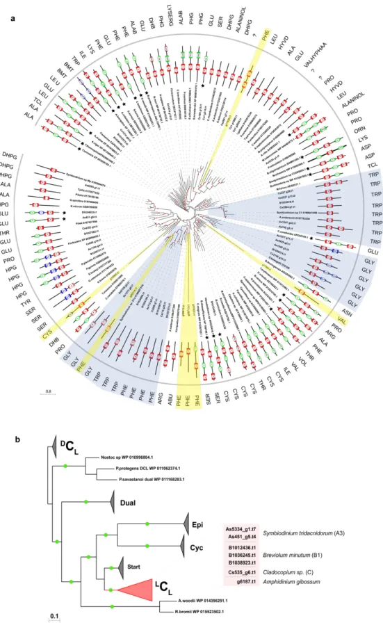
2.3.2 Phylogenetic analysis of adenylation and condensation domain subtypes (
LC
L,
DC
L, Cyc and dual E/C) in NRPS proteins
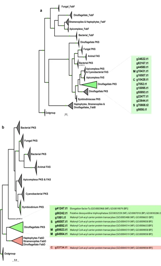
To get a better understanding of freestanding A-domains identified in Symbiodiniaceae genomes, as to whether they obey the same non-ribosomal code of traditional NRPS systems (Stachelhaus et al., 1999), a phylogenetic analysis involving 117 adenylation sequences from several taxa was performed. One significant result was that a freestanding A-domain from Symbiodiniaceae falls into three major groups that utilize tryptophan, glycine, and phenylalanine as substrates, respectively (three highlighted clades in Figure 2.4a). On the contrary, other proteins with di- or multi-domains demonstrated affinity for various substrates.
Phylogenetic analysis of condensation domains was directed by functional categories of C- domains instead of species phylogeny or substrate specificity alone. Four specific functional categories were clearly supported, namely (1) ordinary C-domains, that are composed of
LC
Land
DC
L, (2) heterocyclization (Cyc) domains, (3) dual E/C domains and (4) starter domains, which are found on initiation modules (Figure 2.4b). NaPDOS classification showed that Symbiodiniaceae are rich in
LC
Lsubtypes, which catalyze the condensation of two L -amino acids. Both catalysts possess a conserved His-motif in their active sites with a consensus sequence of HHxxxDG, where x can be any residue. This survey revealed the existence of six condensation domains with the consensus motif being maintained, except for G being substituted with L and N in B1036245.t1 and Cs535_g6.t1, respectively. This analysis also confirms the close relationship between
LC
Land starter C domains and between dual E/C and
D
C
Ldomains, as previously reported in bacterial genomes, adding reliability of this analysis
(Rausch et al., 2007). These results show that NRPS genes are specific for certain amino acids,
thus contributing to a degree of chemical diversity in non-ribosomal peptide biosynthesis.
Chapter 2| Secondary metabolite genes in Symbiodiniaceae 23
Figure 2.4 | Phylogenetic comparison of adenylation (A) and condensation (C) domains of prokaryotic and eukaryotic NRPS. A posterior probability ³ 0.70 generated by Bayesian inference is indicated by dots. (a) Analysis of adenylation domains shows specificity of monofunctional domains from Symbiodiniaceae toward glycine, tryptophan, and phenylalanine (boxed by blue). (b) Condensation domains from Symbiodiniaceae belong to the
L