はじめに M蛋白血症を伴う末梢神経障害には悪性リンパ腫が含ま れているが,馬尾症状が初発症状の悪性リンパ腫で,M 蛋白 血症を伴うものは報告がない.今回われわれは,馬尾症候群 で発症し,M 蛋白血症を呈した悪性リンパ腫を経験したので 報告する. 症 例 症例:63 歳男性 主訴:両下肢筋力低下,感覚障害,膀胱直腸障害 既往歴:11 歳 虫垂炎,48 歳 腰椎椎間板ヘルニア,60 歳 前立腺肥大症 家族歴:父 胃癌 現病歴:2016 年 9 月上旬より左臀部から左下肢背側,左足 背にかけてしびれを自覚するようになった.9 月中旬には左 下肢の疼痛,脱力,排尿障害が出現し,近医整形外科を受診 しプレガバリンを処方され疼痛は軽減した.しかし,排尿障 害と筋力低下は持続し,9 月下旬に泌尿器科を受診して間欠 的自己導尿を開始した.10 月下旬には右下肢筋力低下も出現 し,歩行困難となった.当院整形外科で施行した腰椎 MRI で は椎体や椎間板の変形を認めるが症状を説明しうる所見はな く,当科紹介受診し 11 月上旬に精査目的に入院となった. 入 院 時 現 症: 一 般 身 体 所 見 で は, 体 温 37.0°C, 血 圧 104/58 mmHg,脈 47/min・整,表在リンパ節および腹部腫瘤 は触知しなかった.胸腹部に明らかな異常は認めなかった. 神経学的所見:意識は清明.認知機能障害はなく,脳神経 領域に異常は認められなかった.運動系では,上肢の筋力は, 近位・遠位とも保たれているが,下肢の徒手筋力試験(MRC) は腸腰筋で右 4/ 左 5,大腿屈筋で右 5/ 左 4,前脛骨筋で右 2/ 左 4,腓腹筋で右 5/ 左 4 であった.感覚系では両側 L4 領域 で表在覚鈍麻,L5 領域以下の表在覚は消失し,肛門周囲の触 覚痛覚も消失していた.左下肢優位に振動覚や位置覚の低下 を認めた.腱反射は,両側上腕二頭筋反射,両側上腕三頭筋 反射,両側腕橈骨筋反射は正常であるが,両側膝蓋腱反射と アキレス腱反射が消失し,病的反射は認めなかった.Lasegue 徴候は陰性,つかまり立ちは可能だが自力歩行は不可能で あった.自律神経系では,膀胱直腸障害と便秘を認め,間欠 的自己導尿を要した. 入院時検査所見:血算,赤血球沈降速度,一般尿所見に異 常を認めなかった.血液生化学では,LDH 453 IU/l(正常 110 ~210 U/l),CRP 4.45 mg/dl(0.00~0.14 mg/dl).肝および腎 機能は正常であった.免疫学的検査では免疫グロブリンは IgGが 2,585 mg/dl(861~1,747 mg/dl)と高値で,免疫固定電 気泳動法にて IgG-κ 型の M 蛋白血症を認めた.可溶性 IL-2 受 容体(sIL-2R)4,490 U/ml(121~613 mg/dl),β2 ミクログロ ブリン 3.2 mg/dl(0.9~2.0 mg/dl),クリオグロブリン陰性,尿 ベンスジョーンズ蛋白陰性であった.髄液検査では,総蛋白 189 mg/dl(0~40 mg/dl),細胞数 9/μl(単核球 7/μl,多核球 2/μl),
症例報告
M
蛋白血症を伴った悪性リンパ腫による馬尾症候群の 1 例
小西 宏史
1)*
田口 芳治
1)山本 真守
1)温井 孝昌
1)道具 伸浩
1)中 裕司
1) 要旨: 症例は 63 歳男性.左臀部から左下肢のしびれ,下肢筋力低下,膀胱直腸障害などの馬尾症候群を発症し, 亜急性に症状が進行し入院した.腰仙髄造影 MRI にて馬尾下部領域に造影効果を認め,神経伝導検査では両側脛 骨神経の導出が不良で,腓骨神経は F 波潜時の延長を認めた.M 蛋白血症を認め,多発性骨髄腫を疑ったが,血 清可溶性 IL-2 受容体が 4,490 U/ml と著高で,FDG-PET にて椎体,馬尾の集積増加を認めた.L4 椎弓根生検にてび 漫性大細胞型 B 細胞性リンパ腫と診断し,R-CHOP(rituximab, cyclophosphamide, hydroxydaunorubicin, oncovin, prednisone(prednisolone))療法を行い症状の改善が得られた.M 蛋白血症を伴う馬尾症候群の場合,悪性リンパ 腫の可能性も考慮すべきである.(臨床神経 2018;58:223-228)
Key words: び漫性大細胞型 B 細胞性リンパ腫,neurolymphomatosis,M 蛋白血症,馬尾症候群
*Corresponding author: 富山大学附属病院神経内科〔〒 930-0194 富山県富山市杉谷 2630〕
1)富山大学附属病院神経内科
(Received August 4, 2017; Accepted February 12, 2018; Published online in J-STAGE on March 31, 2018) doi: 10.5692/clinicalneurol.cn-001079
ミエリン塩基性蛋白 579 pg/ml(0~102 pg/ml),sIL-2R 218 U/ml (参考値).髄液細胞診は Class I であった.神経伝導検査では, 両側脛骨神経の運動神経速度,F 波が導出不可能であり,両 側腓骨神経の運動神経伝導速度は正常だが F 波潜時延長と F波導出率の低下を認めた(Table 1).胸腹部単純 CT では, 多発する腫大した大動脈周囲リンパ節を認めた.腰仙髄造影 MRIにて,造影効果を伴う腫大した馬尾を認め(Fig. 1),FDG-PETでは,馬尾や L4 椎体,L5 椎体,L4 椎弓,リンパ節への 集積を認めた(Fig. 2). 入院後経過(Fig. 3):画像検査より,馬尾由来の腫瘍を疑 い,FDG-PET で集積を認めた L4 椎弓の生検を行った.その 結果小型~中型の不整な核を有する腫瘍細胞を認め,核の大 小不同や核小体がめだち,免疫染色では CD20 陽性細胞を び漫性に認めたことから,び漫性大細胞型 B 細胞性リンパ 腫(diffuse large B cell lymphoma; DLBCL)であるとの診断に 至った(Fig. 4).M 蛋白血症を認めたことから多発性骨髄腫 も鑑別に挙げたが,CD138 染色で染色される細胞は軽度で形 質細胞増多は認めず,多発性骨髄腫は否定的であった.デキ サメタゾンにて治療を開始し,病理で確定診断後,R-CHOP (rituximab, cyclophosphamide, hydroxydaunorubicin, oncovin,
prednisone(prednisolone))療法を開始した.その結果,治療 開始 1 か月後には下肢の疼痛や感覚障害,筋力低下,尿閉は 改善傾向となった.治療開始 2 か月後には sIL-2R 493 U/ml (正常 122~496 U/ml),IgG 1,236 mg/dl と著明に低下し,正常 化したが,蛋白分画検査(免疫電気泳動)では M 蛋白様バン ドを認めた.治療開始 5 か月後の神経伝導検査では,脛骨神 経が導出可能となり,右 48.2 m/s,CMAP 0.46 mV,左 42.4 m/s CMAP 0.16 mVとなった.また,脛骨神経の F 波潜時 30.4 ms (右),37.3 ms(左),導出率 100%(右),75%(左)と改善 し,腓骨神経も F 波の導出率が 50%(右),44%(左)と改 善した.蛋白分画検査(免疫電気泳動)では,治療 8 か月後 でも M 蛋白様バンドを認めた.造影 MRI では馬尾の造影効 果は軽減し,また FDG-PET でも馬尾病変や腰椎,大動脈周 囲リンパ節などの集積増加は軽減した. 考 察 本例は,下肢感覚障害,筋力低下,膀胱直腸障害で発症し た DLBCL である.しかし,M 蛋白血症を伴い,当初は M 蛋 白血症を呈する末梢神経障害,特に慢性炎症性脱髄性多発 ニューロパチー,抗ミエリン糖蛋白抗体を伴う脱髄性ニュー ロパチー,多発性骨髄腫,クロウ・深瀬症候群(POEMS 症 候群),原発性アミロイドーシス,サルコイドーシス,肝硬 変,関節リウマチなどとの鑑別を要した1).また,馬尾症候 群を呈した.馬尾症候群は,第 2 腰椎以下の神経根障害によっ て生じる.症状としては,非対称性の運動・感覚障害,下肢 深部腱反射減弱などを呈する2).原因疾患として,腰部脊柱 管狭窄症が最多であるが,感染症や髄膜炎などの内科疾患に 伴う症例も散見される3)4).リンパ腫も鑑別すべき疾患の一つ であり,馬尾症候群を呈したリンパ腫の報告も散見される. 特に最近は血管内リンパ腫(IVL)の報告が認められる.馬 尾症候群を呈したリンパ腫について,渉猟し得た報告を示す (Table 2)5)~13)が,これらの中で M 蛋白血症について記載のあ る報告は認めない.M 蛋白血症を有する polyneuropathy とし Motor NCV (m/s) Distal latency (ms) Amplitude (mV) F-wave latency (ms) F-wave frequency (%) Left tibial Before not evoked
After 42.4 7.3 0.16 37.7 75
Right tibial Before not evoked
After 48.2 7.3 0.46 30.4 100
Left peroneal Before 39.1 8.3 0.33 48.3 6.3
After 36.0 6.2 0.017 38.4 44
Right peroneal Before 40.5 8.3 1.25 50.8 50
Aftrer 42.5 5.3 0.11 30.1 44 Sensory NCV (m/s) Distal latency (ms) Amplitude (μV)
Left sural Before 55.4 3 7.2
After 52.9 1.9 9.5
Right sural Before 52.6 3.8 6.7
After 54.7 1.6 11.7
Motor nerve conduction velocities of both tibial nerves were not evoked. Both side of peroneal nerves were derived, but decline of F-wave suggest an obstacle of a nervous roots.
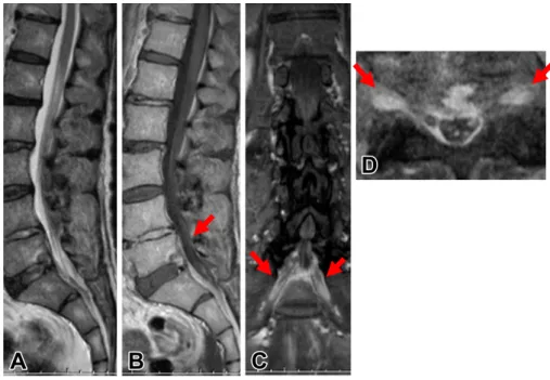
ては,上記の疾患があげられるが,これらの疾患は免疫介在 性機序などによる末梢神経障害が主であり,本例はlymphoma の直接浸潤と考えられ,機序が異なると考えられる.比較的 急速に進行する馬尾障害の際,悪性リンパ腫を考慮する必要 があるが,その際,骨髄腫の鑑別も必要である.悪性リンパ 腫でも M 蛋白を呈することがあり,慎重に生検を行い,確定 診断に至り,治療方針を決定する必要がある. 本例では,治療後の経過観察では,M 蛋白は陽性(免疫電 気泳動法)であるが,IgG は正常化した. 本例の神経伝導検査では,治療 5 か月後の follow にて,特に Fig. 1 Lumbosacral MRI findings.
T2-weighted sagittal image (1.5 T, TR 4,500 ms, TE 98 ms) showed no abnormality (A). Gadlinium-enhanced T1-weighted sagittal image (1.5 T, TR 500 ms, TE 11 ms) showed enhancement of cauda equine (arrow) (B). Gadlinium-enhanced T1-weighted coronal image (1.5 T, TR 676 ms, TE 11 ms) showed enhancement of bilateral L5 narve roots (arrows) (C). Gadlinium-enhanced T1-weighted axial (1.5 T, TR 538 ms, TE 11 ms) and coronal (1.5 T, TR 538 ms, TE 11 ms) images showed enhancement of bilateral L5 narve roots (arrows) (C, D).
A
B
Fig. 2 FDG-PET findings.
Sagittal image showed accumulation localized in the fourth and fifth lumbar vertebra, cauda equine (A). Vertebral image showed accumulation locaied in the cauda equine of fifth lumbar vertebra (B).
Fig. 4 Clinical course.
To diagnosis, we performed a biopsy of the fourth vertebral arch. After diagnosis. the patient received dexamethasone and R-CHOP (rituximab, cyclophosphamide, hydroxydaunorubicin, oncovin, prednisone (prednisolone)) chemotherapy. Neurological symptoms were improved significantly.
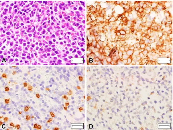
Fig. 3 Biopsy of the fourth vertebral arch findings (Bar = 20 μm).
A plasmocyte wasnʼt admitted by a hematoxylin and eosin stain (A). Some large cells were positive for CD20 staining, (B). Few cell were positive for CD3 staining. (C). A little cells were positive for CD138 staining (D).
脛骨神経にて NCV が改善しているが,CMAP 振幅は低値であ り,軸索障害が残存していると考えられる.軸索障害と脱髄障 害が併存し,脱髄障害の改善を速やかに認めたと考えられる. 本報告の要旨は,第 147 回日本神経学会東海・北陸地方会で発表 し,会長推薦演題に選ばれた. ※著者全員に本論文に関連し,開示すべき COI 状態にある企業,組 織,団体はいずれも有りません. 文 献 1) 桑原 聡.M 蛋白血症と末梢神経障害.日内会誌 2012;101: 2238-2241. 2) 佐藤 功,衣斐 達,中尾直樹ら.脊髄円錐・馬尾症候群― 基礎疾患―.神経内科 1998;49:7-17.
3) Candy S, Chang G, Andronikou S. Acute myelopathy or cauda equine syndrome in HIV-positive adults in a tuberculosis endemic setting: MRI, clinical, and pathologic findings. AJNR Am J Neuroradiol 2014;35:1634-1641.
4) Sailou V, Bailly P, Thomas E, et al. Cauda equine syndrome after Neisseria meningiidis type Cmeningitis. Press Med 2015;44: 567-569.
5) Mauney M, Scioto CG. Primary malignant lymphoma of the cauda equine. Am J Surg Pathol 1983;7:185-190.
6) Knopp EA, Chynn KY, Hughes J. Primary lymphoma of the cauda equine: myelographic, CT myelographic, and MR appearance. AJNR Am J Neuroradiol 1994;8:1187-1189. 7) Giobbia M, Carniato A, Scotton PG, et al. Primary
EBV-associated cauda equina lymphoma. J Neurol 1999;246:739-740. 8) Zagami AS, Granot R. Non-Hodgkin’s lymphoma involving the
cauda equina and ocular cranial nerves: case reports and literature review. J Clin Neurosci 2003;10:696-699.
9) Tajima Y, Sudo K, Matumoto A. Malignant lymphoma originating in the cauda equina mimicking the inflammatory polyradiculo neuro-pathy. Intern Med 2007;46:1029-1032.
10) Khong P, Pitham T, Owler B. Isolated neurolymphomatosis of the cauda equina and filum terminale: case report. Spine (Phila Pa 1976) 2008;33:E807-E811.
11) Morita M, Osawa M, Naruse H, et al. Primary NK/T-cell lymphoma of the cauda equina: a case report and literature review. Spine (Phila Pa 1976) 2009;34:E882-E885.
12) Nishida H, Hori M, Obara K. Primary B-cell lymphoma of the cauda equina, successfully treated with high-dose methotrexate plus high-dose cytarabine: a case report with MRI findings. Neurol Sci 2012;33:403-407.
13) 中島秀樹,本村正勝,山口 将ら.馬尾生検が診断に有用で あった髄膜播種性の悪性リンパ腫の 1 例.臨床神経 2013;53: 803-808.
Table 2 Lymphoma with syndrome of cauda equine.
Auther (year) Age/sex Cell type M-protain Therapy Others
Mauney5) (1983) 68/F B cell N.D. RT
Knopp6) (1994) 69/F N.D. N.D. N.D
Giobbia7) (1999) 30/F B cell N.D. RT/CT EBV positive
Zagami8) (2003) 71/F B cell N.D. CT MTX + CHOP
Tajima9) (2007) 67/F B cell N.D. RT/CT MTX
Khong10) (2008) 16/M B cell N.D. RT/CT CODOX-M
Morita11) (2009) 67/M NK/T cell N.D. RT
Nishida12) (2012) 47/M B cell N.D. CT/RT MTX, Ara-C
Nakashima13) (2013) 59/M B cell N.D. RT/CT MTX
This case 63/M B cell IgGκ CT R-CHOP
RT: radiotherapy, CT: chemotherapy, N.D.: not descrived, EBV: EB virus, MTX: methotrexate, CHOP: cyclophosphamide, hydroxydaunorubicin, oncovin, prednisone (prednisolone), R-CHOP: rituximab, cyclo-phosphamide, hydroxydaunorubicin, oncovin, prednisone (prednisolone), CODOX-M: cyclocyclo-phosphamide, vincristine, doxorubicin, methotrexate, Ara-C: cytarabine.
We searched ceses of lymphoma with syndrome of cauda equine, and there were no cases with M- proteinemia.
A case of neurolymphomatosis presented as cauda equine syndrome
accompanied with M-proteinemia
Hirofumi Konishi, M.D.
1), Yoshiharu Taguchi, M.D., Ph.D.
1), Mamoru Yamamoto, M.D.
1),
Takamasa Nukui, M.D., Ph.D.
1), Nobuhiro Dougu, M.D., Ph.D.
1)and Yuji Nakatsuji, M.D., Ph.D.
1)1)Department of Neurology, Toyama University Hospital