Title
Diffuse Skeletal Muscles Uptake of [18F] Fluorodeoxyglucose on
Positron Emission Tomography in Primary Muscle Peripheral
T-cell Lymphoma( 本文(Fulltext) )
Author(s)
TANAKA, Yuji; HAYASHI, Yuichi; KATO, Jun'ichi;
YAMADA, Megumi; KOUMURA, Akihiro; SAKURAI, Takeo;
KIMURA, Akio; HOZUMI, Isao; HATANO, Yuichiro; HIROSE,
Yoshinobu; TAKAMI, Tsuyoshi; NAKAMURA, Hiroshi;
KASAHARA, Senji; TSURUMI, HIsashi; MORIWAKI, Hisataka
Citation
[Internal Medicine] vol.[50] no.[18] p.[2021]-[2024]
Issue Date
2011
Rights
The Japanese Society of Internal Medicine (一般社団法人日本
内科学会)
Version
出版社版 (publisher version) postprint
URL
http://hdl.handle.net/20.500.12099/47179
Diffuse Skeletal Muscles Uptake of [
18F] Fluorodeoxyglucose
on Positron Emission Tomography in Primary Muscle
Peripheral T-cell Lymphoma
Yuji Tanaka
1, Yuichi Hayashi
1, Jun’ichi Kato
1, Megumi Yamada
1, Akihiro Koumura
1,
Takeo Sakurai
1, Akio Kimura
1, Isao Hozumi
1, Yuichiro Hatano
2, Yoshinobu Hirose
2,
Tsuyoshi Takami
3, Hiroshi Nakamura
4, Senji Kasahara
4, Hisashi Tsurumi
4,
Hisataka Moriwaki
4and Takashi Inuzuka
1Abstract
A 40-year-old man presented with weakness of neck extensor muscles. Cervical magnetic resonance imag-ing showed high-intensity areas in muscles of the left lateral cervical region on T2-weighted images. Fluorodeoxyglucose-positron emission tomography scan demonstrated striking fluorodeoxyglucose uptake by multiple skeletal muscles of the neck, chest, and abdominal region. Muscle biopsy demonstrated peripheral T-cell lymphoma, unspecified. The diagnosis was primary skeletal muscle peripheral T-T-cell lymphoma. Primary skeletal muscle non-Hodgkin’s lymphoma of T-cell immunophenotype is extremely rare and fluorodeoxyglucose-positron emission tomography demonstrated striking fluorodeoxyglucose uptake in multi-ple skeletal muscles and served as a quite useful modality for the diagnosis of this patient.
Key words: primary skeletal muscle lymphoma, non-Hodgkin lymphoma, peripheral T-cell lymphoma,
fluorodeoxyglucose on positron emission tomography, skeletal muscle
(Intern Med 50: 2021-2024, 2011) (DOI: 10.2169/internalmedicine.50.4992)
Introduction
Primary skeletal muscle non-Hodgkin’s lymphoma is ex-tremely uncommon and it is associated with a poor progno-sis (1). Additionally, the majority of these lymphomas dem-onstrate B-cell immunophenotype (1). We report a case with primary skeletal muscle non-Hodgkin’s lymphoma of T-cell immnophenotype, that showed slow progression, character-ized by striking fluorodeoxyglucose uptake involving multi-ple skeletal muscles on positron emission tomography scans.
Case Presentation
In January 1996, a 27-year-old man had swelling of the
right side of the labia and oral mucosa. In April 1996, the patient visited a hospital and was diagnosed with chronic lymphadenitis by a biopsy of the oral mucosa. Oral steroid therapy improved the symptoms. In July 1996, the patient had left ptosis, and left facial weakness. Steroid therapy again improved the symptoms. In September 1998, the pa-tient had diplopia and bilateral facial weakness (manual muscle test (MMT): 1/1). The symptoms were not improved by steroid therapy and remained constant. In May 2000, the patient developed weakness of the neck extensor (MMT: 4/ 4). After several months, the symptoms disappeared sponta-neously. Thereafter, the patient remained free of any new symptoms for the subsequent 9 years.
In May 2009, at the age of 40 years, weakness of neck extensor appeared again. The patient was admitted to our
1Department of Neurology and Geriatrics, Graduate School of Medicine, Gifu University, Japan,2Department of Pathology, Gifu University
Hospital, Japan,3Department of Immunopathology, Graduate School of Medicine, Gifu University, Japan and4The First Department of Internal
Medicine, Graduate School of Medicine, Gifu University, Japan
Received for publication December 13, 2010; Accepted for publication May 13, 2011 Correspondence to Dr. Yuji Tanaka, [email protected]
Intern Med 50: 2021-2024, 2011 DOI: 10.2169/internalmedicine.50.4992
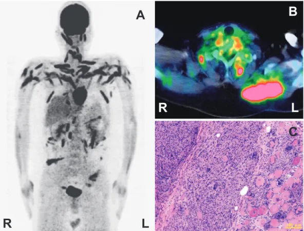
Figure 1. Fluorodeoxyglucose-positron emission tomography (PET) and biopsy of the muscle. (A)
(B) Fluorodeoxyglucose-positron emission tomography (PET): Fluorodeoxyglucose-positron emis-sion tomography (PET) demonstrated striking fluorodeoxyglucose uptake involving the skeletal muscles of the neck, chest, and abdominal region. The maximum standardized uptake value of the left trapezius muscle was 10.32. (C) Biopsy specimen of the muscle: Muscle biopsy of the left trape-zius muscle demonstrated massive infiltration of atypical lymphocytes of varying sizes and clusters of epithelioid histiocytes destroying muscle bundles.
C
L
R
A
B
R
L
hospital. Neurologic evaluation showed weakness of the left pectoral girdle (MMT: 4), restricted bilateral eye movement, and facial weakness. However, no other muscle showed the weakness. The patient did not have myalgia. The patient did not appear to show a loss of weight, fever or sweating such as seen in B symptom. In the serum, creatine phosphokinase (CK) and soluble interleukin-2 receptor were high 311 IU/L (normal range, 40-200 IU/L) (CK-MM 95%) and 625 U/mL (normal range 145-519 U/mL), and continued to rise. Anti-human T-cell lymphotropic virus antibody and anti-Anti-human immunodeficiency virus antibody were negative. Cervical magnetic resonance imaging (MRI) showed high-intensity areas in the muscles of the left lateral cervical region on T2-weighted images. The muscles of other regions did not dem-onstrate abnormal findings on MRI. Fluorodeoxyglucose-positron emission tomography demonstrated striking fluoro-deoxyglucose uptake by multiple skeletal muscles of the neck, chest, and abdominal region, but not of the lower ex-tremities (Fig. 1A, 1B). Maximum standardized uptake value of the left trapezius muscle was 10.32. Ga scintigraphy showed no abnormal uptake. Computed tomography of the whole body revealed no evidence of lymphadenopathies, tu-mor infiltrations, or masses. There were no disease
abnor-malities involving the osseous structure. Bone marrow bi-opsy revealed a normocellular marrow with trilineage he-mopoiesis and no evidence of lymphoma. Biopsy of the left trapezius muscle demonstrated massive infiltration of atypi-cal lymphocytes which varied in size and clusters of epithe-lioid histiocytes destroying muscle bundles (Fig. 1C). Immu-nohistochemical staining revealed that the atypical lympho-cytes were positive for CD3 and CD8, but negative for CD4, CD20, CD56, CD79a, and EBER. Histopathologic and immunocytochemical characteristics were consistent with those of peripheral T-cell lymphoma, unspecified (PTCL-U). The final diagnosis was established as primary skeletal mus-cle peripheral T-cell lymphoma. Chemotherapy with 6 cymus-cles of cyclophosphamide, doxorubicin, vincristin and prednisone completely resolved the weakness of the left pectoral girdle, but it did not improve the eye movement or facial weakness. On serum analyses, CK and IL2R were decreased to the normal range. Fluorodeoxyglucose-positron emission to-mography and magnetic resonance imaging improved after chemotherapy. The maximum standardized uptake value of the left trapezius muscle was reduced. After 6 months of chemotherapy, the symptoms remained the same.
Discussion
A 40-year-old man was diagnosed with primary skeletal muscle peripheral T-cell lymphoma. Multiple neurologic deficits had appeared for 13 years from the initial symp-toms. The patient had been diagnosed with idiopathic or un-known disease because there was no specific finding. Al-though not definite, if those symptoms were related to lym-phoma, this case showed very slow progression. Fluorodeoxyglucose-positron emission tomography demon-strated striking fluorodeoxyglucose uptake by multiple skeletal muscles and was useful in detecting disseminated lymphoma in the skeletal muscles.
Primary skeletal muscle non-Hodgkin’s lymphoma is a rare manifestation of non-Hodgkin’s lymphoma, accounting only for 0.1% of all lymphomas (1). The frequency of pri-mary skeletal muscle non-Hodgkin’s lymphoma (7%) is in-creased in patients with Acquired Immune Deficiency Syn-drome (AIDS)-associated lymphomas (2). It is difficult to differentiate between primary skeletal muscle lymphoma and secondary muscle involvement from bone lymphoma. The following criteria is used to diagnose primary muscular lym-phoma: 1) histopathology proven non-Hodgkin’s lymphoma of the muscle; 2) the absence of systemic/nodal disease at initial presentation; and 3) the presence of a soft tissue mass with normal adjacent marrow or marrow disease much less extensive than soft tissue (3). The present case was diag-nosed as primary skeletal muscle non-Hodgkin’s lymphoma because these three 3 criteria were fulfilled. In cases of pri-mary skeletal muscle non-Hodgkin’s lymphoma the lower extremities and pelvic region are most commonly af-fected (4). Additionally, the majority of reported cases had B-cell immunophenotypes and behaved in an aggressive bio-logic manner (5). T-cell neoplasms constitute 10% to 12% of all non-Hodgkin’s lymphoma (6). PTCL-U represents the most common subtype of T-cell lymphomas, manifesting most often as a nodal disease (7). Extranodal PTCL-U is a rare presentation among neoplasms. Some cases of primary skeletal muscle T-cell lymphomas have been described in the literature (8-12). Furthermore, in a few reported cases fluorodeoxyglucose-positron emission tomography demon-strated striking fluorodeoxyglucose uptake in multiple skele-tal muscles.
Fluorodeoxyglucose-positron emission tomography scans demonstrate multiple sites of hypermetabolic activity within musculature. A recent study suggests that fluorodeoxyglu-cose-positron emission tomography scans are sensitive to de-tect T-cell lymphomas in all regions except for the skin (13). Fluorodeoxyglucose-positron emission tomography is useful in aiding the diagnosis and staging of lymphomas of aggres-sive subtypes, including PTCL-U (14). However, other rea-sons for skeletal muscle fluorodeoxyglucose uptake may stem from a variety of factors such as physical exertion, talking, and chewing as normal physiological phenom-ena (15). As for pathological phenomphenom-ena, elevated
fluorode-oxyglucose muscle may be associated with anxiety-induced neck muscle spasm (15), accessory muscle use in respiratory distress (16), intercostal and diaphragmatic activity in chronic obstructive pulmonary disease (17), abdominal mus-cle contraction in intractable vomiting (18), sepsis-induced shivering (19), hypoglycemia (20), infection, and inflamma-tion of the muscle tissue that increases glycolytic activ-ity (21, 22). In the present case, the patient did not satisfy these conditions. Skeletal muscle fluorodeoxyglucose uptake was induced by T-cell lymphoma, which histopathologically proved PTCL-U.
In conclusion, primary skeletal muscle non-Hodgkin’s lymphoma of T-cell immunophenotype is extremely rare. Fluorodeoxyglucose-positron emission tomography demon-strated striking fluorodeoxyglucose uptake in multiple skele-tal muscles, and served as a useful method for diagnosing this case.
Informed consent was obtained from this patient.
The authors state that they have no Conflict of Interest (COI).
Acknowledgement
The authors are grateful thanks to Rie Tsuburaya, Yukiko K Hayashi, Ichizo Nishino, and Ikuya Nonaka, at the Department of Neuromuscular Research, National Institute of Neuroscience, National Center of Neurology and Psychiatry for histopathologi-cal examination.
References
1. Travis WD, Banks PM, Reiman HM. Primary extranodal soft
tis-sue lymphoma of the extremities. Am J Surg Pathol 11: 359-366, 1987.
2. Raphael M, Gentilhomme O, Tulliez M, Byron PA, Diebold J.
Histopathologic features of high-grade non-Hodgkin’s lymphomas in acquired immunodeficiency syndrome. The French Study Group of Pathology for Human Immunodeficiency Virus-Associated Tu-mors. Arch Pathol Lab Med 115: 15-20, 1991.
3. Lee VS, Martinez S, Coleman RE. Primary muscle lymphoma:
clinical and imaging findings. Radiology 203: 237-244, 1997.
4. Ueno A, Kuribayashi K, Iyama S, et al. Two cases of primary
skeletal muscle lymphoma and a review of the literature. Rinsho Ketsueki 46: 1141-1145, 2005 (in Japanese).
5. Jeffery GM, Golding PF, Mead GM. Non-Hodgkin’s lymphoma
arising in skeletal muscle. Ann Oncol 2: 501-504, 1991.
6. Ralfkieer E, Muller-Hermelink HK, Jaffe ES. T-cell and NK-cell
neoplasms. In: Pathology and Genetics of Tumours of the Haema-topoietic and Lymphoid Tissues: World Health Organization Clas-sification of Tumours. Jaffe E, Harris N, Stein H, Vardiman J, Eds. IARC Press, Lyon, France, 2001: 227-229.
7. Savage KJ, Chhanabhai M, Gascoyne RD, Connors JM.
Charac-terization of peripheral T-cell lymphomas in a single North Ameri-can institution by the WHO classification. Ann Oncol 15: 1467-1475, 2004.
8. Lum GH, Cosgriff TM, Byrne R, Reddy V. Primary T-cell
lym-phoma of muscle in a patient infected with human immunodefi-ciency virus. Am J Med 95: 545-546, 1993.
9. Chim CS, Choy C, Liang R. Primary anaplastic large cell
lym-phoma of skeletal muscle presenting with compartment syndrome. Leuk Lymphoma 33: 601-605, 1999.
10. Chim CS, Loong F, Ooi GC, Srivastava G, Liang R. Primary
Intern Med 50: 2021-2024, 2011 DOI: 10.2169/internalmedicine.50.4992
11. Alekshun TJ, Rezania D, Ayala E, Cualing H, Frank GA. Skeletal
muscle peripheral T-cell lymphoma. J Clin Oncol 26: 501-503, 2008.
12. Broski SM, Bou-Assaly W, Gross MD, Fig LM. Diffuse skeletal
muscle F-18 fluorodeoxyglucose uptake in advanced primary mus-cle non-Hodgkin’s lymphoma. Clin Nucl Med 34: 251-253, 2009.
13. Kako S, Izutsu K, Ota Y, et al. Fluorodeoxyglucose-PET in T-cell
and NK-cell neoplasms. Ann Oncol 18: 1685-1690, 2007.
14. Gill SI, Gibbs SD, Hicks RJ, Seymour JF. Primary skeletal muscle
marginal zone lymphoma with persistent tissue tropism and PET-avidity. Leuk Lymphoma 47: 117-120, 2006.
15. Cook GJ, Fogelman I, Maisey MN. Normal physiological and
be-nign pathological variants of 18-fluoro-2-deoxyglucose positron-emission tomography scanning: potential for error in interpreta-tion. Semin Nucl Med 26: 308-314, 1996.
16. Bural GG, Mavi A, Kumar R, Alavi A. Fluorodeoxyglucose
up-take in intercostal muscles is an indicator of severe respiratory disease. Clin Nucl Med 29: 807-808, 2004.
17. Aydin A, Hickeson M, Yu JQ, Zhuang H, Alavi A. Demonstration
of excessive metabolic activity of thoracic and abdominal muscles
on fluorodeoxyglucose-PET in patients with chronic obstructive pulmonary disease. Clin Nucl Med 30: 159-164, 2005.
18. Abdel-Dayem HM, Naddaf S, El-Zeftawy H. F-18
fluorodeoxy-glucose gastric and anterior abdominal muscle uptake secondary to nausea and vomiting. Clin Nucl Med 23: 769-770, 1998.
19. Ojha B, Muthukrishnan A, Raman AK, Satya R, Mahone T,
Forero-Torres A. Diffuse F-18 fluorodeoxyglucose uptake in skele-tal muscles secondary to chills resulting from sepsis: a cause for underestimation of viable tumor. Clin Nucl Med 29: 600-601, 2004.
20. Shreve PD, Anzai Y, Wahl RL. Pitfalls in oncologic diagnosis with
fluorodeoxyglucose PET imaging: physiologic and benign vari-ants. Radiographics 19: 61-77, 1999.
21. Bakheet SM, Powe J. Benign causes of 18-fluorodeoxyglucose
up-take on whole body imaging. Semin Nucl Med 28: 352-358, 1998.
22. Kaim AH, Weber B, Kurrer MO, et al. (18)F-fluorodeoxyglucose
and (18)F-FET uptake in experimental soft tissue infection. Eur J Nucl Med Mol Imaging 29: 648-654, 2002.
Ⓒ 2011 The Japanese Society of Internal Medicine http://www.naika.or.jp/imindex.html