ESTROGEN IN WOMEN
Dynamic changes in circulating estradiol level, in-cluding increase at menarche and decrease at meno-pause, occur in a woman’s lifetime. Circulating es-tradiol levels decrease drastically during the meno-pausal transition, though the levels differ among races. It has been reported that estradiol levels in both Japanese and Chinese women were lower
than those in Caucasians, Hispanic and African-Americans (1).
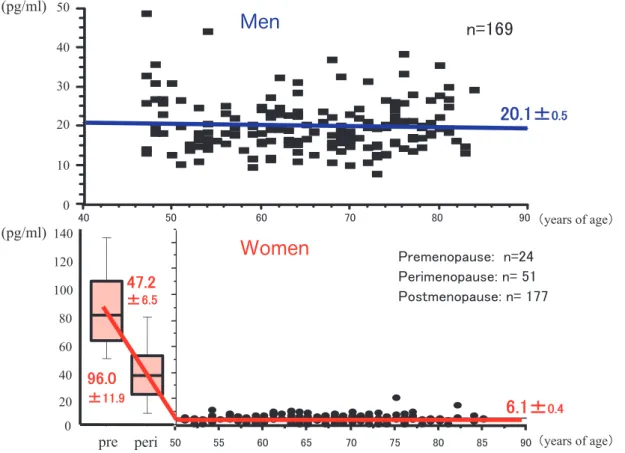
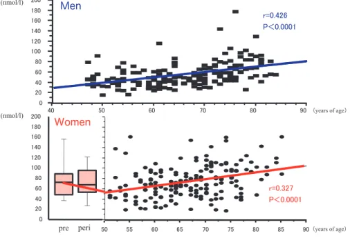
Based on data obtained in a previous study (2), we re-analyzed data for 169 healthy men and 252 healthy women aged from 40 to 85 years in the population-based study with addition of data for premenopausal women. Change in circulating es-tradiol in women from the menopausal transition to postmenopause is characterized as follows : high estradiol level in premenopausal women is drasti-cally decreased and estradiol level in postmeno-pausal women is significantly lower than that in age-matched men. Mean estradiol levels were shown to be 20.1 pg/ml in men and 6.1 pg/ml in postmeno-pausal women (Figure 1). This dynamic decrease in
REVIEW
Androgen in postmenopausal women
Toshiyuki Yasui
1, Sumika Matsui
2, Anna Tani
2, Kotaro Kunimi
2, Satoshi Yamamoto
2,
and Minoru Irahara
21)
Department of Reproductive Technology,2)
Department of Obstetrics and Gynecology, Institute of Health Biosciences, the University of Tokushima Graduate School, Tokushima, Japan
Abstract : Menopausal symptoms, bone loss, changes in lipid profiles and reduction of insu-lin sensitivity due to an abrupt decrease in circulating estrogen level are well known in women during the menopausal transition. On the other hand, the effect of androgen on women’s health has not been fully elucidated. Circulating levels of testosterone and de-hydroepiandrosterone sulfate (DHEA-S) gradually decrease with age in postmenopausal women, although transient increases have been observed during the menopausal transi-tion. High testosterone level has been suggested to be associated with increased risk of cardiovascular disease, increased triglyceride, insulin resistance and increase in the risk of developing breast cancer in postmenopausal women. Circulating DHEA-S level does not affect the risk of cardiovascular disease, mortality or lipid profiles in women. Fe-male androgen insufficiency, which is characterized by the presence of reduced androgen level in circulation, leads to an impairment in sexual drive, reduced libido, depressed mood, and signs and symptoms of limited androgen exposure such as decreased muscle mass, reduced bone density and decreased sense of well-being. An appropriate level of androgen may play important roles in metabolic, psychological and sexual functions in women. In addition, the roles of testosterone and DHEA-S in women’s health may be dif-ferent. J. Med. Invest. 59 : 12-27, February, 2012
Keywords : women, testosterone, DHEA-S, menopause
Received for publication October 19, 2011 ; accepted December 8, 2011.
Address correspondence and reprint requests to Toshiyuki Yasui, Department of Reproductive Technology, Institute of Health Biosciences, the University of Tokushima Graduate School, Tokushima 770 - 8503, Japan and Fax : + 81 - 88 - 631 - 2630.
estradiol level induces menopausal symptoms, such as hot flashes and night sweat, urogenital symptoms, osteoporosis, coronary heart disease, stroke and possibly early onset of Alzheimer’s disease in post-menopausal women as shown in Figure 2.
However, not only estrogen but also other endo-crinological hormones may be involved in the occur-rence of these diseases. Little attention has been paid to roles of endogenous androgens in women despite the results of studies suggesting that an-drogens may play important roles. Anan-drogens are
known to be important for normal physiology in women and to play key roles in the physical, sexual and emotional well-being of women (3). Therefore, it is necessary to take account of androgens as well as estrogen when considering women’s health.
CHANGE IN LEVELS OF ANDROGEN IN
WOMEN
1. Changes in testosterone level in women
In women of reproductive age, daily production of testosterone is shared equally between the ova-ries and adrenal glands and accounts for approxi-mately one-third of the testosterone in circulation. Peripheral conversion of androgen precursor ster-oids to testosterone in non-steroid producing tis-sues accounts for the remaining two-thirds of tes-tosterone in circulation. These ratios change after menopause when the ovaries are in senescence. In women, there is controversy about the direction of circulating testosterone levels across the life span. It has been reported that total and free testoster-one decreased with age between 15 and 60 years (4) and that bioavailable testosterone decreased by approximately 28% between 25 and 85 years of age
Figure 1. Changes in serum estradiol levels in men and women.
upper panel : men, lower panel : women
Figure 2. Increased risks of diseases in postmenopausal
(5). However, it has been shown that testosterone level did not vary during the menopausal transi-tion from 45 to 55 years of age (6). Recent data in-dicate that total testosterone level increased from 43 to 50 years but not thereafter (7). In a previous study, we found that total testosterone level gradu-ally decreased with age in women but that the change was not significant. However, levels of free and bioavailable testosterone showed significant de-creases with age in women (2).
Menopausal transition is characterized by vari-ations in cycle length and elevation in follicle-stimu-lating hormone (FSH) level. Based on these char-acteristics, the American Society for Reproductive Medicine proposed the “Stages of Reproductive Aging Workshop (STRAW) staging system” (8). We also divided 231 healthy women into 7 stages by regularity of menstruation and FSH level : 1) women with regular menstruation cycle and nor-mal FSH level (group A), 2) women with regular menstruation cycle and elevated FSH level (!10 mIU/ml) (late reproductive stage, group B), 3) women with irregular menstruation cycle and ele-vated FSH level (early menopausal transition, group C), 4) women who had irregular menstruation cycle in which the interval of amenorrhea was more than
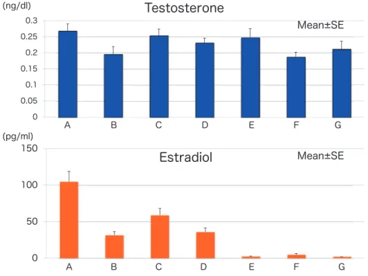
2 months and elevated FSH level (late menopausal transition, group D), 5) women for whom less than 1 year had passed since menopause (group E), 6) women for whom less than 5 years had passed since menopause (group F) and 7) women for whom more than 5 years had passed since menopause (group G). As can be seen in Figure 3, total testos-terone level did not change significantly, though there was a slight increase during the menopausal transition. Changes in free and bioavailable testos-terone showed patterns similar to the pattern of changes in total testosterone. On the other hand, estradiol level was drastically decreased but showed a transient increase in the early menopausal tran-sition, possibly due to an increase in FSH stimula-tion (Figure 3). The ratio of testosterone to estradiol (T/E), as an assessment of the balance of testoster-one and estradiol, gradually increased during the menopausal transition and increased significantly in postmenopausal stages (Figure 4). A relative testos-terone excess was found in postmenopausal women. Torrens et al. reported that a relative androgen ex-cess was found during the menopausal transition and both baseline total T/E ratio and its rate of change were associated with increased incident metabolic syndrome independent of ethnicity (9).
Figure 3. Changes in levels of total testosterone and estradiol during the menopausal transition. upper panel : total testosterone, lower panel : estradiol
Group A : early reproductive stage, Group B : late reproductive stage, Group C : early menopausal transition, Group D : late meno-pausal transition, Group E : women for whom less than 1 year has passed since menopause, Group F : women for whom less than 5 years have passed since menopause, Group G : women for whom more than 5 years have passed since menopause
2. Changes in DHEA-S level in women
Dehydroepiandrosterone (DHEA) and its sulfate (DHEA-S) are steroid hormones mainly produced by the adrenal zona reticularis. The daily production rate of DHEA is 6 to 8 mg, 50% being secreted by the zona reticularis. In women, 20% is secreted by the ovarian theca, while the remaining amount is derived from circulating DHEA-S catalyzed by steroid sulfatase. Changes in DHEA-S levels with age differ among races. Circulating DHEA-S level showed the lowest rate of decline with advancing age in Japanese women and the greatest decline with advancing age in Caucasians (10). In addition, a transient increase in DHEA-S level during late
perimenopause and early postmenopause has been shown. As can be seen in Figure 5, we also showed a transient increase in DHEA-S level in the meno-pausal transition. The rise of DHEA-S during the menopausal transition might be associated with in-crease in luteinizing hormone. Lasley et al. reported that a rise in DHEA-S during the menopausal tran-sition was found in the absence of both ovaries, suggesting that the rise in DHEA-S is most that from the adrenal glands (11). Although its circulat-ing level is the highest of all steroid hormones, little is known about its physiological role. DHEA and DHEA-S were considered to be pro-hormones ex-erting indirect androgenic and estrogenic effects following peripheral conversion into small amounts
Figure 5. Changes in DHEA- S levels during the menopausal transition.
Group A : early reproductive stage, Group B : late reproductive stage, Group C : early menopausal transition, Group D : late meno-pausal transition, Group E : women for whom less than 1 year has passed since menopause, Group F : women for whom less than 5 years have passed since menopause, Group G : women for whom more than 5 years have passed since menopause
Figure 4. Changes in testosterone/estradiol ratio during the menopausal transition.
Group A : early reproductive stage, Group B : late reproductive stage, Group C : early menopausal transition, Group D : late meno-pausal transition, Group E : women for whom less than 1 year has passed since menopause, Group F : women for whom less than 5 years have passed since menopause, Group G : women for whom more than 5 years have passed since menopause
of testosterone and estradiol. However, this concept may change due to the identification of a putative specific DHEA receptor on the plasma membrane of bovine aortic endothelial cells (12).
ACTIONS OF ENDOGENOUS ANDROGENS
1. Female androgen insufficiency
The medical field for testosterone has long ac-cepted the importance of male sexuality, but sexual dysfunction in women and treatment options to ad-dress these concerns have met with great contro-versy. Aging and menopause have been linked to low libido, with 52.4% of naturally menopausal women aged 40-70 years and 36.4% of surgically menopausal women (current age!45 years) who have undergone oophorectomy at less than 45 years of age reporting low sexual desire (13). The decline of androgen levels with ovarian failure and that fol-lowing oophorectomy have sparked the hypothesis that decreased testosterone is related to diminished desire. In 2002, a consensus conference recom-mended that female androgen insufficiency syn-drome be defined by a pattern of clinical symptoms and signs in the presence of decreased free testos-terone and normal estrogen status. Clinical symp-toms of the proposed deficiency state include de-creased libido, sexual receptivity and pleasure ; a diminished sense of well-being ; dysphoric mood and/or blunted motivation ; and persistent unex-plained fatigue. Clinical signs include bone loss, decreased muscle mass and strength, adipose tissue redistribution, decreased sexual hair and changes in cognition or memory (14). There are many causes of low testosterone level in women includ-ing dysfunction of the hypothalamic pituitary axis, surgical or medical oophorectomy, surgical or medi-cal adrenalectomy, premature ovarian failure, Cush-ing syndrome, radiation and /or chemotherapy and thyroid disease. Determination of the root cause of low androgen production in women is important because appropriate treatment can improve the qual-ity of life in many women.
2. Endogenous androgen and symptoms in women
1) Physical functioning
It has been reported that circulating total testos-terone level was associated with physical function in women aged 49-65 years (15), though a signifi-cant association in women aged 42-52 years was not found in another study (16). On the other hand,
DHEA-S showed a modest association with physi-cal functioning (16) and an inverse association with degree of physical disability in women (15). Bell et al. reported that DHEA-S level was associated with greater vitality in premenopausal women, while both testosterone and DHEA-S did not make a contribu-tion to well-being in postmenopausal women (17). 2) Depression
Associations of circulating testosterone and DHEA-S with depressive mood are controversial. Several studies showed no significant association of testosterone with depressive mood (15, 18, 19), although an inverse association of free testosterone with depressive symptoms in elderly women has been found (20). A recent longitudinal study has indicated that higher testosterone levels may con-tribute more severe depressive symptoms in women during the menopausal transition (21). On the other hand, lower DHEA-S level has been shown to be associated with degree of depressive symptoms in women aged 49-65 years (15). Several studies have shown a significant association of low DHEA-S level with the presence of depressive symptoms in older women (20, 22, 23), while discordant results were obtained in other studies (24, 25).
3) Cognitive function
Testosterone may protect the brain from Al-zheimer’s disease by regulating accumulation of β-amyloid protein as well as neuroprotective action (26). However, the association of endogenous tes-tosterone level with cognitive function is still con-troversial, and sex-differential association of testos-terone level with cognitive performance has been found (27, 28). Ryan et al. reported that higher tes-tosterone/estradiol predicted greater semantic mem-ory improvement in postmenopausal women (29). Several studies failed to find a relationship between DHEA-S level and cognitive performance (24, 30), though Hillen et al. reported that lower DHEA-S level was observed in women who subsequently developed Alzheimer’s disease (31).
4) Sexual activity
The most common clinical symptom in women due to androgen deficiency is a pronounced reduc-tion in libido (32). It has been reported that endo-genous testosterone level was associated minimally with higher sexual desire in women aged 42-52 years (16). Several studies showed a correlation be-tween low testosterone level and decrease in libido in premenopausal women who complained of de-creased libido (33, 34). Alarslan et al. suggested that low testosterone level is a predictor of sexual
dysfunction (35). DHEA-S level in postmenopausal women with decreased sexual desire was also lower than that in an age-matched control group (36). It has been reported that levels of total testosterone, free testosterone and DHEA-S in both pre- and post-menopausal women with low libido were lower than those in age-matched healthy volunteers with com-parable BMI and menopause status (37).
3. Endogenous androgen for bone and lipid profiles in women
1) Bone health
Androgens also play an important role in bone physiology. Androgen receptors are found in os-teoblasts, osteoclasts and osteocytes and they are most abundant in the osteoblast (38). Postmeno-pausal women with hip fracture were found to have significantly lower free testosterone level and higher SHBG level than those in age-matched women (39). A cohort study showed that the relative risk of hip fracture was increased by low free testosterone level and high SHBG level in women aged 65 years or older (40).
2) Lipid metabolism, cardiovascular disease and mortality
Accumulating evidence suggests that a high level of endogenous testosterone is associated with unfa-vorable lipid profiles, events of cardiovascular dis-ease and insulin resistance in women. Total testos-terone and free testostestos-terone showed positive correla-tions with total cholesterol, low-density lipoprotein-cholesterol (LDL-C) and triglyceride (TG) and
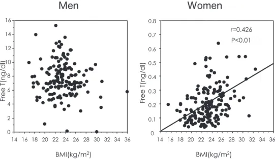
negative correlations with high-density lipoprotein (HDL)-C (41, 42). A high level of free testosterone is associated with increases in events of cardiovas-cular disease (43, 44) and insulin resistance (45, 46). Recently, a high testosterone level has been shown to be associated with subclinical atherosclero-sis in healthy menopausal women (47). In our popu-lation-based study, free testosterone level was posi-tively associated with BMI in women but not in men (Figure 6).
On the other hand, associations of DHEA-S with lipid metabolism, occurrence of cardiovasucular dis-ease and mortality are controversial. Previous stud-ies suggested that DHEA-S level did not affect the risk of fatal cardiovascular disease (48), mortality (49) and lipid profiles (50) in women. However, Trivedi et al. reported that the highest mortality rate was observed in the highest DHEA-S quartile in eld-erly women aged 65-76 years, while the highest mortality rate was found in the lowest DHEA-S quartile in elderly men (51). We showed that DHEA-S level was positively correlated with LDL-C in Japanese women (52). Recent accumulating evi-dence suggests that DHEA-S has a vasculoprotec-tive role. Yoshida et al. reported that DHEA-S was associated with increased carotid blood flow in eld-erly women (53). Lower DHEA-S levels may be re-lated to higher cardiovascular mortality in postmeno-pausal women with CVD risk factors (54). Lower DHEA-S level has been shown to be associated with increased arterial stiffness in menopausal women (47). Casson et al. reported that higher
Figure 6. Associations of free testosterone with BMI in men and women. left panel : men, right panel : women
testosterone level was related to greater maximal aerobic capacity and reduced adiposity and that higher DHEA-S level was correlated with greater in-sulin sensitivity, suggesting that endogenous andro-gens may play a role in the maintenance of benefi-cial patterns of metabolic, morphometric and func-tional parameters in postmenopausal women (55).
4. Endogenous androgen and breast cancer in women
The mechanism by which estrogens can promote the growth of breast cancer has been clearly shown. However, the role of androgens is less clear, al-though it has been shown that androgens can di-rectly stimulate the growth of human breast cancer cell lines (56). It has been reported that high levels of both testosterone and estradiol in serum precede breast cancer in postmenopausal women (57). An analysis of worldwide prospective studies showed strong associations of testosterone and DHEAS with breast cancer risk in postmenopausal women (58). Similar conclusions were obtained from a case-control study (59) and a large multicentric cohort study (60). A recent study has shown that high cir-culating levels of total and free testosterone are as-sociated with the risk of developing breast cancer in postmenopausal women, while circulating estra-diol level is not associated with the risk of breast cancer (61). High total testosterone was also sig-nificantly associated with increased risk of estrogen receptor-positive cancers. However, Danforth et al. reported that there were no significant associations between the score of breast cancer risk and levels of androgens such as testosterone, free testosterone and DHEA-S (62).
SUPPLEMENTATION OF TESTOSTERONE
AND DHEA-S IN WOMEN
For women suffering from androgen deficiency, the option of exogenous testosterone therapy is available. Considerable progress has been made in recent years in the development of different modali-ties of testosterone therapy available to women. Transdermal testosterone patches as well as creams and gels are easily applied to the skin. Goldstat et al. reported that application of testosterone cream for 12 weeks improved well-being, mood, and sex-ual function without any adverse effects in premeno-pausal women with low libido and low testosterone level (63). It has been reported that transdermal
testosterone at a dose of 300μg/day was effective for sexual desire in surgically and naturally meno-pausal women (64) and in women with hypoactive sexual desire disorder after surgically induced menopause (65, 66) without any relevant side ef-fects. Davis et al. also reported that transdermal testosterone treatment resulted in a modest im-provement in sexual function in postmenopausal women (67). Burger et al. reported that implants of estradiol and testosterone in postmenopausal women improved loss of libido, tiredness and lack of concentration (68). The results of Cochrane Re-view meta-analysis showed that hormone therapy (HT) plus testosterone improved libido, sexual func-tion and sexual activity compared to the effects of HT alone (69). Barrett-Connor et al. reported that both estrogen alone and estrogen plus testosterone increased BMD at the hip and spine but that high-dose combination of estrogen and testosterone had the greatest effect in surgically menopausal women (70). Therefore, testosterone has a favorable effect on sense of well-being, increases BMD and im-proves general fatigue, sexual function and sexual activity. On the other hand, it has been reported that short-term testosterone treatment had no effect on verbal fluency and verbal memory in healthy postmenopausal women (71).
There is still considerable controversy regarding the use of testosterone therapy in women. Reported risks and side effects from testosterone therapy in-clude the development of hirsutism, acne, alopecia, liver dysfunction, deepening of the voice, abnor-mal lipid changes, and virilization of a feabnor-male fetus if pregnant. Another major concern of testosterone therapy is whether there is a stimulatory effect on the breast or endometrium. Proposed mechanisms include conversion to estrogen by the aromatase enzyme in breast tissue or direct stimulation of the androgen receptor. A prospective cohort study in the Nurses’ Health Study showed that there was an increased relative risk for breast cancer in women using estrogen plus testosterone in comparison with that in women who had never used estrogen plus testosterone and that the risk was significantly greater than the risk of estrogen alone therapy (72). However, most of the available data do not support the concept of increase in the risk of breast cancer by testosterone but rather suggest that there is no effect or that testosterone reduces the risk of breast cancer by antagonizing the effects of estrogen on mammary tissue (73). On the other hand, there is a lack of data on endometrial safety. Panzer et al.
reported that testosterone replacement therapy for surgically and naturally menopausal women with low sexual desire can be used safely without increased risk of breast or endometrial cancer (74).
There does not appear to be an increase in car-diovascular risk through alterations in blood pres-sure, vascular reactivity, blood viscosity, hemoglo-bin concentrations, coagulation factors, or insulin sensitivity, with the exception of a lowering of HDL with oral testosterone (73). In addition, there does not appear to be an increased risk of hepatotoxicity. In particular, a transdermal patch or gel that avoids first pass has no related hepatic toxicity (75). The current recommendation for testosterone treatment is that therapy should be restricted to short-term therapy until long-term safety issues have been re-solved. The number of adverse events associated with testosterone replacement treatment in women has been limited when protocols of treatment are optimized to achieve physiologic levels of circulat-ing testosterone (67).
The physiological role of DHEA is poorly under-stood. Despite the wide use of DHEA as a dietary supplement, no well-designed study has established the efficacy and safety of DHEA therapy. None of the double-blind placebo-controlled studies have shown beneficial effects of DHEA administration on cognition, attention or memory (76). Wolkowitz et al. only reported that DHEA treatment had signifi-cant beneficial effects on depressive symptoms in patients with major depression (77). Cameron et al. suggested that doses of 30 to 50 mg of oral DHEA produce physiological androgen levels and that 50 mg of DHEA increases serum androgen levels within the physiological range as well as possible improvements of sexual function and mood and decrease in fatigue/exhaustion in women (78). A recent study has revealed that intravaginal admini-stration of DHEA can be used to treat vaginal atro-phy (79). However, since DHEA is converted not only into testosterone but also into small amounts of estradiol, DHEA treatment may have a risk for breast cancer in postmenopausal women. Thus, DHEA therapy should share the contraindications for use of estrogen replacement therapy in women.
EFFECTS OF TESTOSTERONE-DERIVED
PROGESTOGENS IN WOMEN
Hormone replacement therapy (HRT) is available for women with menopausal symptoms. Addition
of progestogen is needed with estrogen for HRT in women with an intact uterus even if the dose of estrogen is low since it has been reported that en-dometrial hyperplasia and enen-dometrial cancer oc-curred in women with an intact uterus when ultra-low dose estrogen alone was used (80). Progesto-gens, such as medroxyprogesterone, levonorgestrel and norethisterone acetate, which are older pro-gestogens, have androgenic activities (81). Estrogen has favorable effects on insulin sensitivity, blood pressure and lipid metabolism, but androgenic ac-tivities of progestogens have unfavorable effects on these favorable effects of estrogen. In particular, testosterone-derived progestogens, such as lev-onorgestrel and norethisterone, have been reported to be associated with increase in the risk of breast cancer (82). Therefore, it is necessary to pay atten-tion to lipid metabolism, insulin sensitivity and risk of breast cancer when using a testosterone-derived progestogen.
EFFECTS OF SHBG ON ANDROGEN
Consideration of only the total testosterone level is inadequate for assessing the androgen environ-ment. Less than 2% of testosterone circulates in an absolute free state in the blood at any one time. Approximately 60-65% of testosterone is carried in peripheral blood bound to SHBG, and testosterone circulates in appreciable amounts bound to albumin (35-40%) and in small amounts bound to corticoster-oid-binding globulin (CBG) (!5%). Since the bind-ing to albumin and CBG is relatively weak, testos-terone can easily disassociate from these proteins to interact with the testosterone receptor. Essen-tially, SHBG has a function as a circulating reser-voir of this potent androgen.
1. Change in SHBG
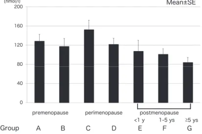
In women, the change in circulating SHBG with age is still controversial. SHBG has been demon-strated to decline steadily with age (7). However, it has been reported that SHBG was virtually un-changed in women, while SHBG in men increased more than 2 fold over the life span (5). Our popula-tion-based study showed that SHBG level in women gradually decreased around menopause and in-creased with age after menopause, while SHBG level was positively correlated with age in men (Figure 7). In addition, SHBG levels show a U-shape pattern in the 7 stages during menopausal
transition as can be seen in Figure 8.
2. Action of SHBG
Many studies have shown associations of SHBG with favorable effects on lipid profiles and insulin sensitivity in men (83, 84). In women, a high SHBG level was also associated with favorable lipid pro-files decrease in the occurrence of cardiovascular
disease and metabolic syndrome. SHBG was shown to be negatively correlated with total cholesterol, LDL-C and TG and to be positively correlated with HDL-C (42, 85). Low SHBG level was associated with the occurrence of cardiovascular disease (43, 44). SHBG level was negatively associated with hyperinsulinemia (86) and risk of metabolic syn-drome (16, 87). We also showed that SHBG level
Figure 7. Changes in SHBG levels in men and women.
upper panel : men, lower panel : women
Figure 8. Changes in SHBG levels during the menopausal transition.
Group A : early reproductive stage, Group B : late reproductive stage, Group C : early menopausal transition, Group D : late meno-pausal transition, Group E : women for whom less than 1 year has passed since menopause, Group F : women for whom less than 5 years have passed since menopause, Group G : women for whom more than 5 years have passed since menopause
was negatively correlated with Homeostasis Model Assessment (HOMA) index in both men and women (88). In addition, SHBG level was negatively corre-lated with TG level in women but not in men. There-fore, SHBG may have biological functions beyond simply regulation of the level of free sex steroid hormones and may play important roles in lipid metabolism and insulin sensitivity.
SITE OF ANDROGEN PRODUCTION IN
POSTMENOPAUSAL WOMEN
Whether ovaries produce androgens in postmeno-pausal women is a matter of debate. It has been re-ported that circulating androgens in postmenopausal women do not originate from the ovaries but from the adrenal gland since levels of androgens in post-menopausal women with natural menopause and those with surgical menopause were not different (89). It was later shown in a cross-sectional study that total and free testosterone levels in women aged 55 years or older with bilateral oophorectomy were significantly lower than those in age-matched women (90). A longitudinal study also showed a 42% decline in testosterone level in postmenopausal women who underwent oophorectomy, suggesting that the postmenopausal ovary is hormonally active and contributes significantly to the circulating pool of testosterone (91). Several studies also showed that women with bilateral oophorectomy had lower testosterone levels than those in natural postmeno-pausal women (92-94). Bui et al. reported that a significant decrease was found in testosterone lev-els after bilateral oophorectomy, whereas no signifi-cant difference was found after natural menopause by using the developed isotope dilution-liquid chro-matography-tandem mass spectrometry (95). How-ever, more recently, it has been shown that levels of testosterone and DHEA-S did not differ signifi-cantly between surgically and naturally menopausal women with a mean age of 52.4 years (96).
MEASUREMENT OF TESTOSTERONE
Ability to measure either total or free testoster-one level accurately is essential for establishing the diagnosis of true androgen deficiency. The nor-mal circulating level of testosterone in both repro-ductive-aged women and postmenopausal women still needs proper validation since methods for
measurement of testosterone have been inadequate in women until now (97). Recent advances with the use of ultrasensitive methods such as mass spec-trometry coupled to either gas or liquid chromatog-raphy have improved the technology for measure-ment of testosterone. Advances in technology may allow clinicians to better define female androgen deficiency and may provide easier treatment options for testosterone replacement therapy.
CONCLUSION
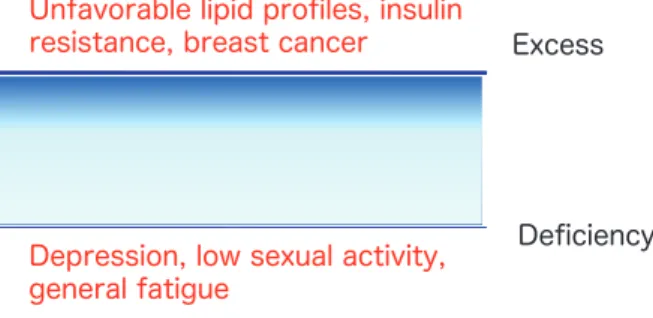
Extreme levels of circulating androgens, whether high or low, may have negative effects on women’s health. An excess endogenous testosterone level may be associated with unfavorable lipid profiles, in-sulin resistance and development of breast cancer in postmenopausal women. On the other hand, in-sufficiency of testosterone leads to an impairment in sexual drive, reduced libido, and depressed mood. For optimal physiological and psychological health in women, circulating testosterone levels should be within normal ranges (Figure 9). An appropriate level of androgen may play important roles in meta-bolic, psychological and sexual functions in women. Although testosterone level was not significantly low in postmenopausal women as shown in Figure 3, various symptoms and diseases due to insufficiency of testosterone in postmenopausal women might be caused by individual difference or SHBG level. In addition, very small changes in testosterone in postmenopausal women may influence on the symp-toms and diseases. According to the estrogen thresh-old hypothesis proposed by Barbieri, tissues vary in their sensitivity to estradiol (98). As well as estro-gen, sensitivity to androgen may be different in vari-ous tissues even if the range is narrow. In addition, the roles of testosterone and DHEA-S in women’s health may be different. Further studies on testos-terone, DHEA-S and SHBG in women are needed.
CONFLICT OF INTERESTS
The authors have no conflicts of interest.
REFERENCES
1. Randolph JF, Sowers M, Bondarenko IV, Harlow SD, Luborsky JL, Little RD : Change in estradiol and follicle-stimulating hormone across the early menopausal transition : Effects of ethnicity and age. J Clin Endocrinol Metab 89 : 1555-1561, 2004
2. Yasui T, Uemura H, Irahara M, Arai M, Kojimahara N, Okabe R, Ishii Y, Tashiro S, Sato H : Differences in sensitivity to cold in Japanese men and postmenopausal women aged!50 years. Gender Medicine 4 : 359-366, 2007 3. Braunstein GD : Androgen insufficiency in
women : summary of critical issues. Fertil Steril 77 : S94-99, 2002
4. Spencer JB, Klein M, Kumar A, Azziz R : The age-associated decline of androgens in repro-ductive age and menopausal black and white women. J Clin Endocrinol Metab 92 : 4730-4733, 2007
5. Khosla S, Melton III LJ, Atkinson EJ, O’Fallon WM, Klee GG, Riggs BL : Relationship of se-rum sex steroid levels and bone turnover mark-ers with bone mineral density in men and women : A key role for bioavailable estrogen. J Clin Endocrinol Metab 83 : 2266-2274, 1996 6. Burger HG, Dudley EC, Cui J, Dennerstein L,
Hopper JL : A prospective longitudinal study of serum testosterone, dehydroepiendrosterone sulfate and sex hormone binding globulin lev-els through the menopause transition. J Clin Endocrinol Metab 85 : 2832-2938, 2000 7. Sowers MFR, Zheng H, McConnell D, Nan B,
Karvonen-Gutierrez CA, Randolph Jr JF : Tes-tosterone, sex hormone-binding globulin and free androgen index among adult women : chronological and ovarian aging. Hum Reprod 24 : 2279-2285, 2009
8. Soules MR, Sherman S, Parrott E, Rebar R, Santoro N, Utian W, Woods N : Executive sum-mary : Stages of reproductive aging workshop (STRAW). Fertil Steril 76 : 874-878, 2001 9. Torrens JI, Sutton-Tyrrell K, Zhao X, Matthews
K, Brockwell S, Sowers M, Santoro N : Relative androgen excess during the menopausal tran-sition predicts incident metabolic syndrome in
mid-life women. Menopause 16 : 257-264, 2009 10. Crawford S, Santoro N, Laughlin GA, Sowers MF, McConnell D, Sutton-Tyrrell K, Weiss G, Vuga M, Randolph J, Lasley B : Circulating de-hydroepiandrosterone sulfate concentrations during the menopausal transition. J Clin Endo-crinol Metab 94 : 2945-2951, 2009
11. Lasley BL, Crawford SL, Laughlin GA, Santoro N, McConnell DS, Crandall C, Greendale GA, Polotsky AJ, Vuga M : Circulating dehydroepi-androsterone sulfate levels in women who un-derwent bilateral salpingo-oophorectomy dur-ing the menopausal transition. Menopause 18 : 494-498, 2011
12. Liu D, Dillon JS : Dehydroepiandrosterone ac-tivities endothelial cell nitric-oxide synthase by a specific plasma membrane receptor couple to Gαi2,3. Bio Chem 277 : 21379-21388, 2002
13. West SL, D’Aloisio AA, Agans RP, Kalsbeck WD, Borisov NN, Thorp JM : Prevalence of low sexual desire and hypoactive sexual desire dis-order in a nationally representative sample of US women. Arch Intern Med : 168 : 1441-9, 2008
14. Rivera-Woll LM, Papalia M, Davis SR, Burger HG : Androgen insufficiency in women : diag-nostic and therapeutic implications. Hum Re-prod Update 10 : 421-432, 2004
15. Haren MT, Malmstrom TK, Banks WA, Patrick P, Miller DK, Morley JE : Lower serum DHEAS levels are associated with a higher degree of physical disability and depressive symptoms in middle-aged to older African American women. Maturitas 57 : 347-360, 2007
16. Santoro N, Torrens J, Crawford S, Allsworth JE, Finkelstein JS, Gold EB, Korenman S, Lasley WL, Luborsky JL, McConnell D, Sowers MF, Weiss G : Correlates of circulating androgens in mid - life women : the study of women’s health across the nation. J Clin Endocrinol Metab 90 : 4836-45, 2005
17. Bell RJ, Donath S, Davison SL, Davis SR : En-dogenous androgen levels and well-being : dif-ferences between premenopausal and post-menopausal women. Menopause 13 : 65-71, 2006
18. Woods NF, Smith-Dijulio K, Percival DB, Tao EY, Mariella A, Mitchell ES. Depressed mood during the menopausal transition and early postmenopause : observations from the Seattle Midlife Women’s Health Study. Menopause 15 : 223-232, 2008
19. Gallicchio L, Schilling C, Miller SR, Zacur H, Flaws JA. Correlates of depressive symptoms among women undergoing the menopausal transition. J Psychosom Res 63 : 263-268, 2007 20. Morsink LF, Vogelzangs N, Nicklas BJ, Beekman AT, Satterfield S, Rubin SM, Yaffe K, Simonsick E, Newman AB, Kritchevsky SB, Penninx BW ; Health ABC Study. Associations between sex steroid hormone levels and de-pressive symptoms in elderly men and women : results from the Health ABC Study. Psycho-neuroendocrinol 32 : 874-883, 2007
21. Bromberger JT, Schott LL, Kravitz HM, Sowers M, Avis NE, Gold EB, Randolph JF Jr. Matthews KA : Longitudinal change in repro-ductive hormones and depressive symptoms across the menopausal transition : results from the Study of Women’s Health Across the Na-tion (SWAN). Arch Ben Psychiatry 67 : 598-607, 2010
22. Barrett-Connor E, von Muhlen D, Laughlin GA, Kripke A : Endogenous levels of dehy-droepiandrosterone sulfate, but not other sex hormones, are associated with depressed mood in older women : the Rancho Bernardo Study. J Am Geriatr Soc 47 : 685-691, 1999
23. Berr C, Lafont S, Debuire B, Dartigues JF, Baulicu EE : Relationships of dehydroepien-drosterone sulfate in the elderly with functional, psychological, and mental status, and short-term mortality : a French community-based study. Proc Natl Acad Sci USA 93 : 13410-13415, 1996
24. Rigaud AS, Pellerin J : Neuropsychic effects of dehydroeiandrosterone. Ann Med Interne 152 [Suppl 3] : IS43-49, 2001
25. Wolf OT, Kirschbaum C : Actions of dehy-droepiandrosterone and its sulfate in the cen-tral nervous system : effects on cognition and emotion in animals and humans. Brain Res Rev 30 : 264-288, 1999
26. Spencer JL, Waters EM, Romeo RD, Wood GE, Milner TA, McEwen BS : Uncovering the mechanisms of estrogen effects on hippocampal function. Front Neuroendocrinol 29 : 219-237, 2008
27. Hogervorst E, Matthews FE, Brayne C : Are op-timal levels of testosterone associated with bet-ter cognitive function in healthy older women and men? Biophys Biochim Acta 1800 : 1145-1152, 2010
28. Thilers PP, Macdonald SW, Herlitz A : The
association between endogenous free testoster-one and cognitive performance : a population-based study in 35 to 90-year-old men and women. Psychoneuroendocrinol 31 : 565-576, 2006
29. Ryan J, Stanczyk FZ, Dennerstein L, Mack WJ, Clark MS, Szoeke C, Kildea D, Henderson VW : Hormone levels and cognitive function in postmenopausal midlife women. Neurobiol Aging : Dec 14. [Epub ahead of print], 2010 30. Vallee M, Mayo W, Le Moal M : Role of
pregnenolone, dehydroeiandrosterone and their sulfate esters on leaning and memory in cog-nitive aging. Brain Res Rev 37 : 301-312, 2001 31. Hillen T, Lun A, Reischies FM, Borchelt M,
Steinhagen-Thiessen E, Schaub RT : DHEA-S plasma levels and incidence of Alzheimer’s disease. Biol Psychiatr 47 : 161-163, 2000 32. Karpf JM, Simon JA : The role of testosterone
in the management of hypoactive sexual desire disorder in postmenopausal women. Maturitas 63 : 213-219, 2009
33. Riley A, Riley E : Controlled studies on women presenting with sexual disorders : I. Endocrine status. J Sex Marital Ther 26 : 269-283, 2000 34. Guay AT : Decreased testosterone in regularly
menstruating women with decreased libido : a clinical observation. J Sex Marital Ther 27 : 513-519, 2001
35. Alarslan D, Sarandol A, Congiz C, Develioglu OH. Androgens and sexual dysfunction in natu-rally and surgically menopausal women. J Ob-stet Gynecol Res 37 : 1027-1034, 2011
36. Guay AT, Jacobson J : Decreased free testos-terone and dehydroepiandrostestos-terone-sulfate (DHEA-S) levels in women with decreased li-bido. J Sex Marital Ther 28, Supple 1 : 129-142, 2002
37. Turna B. Apaydin E, Semerci B, Altay B, Cikili N, Nazli O : Women with low libido : correla-tion of decreased androgen levels with female sexual function index. Intern J Impotence Res 17 : 148-153, 2005
38. Abu EO, Horner V, Kusec V, Triffitt JT, Compston JE : The localization of androgen receptors in human bone. J Clin Endocrinol Metab 82 : 3493-3497, 1997
39. Davidson BJ, Ross RK, Paganini - Hill A, Hammond GD, Siiteri PK, Judd HL : Total and free estrogens and androgens in postmeno-pausal women with hip fractures. J Clin Endo-crinol Metab 54 : 115-120, 1982
40. Cummings SR, Browner WS, Bauer DC, Stone K, Ensrud KE, Jamal S, Ettinger B : Endoge-nous hormones and the risk of hip and verte-bral fractures among older women. Study of Osteoporosis Fractures Research Group. N Eng J Med 339 : 733-738, 1998
41. Shelly JM, Green A, Smith AMA, Dudley E, Dennerstein L, Hopper J, Burger H : Relation-ship of endogenous sex hormones to lipids and blood pressure in mid-aged women. Ann Epi-demiol 8 : 39-45, 1998
42. Lambrinoudaki I, Christodoulakos G, Rizos D, Economou E, Argeitis J, Vlachou S, Creatsa M, Kouskouni E, Botsis D : Endogenous sex hor-mones and risk factors for atherosclerosis in healthy Greek postmenopausal women. Eur J Endocrinol 154 : 907-16, 2006
43. Rexrode KM, Manson JE, Lee IM, Ridker PM, Sluss PM, Cook NR, Buring JE : Sex hormone levels and risk of cardiovascular events in post-menopausal women. Circulation 108 : 1688-1693, 2003
44. Sutton-Tyrrell K, Wildman RP, Matthews KA, Chae C, Lasley BL, Brockwell S, Pasternak RC, Lloyd-Jones D, Sowers MF, Torrens JI : Sex hormone-binding globulin and the free andro-gen index are related to cardiovascular risk fac-tors in multiethnic premenopausal and peri-menopausal women enrolled in the study of women across the nation (SWAN). Circulation 111 : 1242-1249, 2005
45. Kalish, GM, Barrett-Connor E, Laughlin GA, Gulanski BI : Association of endogenous sex hormones and insulin resistance among menopausal women : Results from the post-menopausal estrogen/progestin intervention trial. J Clin Endocrinol Metab 88 : 1646-1652, 2003
46. Lee CC, Kasa-Vubu JZ, Supiano MA : Andro-genicity and obesity are independently associ-ated wit insulin sensitivity in postmenopausal women. Metabolism 53 : 507-512, 2004 47. Creatsa M, Armeni E, Stamatelopoulos K, Rizos
D, Georgipoulos G, Kazani M, Alexandrou A, Dendrinos S, Augoulea A, Papamichael C, Lambrinoudaki I. Circulating androgen levels are associated with subclinical atherosclerosis and arterial stiffness in healthy recently meno-pausal women. Metabolism 2011 [Epub ahead of print]
48. Barrett-Connor E, Goodman-Gruen D : Dehy-droepiandrosterone sulfate dose not predict
cardiovascular death in postmenopausal women : The Rancho Bernardo Study. Circulation 91 : 1757-1760, 1995
49. Mazat L, Lafont S, Berr C, Debuire B, Tessier JF, Dartigues JF, Baulieu EE : Prospective measurements of dehydroepiandrosterone sul-fate in a cohort of elderly subjects : relationship to gender, subjective health, smoking habits, and 10-year mortality. Proc Natl Acad Sci USA 98 : 8145-8150, 2001
50. Osmanagaoglu MA, Okumus B, Osmanagaoglu T, Bozkaya H : The relationship between serum dehydroepiendrosterone sulfate concentration and bone mineral density, lipids, and hormone replacement therapy in premenopausal and postmenopausal women. J Women’s Health 13 : 993-999, 2004
51. Trivedi DP, Khaw KT : Dehydroepiandroster-one sulfate and mortality in elderly men and women. J Clin Endocrinol Metab 86 : 4171-4177, 2001
52. Yasui T, Uemura H, Irahara M, Arai M, Kojimahara N, Okabe R, Ishii Y, Tashiro S, Sato H : Associations of endogenous sex hor-mones and sex hormone-binding globulin with lipid profiles in aged Japanese men and women. Clinica Chimica Acta 398 : 43-47, 2008
53. Yoshida S, Aihara K, Azuma H, Uemoto R, Sumitomo-Ueda Y, Yagi S, Ikeda Y, Iwase T, Nishio S, Kawano H, Miki J, Yamada H, Hirata Y, Akaike M, Sata M, Matsumoto T. Dehy-droepiandrosterone sulfate is inversely associ-ated with sex-dependent diverse carotid athe-rosclerosis regardless of endothelial function. Atherosclerosis 212 : 310-315, 2010
54. Shufelt C, Bretsky P, Almeida CM, Johnson BD, Shaw LJ, Azziz R, Braunstein GD, Pepine CJ, Bittner V, Vldo DA, Stanczyk FZ, Bairey Merz CN. DHEA-S levels and cardiovascular disease mortality in postmenopausal women : results from the National Institutes of Health-National Heart, Lung, and Blood Institute (NHLBI)-sponsored Women’s Ischemia Syn-drome Evaluation (WISE). J Clin Endocrinol Metab 95 : 4985-92, 2010
55. Casson PR, Toth MJ, Johnson JV, Stanczyk FZ, Casey CL, Dixon ME : Correlation of serum androgens with anthropometric and metabolic indices in healthy, nonobese postmenopausal women. J Clin Endocrinol Metab 95 : 4276-4282, 2010
androgens and antiandrogens on hormone-responsive human breast cancer in long-term tissue culture. Cancer Res 36 : 4610-4618, 1976 57. Berrino F, Muti P, Micheli A, Bolelli G, Krogh V, Sciajno R, Pisani P, Panico S, Secreto G : Serum sex hormone levels after menopause and subsequent breast cancer. J Natl Cancer Inst 88 : 291-296, 1996
58. Key T, Appleby P, Barnes I, Reeves G : Endo-genous sex hormones and breast cancer in postmenopausal women : reanalysis of nine pro-spective studies. J Natl Cancer Inst 94 : 606-616, 2002
59. Missmer SA, Eliassen AH, Barbieri RL, Hankinson SE : Endogenous estrogen, andro-gen, and progesterone concentrations and breast cancer risk among postmenopausal women. J Natl Cancer Inst 96 : 1856-65, 2004 60. Kaaks R, Rinaldi S, Key TJ, Berrino F, Peeters
PH, Biessy C, Dossus L, Lukanova A, Bingham S, Khaw KT, Allen NE, Bueno-de-Mesquita HB, van Gils CH, Grobbee D, Boeing H, Lahmann PH, Nagel G, Chang-Claude J, Clavel-Chapelon F, Fournier A, Thiébaut A, González CA, Quirós JR, Tormo MJ, Ardanaz E, Amiano P, Krogh V, Palli D, Panico S, Tumino R, Vineis P, Trichopoulou A, Kalapothaki V, Trichopoulos D, Ferrari P, Norat T, Saracci R, Riboli E : Post-menopausal serum androgens, oestrogens and breast cancer risk : the European Prospective Investigation into Cancer and Nutrition. Endocr Relat Cancer 12 : 1071-82, 2005
61. Sieri S, Krogh V, Bolelli G, Abagnato CA, Grioni S, Pala V, Evangelista A, Allemani C, Micheli A, Tagliabue G, Schunemann HJ, Menard S, Berrino F, Muti P : Sex hormone levels, breast cancer risk, and cancer receptor status in postmenopausal women : the ORDET cohort. Cancer Epidemiol Biomarkers Prev 18 : 169-176, 2009
62. Danforth KN, Eliassen AH, Tworoger SS, Missmer SA, Barbieri RL, Rosnet BA, Colditz GA, Hankinson SE : The association of plasma androgen levels with breast, ovarian and en-dometrial cancer risk factors among postmeno-pausal women. Int J Cancer 126 : 199-207, 2010
63. Goldstat R. Briganti E, Tran J, Wolfe R, Davis SR : Transdermal testosterone therapy im-proves well-being, mood, and sexual function in premenopausal women. Menopause 10 : 390-398, 2003
64. Nappi RE, Albani F, Santamaria V, Tonani S, Martini E, Terreno E, Brambilla E, Polatti F : Menopause and sexual desire : the role of tes-tosterone. Menopause Int 16 : 162-168, 2010 65. Simon J, Braunstein G, Natchtigall L, Utian
W, Katz M, Miller S, Waldbaum A, Bouchard C, Derzko C, Buch A, Rodenberg C, Lucas J, Davis S : Testosterone patch increases sexual activity and desire in surgically menopausal women with hypoactive sexual desire disorder. J Clin Endocrinol Metab 90 : 5226-5233, 2005 66. Braunstein GD, Sundwall DA, Katz M, Shifren
JL, Buster JE, Simon JA, Bachman G, Aguirre OA, Lucas JD, Rodenberg C, Buch A, Watts NB : Safety and efficacy of a testosterone patch for the treatment of hypoactive sexual desire disorder in surgically menopausal women. Arch Intern Med 165 : 1582-1589, 2005
67. Davis SR. Moreau M. Kroll R, Bouchard C, Panay N, Gass M, Braunstein GD, Hirschberg AL, Rodenberg C, Pack S, Koch H, Moufarege A, Studd J ; APHRODITE Study Team : Testos-terone for low libido in postmenopausal women not taking estrogen. N Eng J Med 359 : 2005-17, 2008
68. Burger HG, Hailes J, Menelaus M. Nelson J, Hudson B, Balazs N : The management of per-sistent symptoms with estradiol-testosterone implants : clinical, lipid and hormonal results. Maturitas 6 : 351-358, 1984
69. Somboonporn W, Davis S, Seif M, Bell R : Tes-tosterone for peri- and postmenopausal women. The Cochrane Database of Systematic Reviews 2005 : 4 [article no : CD004509].
70. Barrett-Connor E, Young R, Notelovitz M, Sullivan J, Wiita B, Yang HM, Nolan J : A two-year, double-blind comparison of estrogen-androgen and conjugated estrogens in surgi-cally menopausal women. Effects on bone min-eral density, symptoms and lipid profiles. J Re-prod Med 44 : 1012-1020, 1999
71. Kocoska-Maras L, Zethraeus N, Radestad AF, Ellingsen T, von Schoultz B, Johannesson M, Hirschberg AL : A randomized trial of the ef-fect of testosterone and estrogen on verbal fluency, verbal memory, and spatial ability in healthy postmenopausal women. Fertil Steril 95 : 152-157, 2010
72. Tamimi RM, Hankinson SE, Chen WY, Rosner B, Colditz GA : Combined estrogen and testos-terone use and risk of breast cancer in post-menopausal women. Arch Intern Med 166 :
1483-9, 2006
73. Braunstein GD : Safety of testosterone treat-ment in postmenopausal women. Fertil Steril 88 : 1-17, 2007
74. Panzer C, Guay A : Testosterone replacement therapy in naturally and surgically menopausal women. J Sex Med 6 : 8-18, 2009
75. Shufelt CL, Braunstein GD : Safety of testoster-one use in women. Maturitas 63 : 63-66, 2009 76. Buvat J : Androgen therapy with
dehydroepi-endrosterone. World J Urol 21 : 346-355, 2003 77. Wolkowitz OM, Reus VI, Keebler A, Nelson N, Friedland M, Brizendine L : Double blind treat-ment of major depression with dehydroepian-drosterone. Am J Psychiatr 156 : 646-649, 1999 78. Cameron DR, Braunstein GD : The use of de-hydroepiandrosterone therapy in clinical prac-tice. Treat Endocrinol 4 : 95-114, 2005
79. Panjari M, Davis SR. Vaginal DHEA to treat menopause related atrophy : a review of the evidence. Maturitas 70 : 22-25, 2911
80. Weiderpass E Baron JA, Adami HO,
Magnusson C, Lindgren A, Bergström R, Correia N, Persson I : Low-potency oestrogen and risk of endometrial cancer : a case-control study. Lancet 29 : 1824-8, 1999
81. Nath A, Sitruk-Ware R : Different cardiovascu-lar effects of progestins according to structure and activity. Climacteric 12 Suppl 1 : 96-101, 2009
82. Flesch-Janys D, Slanger T, Mutschelknauss E, Kropp S, Obi N, Vettorazzi E, Braendle W, Bastert G, Hentschel S, Berger J, Chang-Claude J : Risk of different histological types of postmenopausal breast cancer by type and regimen of menopausal hormone therapy. Int J Cancer 123 : 933-41, 2008
83. Bataille V, Perret B, Evans A, Amouyel P, Arveiler D, Ducimetiere P, Bard JM, Ferrieres J : Sex hormone-binding globulin is a major determinant of the lipid profile : the PRIME study. Atherosclerosis 179 : 369-373, 2005 84. Kupelian V, page ST, Araujo AB, Travison TG,
Bremner WJ, McKinlay JB : Low sex hormone-binding globulin, total testosterone, and symp-tomatic androgen deficiency are associated with development of the metabolic syndrome in nonobese men. J Clin Endocrinol Metab 91 : 843-850, 2006
85. Mudai S, Dobs AS, Ding J, Cauley JA, Szklo M, Golden SH : Endogenous postmenopausal hor-mones and serum lipids : The atherosclerosis
risk in communities study. J Clin Endocrinol Metab 90 : 1202-1209, 2005
86. Preziosi, P, Barrett-Connor E, Papoz L, Roger M, Saint-Paul M, Nahoul K, Simon D : Interre-lation between plasma sex hormone-binding globulin and plasma insulin in healthy adult women : the telecom study. J Clin Endocrinol Metab 76 : 283-7, 1993
87. Hajamor S, Despres JP, Couillard C, Lemieux S, Tremblay A, Prud’homme D, Tchernof A : Relationship between sex hormone-binding globulin levels and features of the metabolic syndrome. Metabolism 52 : 724-730, 2003 88. Yasui T, Tomita J, Miyatani Y, Yamada M,
Uemura H, Irahara M, Arai M, Kojimahara N, Okabe R, Ishii Y, Tashiro S, Sato H : Asso-ciation of adiponectin with sex hormone-bind-ing globulin levels in aghormone-bind-ing male and female populations. Clinica Chimica Acta 386 : 69-75, 2007
89. Couzinet B, Meduri G, Lecce MG, Young J, Brailly S, Loosfelt H, Lilgron E, Schaison G : The postmenopausal ovary is not a major an-drogen-producing gland. J Clin Endocrinol Me-tab 86 : 5060-5066, 2001
90. Davison SL, Bell R, Donath S, Montalto JG, Davis SR : Androgen levels in adult females : changes with age, menopause and oophorec-tomy. J Clin Endocrinol Metab 90 : 3847-3853, 2005
91. Fogle RH, Stanczyk FZ, Zhang X, Paulson RJ : Ovarian androgen production in postmeno-pausal women. J Clin Endocrinol Metab 92 : 3040-3043, 2007
92. McTiernan A, Wu L, Barnabei VM, Chen C, Hendrix S, Modugno F, Rohan T, Stanczyk FZ, Wang CY : Relation of demographic factors, menstrual history, reproduction and medication use to sex hormone levels in postmenopausal women. Breast Cancer Res Treat 108 : 217-231, 2008
93. Korse CM, Bonfrer JMG, van Beurden M, Verheijen RHM, Rookus MA : Estradiol and testosterone levels are lower after oophorec-tomy than after natural menopause. Tumor Biol 30 : 37-42, 2009
94. Endogenous Hormones and Breast Cancer Col-laborative Group : Circulating sex hormones and breast cancer risk factors in postmeno-pausal women : reanalysis of 13 studies. British J Cancer 105 : 709-722, 2011
Thienpont LM, Kenemans P, Verhoeven MO, Jakobs C, Dijstelbloem HM, Blankenstein MA : Serum testosterone levels measured by isotope dilution-liquid chromatography-tandem mass spectrometry in postmenopausal women ver-sus those in women who underwent bilateral oophorectomy. Ann Clin Biochem 47 : 248-252, 2010
96. Alarslan D, Sarandol A, Cengiz C, Develioglu
OH. Androgen and sexual dysfunction in natu-rally and surgically menopausal women. J Ob-stet Gynaecol Res 37 : 1027-1034, 2011 97. Demers LM : Androgen deficiency in women :
role of accurate testosterone measurements. Maturitas 67 : 39-45, 2010
98. Barbieri RL : Hormone treatment of endometri-osis : The estrogen threshold hypothesis. Am J Obstet Gynecol 166 : 740-745, 1992