The effects of colostral antibodies from immunized dairy cows on inhibition of absorption into the blood of Verotoxin 2 derived from enterohemorrhagic Escherichia
coli O157:H7 and on eradication of Helicobacter pylori in experimental animals
麻布大学大学院 環境保健学研究科 環境保健科学専攻 生体防御学
DE1101 清田
哲郎The effects of colostral antibodies from immunized dairy cows on inhibition of absorption into the blood of Verotoxin 2 derived from enterohemorrhagic Escherichia
coli O157:H7 and on eradication of Helicobacter pylori in experimental animals
DE1101 Tetsuro Seita
Laboratory of Immunology
The Graduate School of Environmental Health Sciences
Azabu University
実験動物での腸管出血性大腸菌
O157:H7
由来ベロ毒素2
の吸収抑制 ならびにHelicobacter pylori
の除菌におけるウシ免疫初乳抗体の効果麻布大学大学院 環境保健学研究科 環境保健科学専攻 生体防御学
DE1101 清田
哲郎Contents
要旨
( Abstract in Japanese ) ··· 3
New application of indirect fluorescent antibody (IFA) technique using latex particles coupled with verotoxin 2 from Escherichia coli O157:H7 in order to determine colostral antibody titers in immunized dairy cows ··· 7
Abstract ··· 8
Introduction ··· 10
Materials and Methods ··· 11
Results ··· 17
Discussion ··· 18
Acknowledgements ··· 20
Reference ··· 22
Figure and Table ··· 29
Inhibition of the absorption to systemic blood of verotoxin (VT) 2 from intestine by repeatedly administration of bovine immune colostral antibody against VT 2 in mice ··· 34
Abstract ··· 35
Introduction ··· 37
Materials and Methods ··· 38
Results ··· 42
Discussion ··· 44
Acknowledgment ··· 49
References ··· 50
Figure and Table ··· 56
Eradication effects of Helicobacter pylori in Mongolian gerbils by colostral antibody obtained from dairy cows immunized with H. pylori and its antibody with (bovine) complement compared
with antibiotics ··· 60
Introduction ··· 61
Materials and methods ··· 64
Result ··· 69
Discussion ··· 70
Reference ··· 76
Figure and Table ··· 84
Acknowledgements ··· 88
要旨
腸管出血性大腸菌
O157:H7
(E. coli O157:H7
)は、死者の発生を伴う食中毒 の原因菌として、Helicobacter pylori ( H. pylori )
は、胃潰瘍、胃がんなどを誘 発する細菌としてよく知られており、これらの消化器感染症に対する有効な対応 策の確立が待たれている。そこで、乳牛で作製した免疫初乳抗体を用いてこれらの消化器感染症における 受動免疫の有効性を動物モデルで明らかにした。腸管感染症モデルでは、
E. coli O157:H7
の産生するベロ毒素2
(VT2
)に対する免疫初乳抗体を用いてマウスに おけるVT2
の吸収阻止効果を明らかにした。胃感染症モデルでは、H. pylori
に 対する免疫初乳抗体及びその抗体と補体(新鮮ウシ血清)とを用いてスナネズミ における除菌効果を実証した。これらの蛋白質分解酵素に強い抵抗性を有する免 疫初乳抗体は、乳牛でそれぞれ作製した。Ⅰ. 可溶性
VT2
に対する抗体測定が可能な間接蛍光抗体法(IFA)の開発IFA
用のVT2
感作ラテックスの調製及び乳牛への免疫原に用いたVT2
は、マ ウス抗VT2
モノクローナル抗体感作Sepharose4B
カラムを用いたアフィニティ ークロマトグラフィーによってE. coli O157:H7 VT2
産生株の培養液から分離し た。VT2
に対する免疫初乳抗体は、分娩4
ヵ月前の乳牛へ毎週1
回VT2
を免疫し て作製した。分娩3
日後までの初乳を採取し、低速遠心による脱脂及びレンネッ トによる脱カゼインを行って乳清を分離した。これを免疫初乳抗体として供試し た。VT2
感作ラテックスは、粒径6 μm
の2.5 %
ラテックス粒子0.5 ml
へ30 μg/ml
のVT2 1.0ml
を感作して調製した。それを20 %グリセリン、1 %
卵白アルブミ ン(OVA
)を含む食塩加リン酸緩衝液(PBS
)に分散させ、この5 μl
をIFA
用 スライドガラスのwell
に塗抹してIFA
スライドグラスを作製した。これに1:2
~1:512
に希釈した免疫初乳抗体(10 μl / well
)を加えて室温で1時間反応させた。次に、至適濃度の
FITC
標識抗ウシγグロブリン、IgG、IgAあるいはIgM
抗体ナトリウムを含む
PBS
で洗浄することによって、非特異反応を完全に排除する ことができた。なお、ラテックス粒子に自家蛍光は認めなかった。一般的なVT2
に対する抗体測定法であるベロ細胞を用いた中和試験では、免疫グロブリンクラ ス別の抗体測定が不可能であったが、このIFA
によってその問題が解決された。この
IFA
で測定した免疫初乳抗体の抗体価は、免疫に用いた乳牛の血清抗体価の 約4
倍高力価であった。免疫初乳抗体のIFA
価は、分娩直後に採取した初乳が最 も高い1:512
を示し、分娩3
日後までの初乳も1:128
~1:256
と比較的高力価を 示した。Ⅱ
.
マウスにおける免疫初乳抗体によるVT2
の血中への吸収阻止作用マウスの血中に吸収された
VT2
の濃度は、0.2 ng/ml まで測定可能な蛍光ELISA
によって測定した。マウス(146
匹)を用いて免疫初乳抗体によるVT2
の血中への吸収阻止効果を検討した。
吸収に至適な
477.8 ng/ml
のVT2 0.3ml
をゾンデを用いて投与し、その1
時 間後からVT2
に対する免疫初乳抗体0.3ml
を1
時間間隔で計3
回投与したとこ ろ、血中へのVT2
の吸収は、わずか0.3~2.6 ng/ml
と微量であった。それに対 して、免疫初乳抗体の代わりにVT2
に対する抗体を含まない初乳乳清を投与した 対照群では、VT2
投与12
時間後に15.4
±5.0 ng/ml
、16
時間後に4.3
±1.6
ng/ml
まで上昇した。免疫初乳抗体を投与したマウスのVT2
濃度は、対照群に比べて有意に低値を示した。これは、腸管内において免疫初乳抗体が
VT2
と結合 してVT2
の毒素活性部位をブロックするとともに大きな免疫複合体を形成して 糞便中へ排泄されたために吸収量が少なかったと推察された。多量(
955.6 ng/ml
)のVT2
を投与した場合には、16
時間後にマウスの血中へ わずか8.2 ng/ml
しか吸収されなかった。これは多量のVT2
によって腸管粘膜が 強く傷害され、吸収機能が低下したためと考えられた。Ⅲ
.
スナネズミにおける免疫初乳抗体によるH. pylori
の除菌効果H. pylori
に対する免疫初乳抗体は、分娩3
ヵ月前の乳牛へ毎週1
回H. pylori
を免疫して作製した。分娩後3
日分の初乳を採取し、VT2
に対する免疫初乳抗体 と同様の方法で、乳清を分離した。この免疫初乳抗体は、H. pylori
の菌体及び鞭試した。
H. pylori
の除菌に関する実験には、5~10
週齢のスナネズミ(101
匹)を用いた。スナネズミへの
H. pylori
の接種は、ゾンデを用いて0.1%
重曹0.3ml
を投与 後、5
×10
7CFU
のH. pylori
を1
日に1
回、2
日間接種し、2
週間後にELISA
によって
H. pylori
に対する血中のIgM
及びIgG
抗体価の上昇から感染の成立を確認して本実験に用いた。なお、この
ELISA
での抗体価の上昇が、H. pylori
感染 成立の指標となることは、別な実験で確認済みである。除菌処置を施したスナネズミは、除菌処置終了
1
ヵ月後に安楽死させ、胃のホ モジネート10 μl
をウマ血清加BHI
培地に塗抹して37
℃、微好気環境下で7
日 間培養した後に、H. pylori
のコロニー形成の有無によって除菌効果を判定した。H. pylori
を感染させたスナネズミへヒトの治療で最も一般的に用いられているオメプラゾール、クラリスロマイシン及びアモキシシリンをヒトにおける用量 の約
1.3
倍量に相当する10mg/kg
及び約2
倍量に相当する20mg/kg
を1
日2
回、7
日間経口投与した。その結果、H. pylori
の除菌率は10mg/kg
投与群で92%
(11/12例)、20 mg/kg 投与群では
100%(12/12
例)であった。免疫初乳抗体による除菌実験では、スナネズミへ
0.1 %
重曹0.3ml
を投与して 胃内のpH
を中性付近に調整した後に、0.5 ml
の免疫初乳抗体を1
日2
回、1
ヵ 月間または2
ヵ月間経口投与した。対照群へは、免疫初乳抗体の代わりに、H.
pylori
に対する抗体を含まない初乳乳清を同量投与した。その結果、H. pylori
の除菌率は、免疫初乳抗体
1
ヵ月間投与群で83%
(10/12
例)、2
ヵ月間投与群では、薬剤
10 mg/kg
投与群と同じ、92%
(11/12
例)であった。これらの対照群の除 菌率はいずれも0%
(0/6
例)であった。本実験に用いた免疫初乳抗体は、H. pylori
の菌体と鞭毛の両方に対する抗体活性を有していることから、抗体分子がH.
pylori
の菌体や鞭毛へ結合することによってH. pylori
の運動性や定着の阻害に加えて
H. pylori
との免疫複合体が形成されて排泄が促進され、除菌効果が発現されたものと考えられた。
免疫初乳抗体と補体とによる除菌実験では、スナネズミへ
0.1 %
重曹0.3 ml
投 与後、免疫初乳抗体及び補体をそれぞれ0.5 ml、1
日2
回、2~3日間経口投与し化した補体を実験群と同じ条件で投与した。その結果、免疫初乳抗体の
2
日間投 与群では83 %
(10/12
例)、3
日間投与群では100 %
(12/12
例)の除菌効果が認 められた。in vitro
で、H. pylori
へ免疫初乳抗体と補体とを作用させた場合に、H. pylori
が強く傷害(溶菌)される現象が確認されたことから、胃内においても、in vitro
と同様に、活性化された補体によってH. pylori
の菌体が傷害された結果、短期間で強い除菌効果が発現したと考えられた。これらの対照群では、いずれも
8%
(1/12
例)の除菌率を示した。これは各除菌処置日における初回投与前に不 活化した補体の機能が時間の経過に伴って復活し、2
回目の抗体・不活化補体投 与時に補体が活性化されてH. pylori
を傷害したためと考えられた。ヒトの
H. pylori
の除菌治療においては、薬剤耐性菌の出現及び薬剤に対する過敏症患者への投与が問題となっている。それに対して、免疫初乳抗体あるいは 免疫初乳抗体と補体とを用いる方法は、牛乳アレルギーを有するヒト以外の患者 へ、耐性菌の問題が全くなく、反復投与ができる有用な
H. pylori
の感染予防法 あるいは除菌法として応用が可能と考えられた。ことに、胃内で補体を活性化さ せる方法は、きわめて短期間で除菌が可能な画期的な手法になりうると考えられ た。Original articles
New application of indirect fluorescent antibody (IFA) technique using latex particles coupled with verotoxin 2 from Escherichia coli O157:H7 in order to determine colostral antibody titers in immunized dairy cows
Tetsurou Seita
1, Takashi Kuribayashi
1, Seiji Yamaguchi
2, Katsunori Furuhata
3and Shizuo Yamamoto
1*Laboratories of
1)Immunology and
3)Microbiology, Graduate School of Environmental Health Science, Azabu University, 1-17-71 Fuchinobe, Chuo-ku, Sagamihara, Kanagawa 252-5201, Japan.
2)
Department of Pediatrics, Shimane University Faculty of Medicine, 89-1 Enya, Izumo, Shimane 693-8501, Japan.
Running tirle: New application of IFA technique using latex particles
*
Corresponding author: Shizuo Yamamoto, DVM, PhD, Laboratory
of Immunology, Graduate School of Environmental Health Science,
Azabu University, 1-17-71 Fuchinobe, Chuo-ku, Sagamihara,
Kanagawa 252-5201, Japan
Abstract
A simple and novel assay method for determining colostral
and serum against soluble verotxin 2 (VT2) titers by indirect
fluorescent antibody (IFA) assay using latex sensitized with
VT2 was devised. The latex particles did not auto-fluoresce,
and nonspecific reactions disappeared after washing with
phosphate buffered saline containing 3 M Nacl. The highest titer
measured by neutralizing test was observed at 1 day after
delivery. The highest titer for each immunoglobulin class
measured by enzyme linked immunosorbent assay (ELISA) or IFA
using latex sensitized with VT2 was also observed at 1 day after
delivery. The changes in titer measured by each method showed
similar patterns. Furthermore, the titers for IgG antibody were
higher than those for IgM or IgA antibodies. Thus, the titers
of bovine immune colostral antibody and each immunoglobulin
class could be measured by IFA using latex sensitized with VT2.
Introduction
We have employed a neutralizing test using vero cells
[1]to determine the neutralizing antibody titers of colostral and
serum antibodies against vero toxin 2 (VT2) obtained from cows
immunized with VT2.
[2-4]However, this neutralizing test has some
unresolved issues. For example, it is necessary to maintain vero
cells for the neutralizing test, and the test itself requires
at least 3 days to obtain the results. In addition, it is very
difficult and/or impossible to determine the neutralizing
antibody titers of each immunoglobulin class in the neutralizing
test. Therefore, the protease resistance activity of each
immunoglobulin class of colostral antibody recovered from the
intestinal tract of beagle dogs was observed by enzyme-linked
immunosorbent assay (ELISA).
[4]The authors have already reported a simple and novel
indirect fluorescent antibody (IFA) technique using latex
particles (latex) as a carrier for free soluble antigen to
determine antibody titers.
[4]This technique was considered to
for application to various measurement methods. The aim of this
study was to develop a novel assay method for determining
colostral and serum antibodies against soluble VT2 and its
immunoglobulin class titers by IFA using latex sensitized with
VT2.
Materials and Methods
Escherichia coli O157:H7
A strain of Escherichia coli O157:H7 producing only VT2 was used.
[2]
Quantification of proteins
Purified VT2 was quantified by the Bradford method
[23]using
Coomassie brilliant blue G-250. Concentrations of colostral whey
and each immunoglobulin class isolated from bovine colostrum
were quantified using a Bio-Rad Protein Assay Kit (Bio-Rad
Laboratories, Hercules, CA).
Preparation of bovine colostral anti-VT2 antibody
Three dairy cows were immunized with purified or crude VT2
according to the method of Kuribayashi et al.
[2]Latex particles
Polybead polystyrene microspheres (2.5%(w/v)) (Polyscience,
Inc., Warrington, PA) having grain sizes of 6.0 μm were used.
[4]
Isolation of VT2 by affinity chromatography
Mouse anti-VT2 monoclonal antibody (Capricorn Products, LLC,
Portland, ME, USA) was coupled to CNBr-activated Sepharose 4B
(GE Healthcare UK Ltd., Little Chalfont, UK) as an
immunoadsorbent and was then packed into column for affinity
chromatography
[21, 22]. Culture medium containing VT2 from E. coli
O157:H7 was applied to the affinity column, which was washed
with 0.01 M phosphate buffered saline (PBS) containing 0.14 M
NaCl. After washing, VT2 was dissociated from the
immunoadsorbent using 0.14 M glycine-HCl buffer (pH 2.3) and
was immediately adjusted to pH 7.0 with Tris-HCl buffer (pH8.9).
Neutralizing test
Neutralizing test of colostral antibody titer was conducted
according to the methods of Konowalchuk et al.
[24]Preparation of VT2-sensitized latex for IFA
The preparative procedure of VT2 sensitized latex was carried
out according to the method of Kuribayashi et al .
[4]Briefly,
VT2 (30 g/ml) was dialyzed against 0.1 M borate buffer (pH 8.5)
for 48 h and was used for sensitization to 0.5 ml of 2.5% latex.
Latex was spread in PBS containing 20% glycerin, 1% ovalbumin
(OVA) and 0.01% NaN
3. Five microliters of latex coupled with VT2
was placed onto the each well on the slide glass for fixation
onto the slide glass for 12 h at room temperature.
IFA technique
IFA technique for bovine colostral antibody and each
immunoglobulin class of colostral antibody was performed using
a modification of the method reported by Killinger et al .
[25]FITC-conjugated anti-bovine γ-globulin, IgG (Rockland
Immunochemicals Inc., Gilbertsville, PA, USA), IgM (Bethyl
Laboratories Inc., Montgomery, TX, USA) and IgA (Bethyl
Laboratories Inc.) antibodies were diluted 2~512 times and then
placed into each well to determine optimal dilutions. Slide
glasses were washed with PBS containing 3 M NaCl and/or 0.14
M NaCl for 15 min after the reaction. The IFA patterns were
examined under a fluorescence microscope and the strength of
fluorescence of VT2 sensitized latex was classified as 3+ ~ -.
IFA titers of antibodies were read as the maximum dilution at
which a positive reaction was observed.
Enzyme linked immunosorbent assay (ELISA)
VT2 diluted with carbonate buffer (pH 9.6) was fixed to
immunoplates for ELISA (Thermo Fisher Scientific Inc., Waltham,
MA, USA) for 1 h and 1% ovalbumin dissolved in carbonate buffer
(pH 9.6) was used to block unabsorbed sites. One hundred
microliters of horseradish peroxide-labeled anti-bovine IgG,
IgA or IgM was then added, and after 1 h, 2-fold diluted bovine
colostral antibodies were added. Absorption was measured using
a microplate reader equipped with 415 nm and 492 nm filters
(Corona Electric Co., Ltd., Ibaragi, Japan).
Results
IFA patterns
The VT2-sensitized latex particles fixed onto the slide
glass remained fixed during immersion, as described by
Kuribayashi et al .
[4]The latex particles did not show
auto-fluorescence, and nonspecific reactions disappeared after
washing with PBS containing 3 M NaCl (Figure 1). IFA patterns
are shown in Figure 2. Titers of bovine colostral antibodies
obtained from three cows measured by neutralizing test and IFA
using VT2-sensitized latex particles and ELISA are shown Figure
3.
The highest titer measured by neutralizing test was observed
at first milking after delivery. The titers of each
immunoglobulin class measured by ELISA or IFA using
VT2-sensitized latex particles also showed the highest titer(s)
in colostrum at first milking after delivery. The titers measured
by each method decreased gradually with time. Furthermore, the
decrease in titers of bovine colostral antibodies measured by
each method was similar. The titers for IgG antibody measured
by ELISA and IFA using VT2-sensitized latex particles were higher
than those for IgM and IgA antibodies.
Discussion
We believe that it is possible to use IFA assay to determine
soluble antibody titers using latex particles as a carrier of
soluble antigen.
[4]We therefore investigated whether antibody
titers could be measured by IFA using VT2-sensitized latex
particles. When washing with PBS, some antibody observed showed
weak nonspecific reactions. However, these nonspecific
reactions disappeared after washing with PBS containing 3 M NaCl.
This is likely due to the F/P ratio, which is 3.0~5.0 for
FITC-conjugated antibodies
The titers of bovine colostral antibodies measured by each method
showed similar changes. Furthermore, the titers of each
immunoglobulin class could be measure by IFA using
VT2-sensitized latex particles. This technique was very useful,
as it allows the titers of free soluble antigens to be easily
and more rapidly determined. Furthermore, this technique can
measure titers for each immunoglobulin class.
We believed that it was possible to use IFA assay to measure
free soluble antigen based on a preliminary study.
[4]Titers for
bovine colostral antibody against VT2 could be measured using
this method, but the most useful characteristic of this method
is that the titer of each immunoglobulin class can be determined.
In contrast, neutralizing tests using vero cells are unable to
determine the titer of each immunoglobulin class. This method
is thus considered to be very useful, and we plan to apply it
to other free soluble antigens.
Acknowledgements
This research was partially supported by a research project grant
awarded by the Azabu University.
This paper is made published and posted below.
Tetsurou Seita, Takashi Kuribayashi, Seiji Yamaguchi, Katsunori
Furuhata and Shizuo Yamamoto. New application of indirect
fluorescent antibody (IFA) technique using latex particles
coupled with Verotoxin 2 from Escherichia coli O157:H7 in order
to determine colostral antibody titers in immunized dairy cows.
Journal of immunoassay and immunochemistry, 35:314–321, 2014
Reference
1 Konowalchuk, J.; Speirs J.I.; Stavric, S. Vero response to
a cytotoxin of Escherichia coli . Infec. Immun. 1977, 18,
775-779.
2 Kuribayashi, T.; Seita, T.; Fukuyma, M.; Furuhata, M.; Honda,
M.; Matsumoto, M.; Seguchi, H.; Yamamoto, S. Neutralizing
activity of bovine colostral antibody against verotoxin
derived from enterohemorrhagic Escherichia coli O157:H7 in
mice. J. Infect. Chemother. 2006, 12, 251-256.
3 Kuribayashi, T.; Seita, T.; Matsumoto, M.; Furuhata, K.;
Tagata, K.; Yamamoto, S. Bovine colostral antibody against
verotoxin 2 derived from Escherichia coli O157:H7:
resistance to protease and effects in beagle dogs. Comp. Med.
2009, 59, 163-167.
4 Seita, T., Kuribayashi, T., Honjo, T., Yamamoto, S.
Comparison of efficacies of bovine immune colostral antibody
and each immunoglobulin class against verotoxin 2, flagellum
and somatic cells of Escherichia coli O157:H7 in mice. J.
Microbiol. Immunol. Infect. 2013, 46, 73-79.
5 Kuribayashi, T.; Seita, T.; Honjo, T.; Kawato, K.; Takada,
K.; Yamamoto, S. Determination of antibody titers agaist
soluble antigen by indirect fluorescent antibody (IFA) assay
using latex particles coupled with soluble antigen: A
preliminary antigen study. J. Immunoassay. Immunochem . 2013,
34, 39-48.
6 Watanabe, H.; Wada, A.; Inagaki, Y.; Itoh, K.; Tamura, K.
Outbreaks of enterohaemorrhagic Escherichia coli O157
:H7 infection by two different genotype strains in Japan,
1996. Lancet 1996, 348, 831-832.
7 Fukushima, H.; Hashizume, T.; Morita, Y.; Tanaka, J.; Azuma,
K.; Kaneno, M. et al. Clinical experiences in Sakai City
Hospital during the massive outbreak of enterohemorrhagic
Escherichia coli O157 infections in Sakai City, 1996.
Pediatr. Int . 1999, 41, 213-217.
8 Kitajima, H.; Ida, S; Fujimura, M. Daily bowel movements and
Escherichia coli O157 infection. Arch. Dis. Child. 2002, 87,
335-336.
9 Michino, H.; Araki, K.; Minani, S.; Takaya, S.; Sakai, N.;
Miyazaki, M.; et al. Massive outbreak of Escherichia coli
O157:H7 infection in schoolchildren in Sakai City, Japan,
associated with consumption of white radish sprouts. Am. J.
Epidemiol. 1999, 150, 787-796.
10 Yukioka, H.; Kurita, S. Escherichia coli O157 infection
disaster in Japan. Eur. J. Emerg. Med. 1997, 4, 165.
11 Infectious Agents Surveillance Report. VTEC isolation, 2008
– 2012, http://www.nih.go.jp/ Accessed December 10, 2012
12 Matulkova, P.; Gobin, M.; Taylor, J.; Oshin, F.; O'Connor,
K,; Oliver, I. Crab meat: a novel vehicle for E. coli O157
identified in an outbreak in South West England, August 2011.
Epidemiol. Infect . 2012, 6, 1-8.
13 Wikswo, M.E.; Hall, A.J. Outbreaks of Acute Gastroenteritis
Transmitted by Person-to-Person Contact - United States,
2009-2010. MMWR Surveill. Summ. 2012, 61, 1-12.
14 Beutin, L.; Martin, A. Outbreak of Shiga toxin-producing
Escherichia coli (STEC) O104:H4 infection in Germany causes
a paradigm shift with regard to human pathogenicity of STEC
strains. J. Food. Prot. 2012, 75, 408-418.
15 Karmali, M.A,; Petric, M.; Lim, C.; Fleming, P.C.; Arbus,
G.S.; Lior, H. The association between idiopathic hemolytic
uremic syndrome and infection by verotoxin-producing
Escherichia coli . J. Infect. Dis. 1985, 151, 775-782.
16 Karmali, M.A.; Steele, B.T.; Petric, M.; Lim, C. Sporadic
cases of haemolytic-uraemic syndrome associated with faecal
cytotoxin and cytotoxin-producing Escherichia coli in
stools. Lancet 1983, 19, 619-620.
17 Smith, M.J.; Teel, L.D.; Carvalho, H.M.; Melton-Celsa, A.R.;
O'Brien, A.D. Development of a hybrid Shiga holotoxoid
vaccine to elicit heterologous protection against Shiga
toxins types 1 and 2. Vaccine 2006, 24, 4122-4129.
18 Gyles, C.L. Escherichia coli cytotoxins and enterotoxins.
Can. J. Microbiol. 1992, 38, 734-746.
19 Karmali, M.A.; Gannon, V.; Sargeant, J.M.
Verocytotoxin-producing Escherichia coli (VTEC). Vet
Microbiol 2010, 140, 360-370.
20 Louise, C.B.; Obrig, T.G. Specific interaction of
Escherichia coli O157:H7-derived Shiga-like toxin II with
human renal endothelial cells. J. Infect. Dis. 1995, 172,
1397-1401.
21 David, G.S.; Chino, T.H.; Reisfeld, R.A. Binding of proteins
to CNBr-activated sepharose 4B. FEBS 1974, 43, 264-266.
22 Van Eijk, H.G.; Van Noort, W.L. Isolation of rat transferrin
using CNBr-activated sepharose 4B. J. Clin. Chem. Clin.
Biochem . 1976, 14, 475-478.
23 Bradford, M.M. A rapid and sensitive method for the
quantitation of microgram quantities of protein utilizing
the principle of protein-dye binding. Anal. Biochem . 1976,
72, 248-254.
24 Konowalchuk, J.; Speirs, J.I.; Stavric, S. Vero response to
a cytotoxin of Escherichia coli . Infect. Immun . 1977, 18,
775-779.
25 Killinger, A.H.; Weisiger, R.M.; Helper, L.C. Mansfield ME.
Detection of Moraxella bovis antibodies in the SIgA, IgG and
IgM classes of immunoglobulin in bovine lacrimal secretions
by an indirect fluorescent antibody test. Am. J. Vet. Res.
1978, 39, 931-934.
A B
Figure and Table Figure 1: Nonspecific reactions of IFA disappeared after washing with 3 M NaCl.
A, Washing by phosphate buffer saline containing 0.14M NaCl; B, Washing by phosphate buffer saline containing 3M
NaCl.
3+ 2+
Negative
1+
Inhibition of the absorption to systemic blood of verotoxin (VT) 2 from intestine by repeatedly administration of bovine immune colostral antibody against VT 2 in mice
Seita Testrou
1, Takashi Kuribayashi
1, Masafumi Fukuyama
2, Seiji Yamaguchi
3, Shizuo Yamamoto
11
Graduate School of Environmental and Health Sciences,
2
Laboratory of microbiology, Faculty of Life and Environmental
Science, Azabu University, 1-17-71 Fuchinobe, Chuou-ku
Sagamihara, Kanagawa 252-5201, Japan,
3Department of Pediatrics,
Shimane University School of Medicine, 89-1 En-ya-cho, Izumo,
Shimane, 693-8501, Japan
Abstract
Whether absorption of verotoxin (VT) 2 from the intestine in
mice is inhibited by administration bovine immune colostral
antibody to VT2 was investigated. Three-week-old mice were
administered VT2 solution at 477.8 ng/ml or 955.6 ng/ml, and
bovine immune colostral antibody against VT2 was then
administered three times. Whey without antibody against VT2 was
administered to control mice. Serum levels of VT2 were measured
by fluorescence enzyme immunoassay. Serum levels of VT2 in mice
administered VT2 solution at 477.8 ng/ml and bovine immune
colostral antibody against VT2 scarcely changed. On the other
hand, serum levels of VT2 in control mice increased and peaked
at 12 hours after administration. Peak values were 15.4
±5.04
ng/ml. Furthermore, serum levels of VT2 at 12 and 16 hours in
control mice were significantly higher than in mice administered
bovine colostral antibody against VT2. Serum levels of VT2 in
mice administered antibody at 955.6 ng/ml showed no significant
differences between repeated administration of bovine immune
colostral antibody and controls. These results suggest that
absorption of VT2 from the intestine was inhibited by repeated
administration of bovine immune colostral antibody against VT2
at early stages of E. coli O157:H7 infection, while VT2 in the
intestine remained at low levels.
Key waords: mice, absorption, intestine, VT2, bovine colostral
antibody
Introduction
Food poisoning caused by Escherichia coli ( E. coli ) O157:H7
continues to occur in Japan (Koseki et al. , 2011; Asano et al. ,
2013). Treatment for this type of infection generally does not
involve antibiotics (Carter et al ., 1987; Karch, et al .,2005),
as verotoxin 2 (VT2) released from E. coli O157:H7 killed by
antibiotics induces serious complications, such as hemolytic
uremic syndrome (HUS), thrombotic thrombocytopenic purpra (TPP)
and brain damage (Ito et al ., 1997; Wong et al. , 2000; Baum et
al. , 2005). The authors have reported the neutralizing efficacy
of bovine immune colostral antibody against VT2 in mice and
beagle dogs (Kuribayashi et al . 2006 and 2009; Seita et al .,
2013). We compared serum levels of VT2 between co-administration
of immune colostral antibody against VT2 and saline in mice
administered VT2 (Seita et al ., 2013). Serum levels of VT2 were
lower than in control mice after single administration of immune
bovine colostral antibody. In particular, serum levels of VT
at 8 and 12 hours after administration of VT2 were significantly
lower than in control mice (Seita et al ., 2013). However, the
absorption of VT2 was not completely inhibited in this experiment.
Thus, several administrations of bovine immune colostral
antibody are necessary to inhibit the absorption of VT2 from
the intestine. This study therefore investigated serum levels
of VT2 in mice administered immune bovine colostral antibody
repeatedly after administration of VT2.
Materials and Methods
Verotoxin 2
VT 2 was used supernatant of culture the E. coli O157:H7
produced VT2 isolated from human.
Mice
Male SPF ICR mice (age, 3 weeks) were purchased from
Charles River Inc. (Yokohama, Japan). Mice were kept in cages
at a temperature of 23 ± 2°C, and a relative humidity of 55%
± 10%, on a 12/12 dark (18:00-6:00)/light (6:00-18:00) cycle
with the air exchanged 12 times or more per hour. Mongolian
gerbils were fed MF (Oriental Yeast Co., Ltd., Tokyo, Japan),
and were allowed free access to water. All experiments were
approved by the Institutional Review Board of Azabu University
and were conducted in accordance with the institute’s Animal
Experimentation guidelines (Japanese Association for
Laboratory Animal Science, JALAS, 1987).
Animal experiments
First, the toxicity of VT2 administration was estimated.
Four VT2 concentrations (955.6, 477.8, 318.5 or 238.9 ng/ml)
were assessed, and four mice were administered VT2 at these
concentrations. Mice were sacrificed at 16 hours after
administration. Serum VT2 concentrations, hemoglobin and red
blood cell counts were measured. Hemoglobin and red blood cell
counts were measured by Celltac (Nihon Kohden Corporation, Tokyo, Japan).
Mice were orally administered VT2 solution at 477.8 ng/ml
or 955.6 ng/ml. Bovine colostral antibody against VT2 was given
at 1 hour after administration, 3 times at 1-hour intervals
(bovine immune colostral antibody group). The control group was
administered whey without antibody against VT2 instead of bovine
colostral antibody against VT2. Blood was collected before and
at 4, 8, 12, 16, 24, 36 and 48 hours after administration. Three
mice were sacrificed for blood collection blood at each time
point. Sera were obtained by centrifugation of blood at 1,610
× g for 10 min. Sera were stored at -80°C until measurement.
Measurement method for serum concentration of VT2
Serum concentrations of VT2 were measured by fluorescence enzyme
immunoassay according to the procedure of Seita et al. (Seita
et al. , 2013)
Statistical analysis
Data are presented as means ± standard deviation for three mice
at each time point. Statistical analysis of serum concentrations
of VT2, hemoglobin and red blood cell count were performed by
unpaired Student- t test. Differences were considered to be
significant at p <0.05.
Results
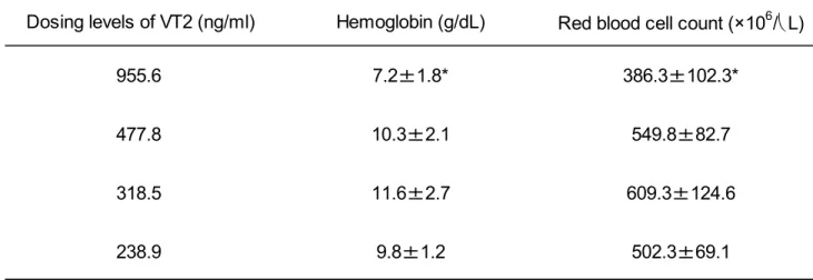
Determination of VT2 doses
Serum levels of VT2 were 8.2 ng/ml, 40.5 ng/ml, 2.9 ng/ml and
2.3 ng/ml at 16 hours after administration of the various test
concentrations (Figure 1). Mean hemoglobin and red blood cell
counts are shown in Table 1. Hemoglobin in mice administered
VT2 solution at 955.6 ng/ml was significantly lower when compared
to mice administered other VT2 concentrations. Red blood cell
counts in mice administered VT2 solution at 955.6 ng/ml were
also significantly lower when compared to mice administered VT2
solutions at 477.8 or 318.5 ng/ml.
Changes in VT2 levels in mice
Serum levels of VT2 in mice administered VT2 solution at 955.6
ng/ml and 477.8 ng/ml are shown in Figures 2 and 3, respectively.
Serum levels of VT2 in mice administered VT2 solution at 955.6
ng/ml did not show significant differences between repeated
administration of bovine immune colostral antibody and controls.
On the other hand, serum levels of VT2 in mice administered VT2
solution at 477.8 ng/ml showed little changes in the repeated
bovine immune colostral antibody administration group. Serum
levels in control mice increased after administration of VT2
and peak levels were observed at 12 hours after administration
(Figure 2). Peak levels were 15.4 ± 5.04 ng/ml. Serum levels
in control mice at 12 and 16 hours were significantly higher
than those in mice repeatedly administered bovine immune
colostral antibody.
Discussion
VT2 derived from E. coli O157:H7 in the intestine is known to
induced serious complications, including HUS and brain damage,
in patients infected with E. coli O157:H7 (Pavia, et al ., 1990;
Clary et al ., 2004; Phillips et al . 2005; Tarr et al ., 2005).
In infection models, mice showing intestinal bleeding died, but
those not showing intestinal bleeding did not die (Kuribayashi
et al ., 2006; Seita et al ., 2013). The cause of death was presumed
toxicity of VT2 absorbed from the intestine. The authors have
reported that serum levels of VT2 continue to increase for 24
hours in mice administered VT2 (Seita et al ., 2013). Serum levels
of VT2 in mice administered bovine immune colostral antibody
against VT2 were lower than those in control mice (Seita et al .,
2013). However, absorption of VT2 was not fully inhibited by
single administration of bovine immune colostral antibody. We
presumed that serious complications were prevented by inhibiting
absorption of VT2 from the intestine. Bovine immune colostral
antibodies were thus administered repeatedly in this study.
Serum levels at 16 hours after administration of VT2 were highest
in mice administered VT2 solution at 477.8 ng/ml. On the other
hand, hemoglobin and red blood cell counts in mice administered
the VT2 solution at 955.6 ng/ml were significantly lower than
in mice administered VT2 solution at other concentrations. These
results suggest that severe intestinal bleeding occurred and
this interfered with intestinal function. Thus, the doses of
VT2 used were 955.6 ng/ml and 477.8 ng/ml in order to evaluate
the inhibition of VT2 absorption in mice.
Serum concentrations of VT2 peaked at 12 hours after
administration of VT2 and decreased in control mice administered
VT2 solution at 477.8 ng/ml. In particular, serum levels of VT2
at 12 and 16 hours in control mice were significantly higher
than in mice administered bovine immune colostral antibody
repeatedly. These results suggest that absorption of VT2 from
the intestine was inhibited by three-time administration of
bovine immune colostral antibody. However, serum levels of VT2
were not significantly different between repeated
administration in the bovine colostral antibody group and the
control group with VT2 administered at 955.6 ng/ml. It was
assumed that VT2 was unable to be absorbed by the intestine to
systemic circulation due to severe intestinal damage. The cause
of death in mice with severe intestinal damage was considered
to be bleeding.
Kita et al . estimated the serum levels of Shiga toxin (Stx) 1
in mice after inoculation with E. coli O157:H7 producing Stx
1 and 2. Peak levels of 34.8
±4.6 pg/ml were observed at 4 days
after inoculation (Kita et al . 2000). Higher levels of VT2 were
inhibited by repeated administration of bovine immune colostral
antibody in this study. Furthermore, treatment of E. coli O157:H7
infection with fosfomycin at early stages prevents progression
to serious symptoms (Sawamura, et al. 1999;Takada et al. 2003).
Thus, adsorption of VT2 appeared to be inhibited by
administration of bovine immune colostral antibody at early
stages after infection with E. coli O157:H7, despite VT2 levels
in intestine increasing from disruption of E. coli O157:H7 by
antibiotics. Furthermore, serious complications such as HUS or
encephalopathy caused by VT2 were prevented.
Unfortunately, the mechanism of inhibition for absorption of
VT2 by administration of bovine immune colostral antibody was
not clarified in this study. Further study will therefore be
necessary.
In conclusion, the absorption of VT2 was inhibited by repeated
administration of bovine immune colostral antibody against VT2
in mice.
Acknowledgment
This research was partially supported by a research project grant
awarded by Azabu University.
References
1. Asano Y, Karasudani T, Tanaka H, Matsumoto J, Okada M,
Nakamura K, et al . Characterization of the Escherichia coli
O157:H7 Outbreak Strain Whose Shiga Toxin 2 Gene Is
Inactivated by IS1203v Insertion. Jpn J Infect Dis 2013;
66:201-6.
2. Koseki S, Mizuno Y, Kawasaki S, Yamamoto K. A Survey of
Iceberg Lettuce for the Presence of Salmonella , Escherichia
coli O157:H7, and Listeria monocytogenes in Japan. J Food
Prot 2011;74:1543–6.
3. Nataro J and Kaper JB. Diarrheagenic Escherichia coli. Clin
Microbiol Rev 1998;11:142-201.
4. Carter AO, Borczyk AA, Carlson JAK, Harvey B, Hockin JC,
Karmali MA, et al . A severe outbreak of Escherichia coli
O157:H7-associated hemorrhagic colitis in a nursing home.
N Engl J Med 1987;317:1496-500.
5. Karch H. Tarr PI, Bielaszewska M. Enterohaemorrhagic
Escherichia coli in human medicine. Int J Med Microbiol
2005;295:405-18.
6. Wong CS, Jelacic S, Hareeb RL, Watkins SL, Tarr PI. The risk
of the hemolytic-uremic symdrome after antibiotic treatment
of Escherichia coli O157: H7 infections. N Engl J Med
2000;342:1930-6.
7. Baum H, Marre R. Antimicrobial resistance of Escherichia
coli and therapeutic implications. Int J Med Microbiol
2005;295:503-11.
8. Ito T, Akino E, Hiramatsu K. Evaluation of antibiotics used
for enterohemorrhagic Escherichia coli O157 enteritis
-effect of various antibiotics on extracellular release of
verotoxin-. Kansennshoshi 1997;71:130-5 (in Japanese with
English summary).
9. Kuribayashi, T. Seita, T., Fukuyma, M., Furuhata, M., Honda,
M, Matsumoto, M, et al . Neutralizing activity of bovine
colostral antibody against verotoxin derived from
enterohemorrhagic Escherichia coli O157:H7 in mice. J
Infect Chemother 2006;12:251-6.
10. Kuribayashi T, Seita T, Matsumoto M, Furuhata K, Tagata K,
Yamamoto S. Bovine colostral antibody verotoxin 2 derived
from Escherichia coli O157:H7: Resistance to proteases and
effects in beagle dogs. Comp Med 2009;59:163-7.
11. Seita T, Kuribayashi T, Honjo T, Yamamoto S. Comparison of
efficacies of bovine immune colostral antibody and each
immunoglobulin class against verotoxin 2, flagellum and
somatic cells of Escherichia coli O157:H7 in mice. J
Microbiol Immunol Infect 2013;46:73-9.
12. Kita E, Yunou Y, Kurioka T, Harada H, Yoshioka S, Mikasa
K, Higashi N. Pathogenic mechanism of mouse brain damage
caused by oral infection with Shiga toxin-producing
Escherichia coli O157:H7. Infect Immun 2000;68,1207-14.
13. Clary TG. The role of Shiga-toxin-producing Escherichia
coli in hemorrhagic colitis and hemolytic uremic syndrome.
Semin Pediatr Infect Dis 2004;14:260-5.
14. Pavia AT, Nicols CR, Green DP, Taxue RV, Mottice S, Green
KD, et al . Hemolytic-uremic syndrome during an outbreak of
Escherichia coli O157:H7 infectious for mentally retarded
persons: clinical and epidemiologic observations. J Pediatr
1990;116:544-51.
15. Phillips B, Tryerman K, Whiteley SM. Use of antibiotics in
suspected haemolytic-uraemic syndrome. BMJ
2005;330:409-11.
16. Tarr PI, Gordon CA, Chandler W. Shiga-toxin-producing
Escherichia coli and haemolytic-uremic syndrome. Lancet
2005;365:1073-86.
17. Sawamura S, Tanaka K, Koga Y. Therapeutic effects of
antibiotics against enterohemorrhagic Escherichia coli
(EHEC) O157:H7 (O157) infection: In vivo analysis using
germfree mice. Kansenshoshi 1999;73:1054-63 (in Japanese
with English summary).
18. Takata T, Tabata T, Tsuruoka T, Watanabe H. Activity of
fosfomycin against Escherichia coli O157:H7-morphological
changes and production of Shiga toxins. Jpn J Antibiot
2003;56:691-6.
0.0 20.0 40.0 60.0
1 2 3 4
S e ru m le ve ls o f VT 2 ( n g /ml)
Dosing solution No.
0.0 5.0 10.0 15.0 20.0 25.0
0 12 24 36 48
Bovine immune colostral group Control group
Se ru m l e vel s o f VT 2 ( n g /ml )
Hours after administration
0.0 5.0 10.0 15.0 20.0 25.0
0 12 24 36 48
Control group
Bovine immune colostral group
Se ru m le v e ls o f V T 2 ( n g /m l)
Hours after administration
*
*
Table 1 Hemoglobin and Red blood cell count at 16 hours after administered with different VT2 concentrations dosing solution to mice. Each value represented mean standard deviation (n=4). *Value differs significantly from mice administered with 477.8 of VT2 (p<0.05).
Dosing levels of VT2 (ng/ml) Hemoglobin (g/dL) Red blood cell count (×10
6/ L)
955.6 7.2±1.8* 386.3±102.3*
477.8 10.3±2.1 549.8±82.7
318.5 11.6±2.7 609.3±124.6
238.9 9.8±1.2 502.3±69.1
Eradication effects of Helicobacter pylori in Mongolian gerbils by colostral antibody obtained from dairy cows immunized with H. pylori and its antibody with (bovine) complement compared with antibiotics
Seita Testrou
1, Takashi Kuribayashi
1, Seiji Yamaguchi
2, Shizuo Yamamoto
11
Graduate School of Environmental and Health Sciences, Azabu
University, 1-17-71 Fuchinobe, Chuou-ku Sagamihara, Kanagawa
252-5201, Japan,
2Department of pediatrics, Shimane University
School of Medicine, 89-1 En-ya-cho, Izumo, Shimane, 693-8501
Japan
Introduction
Helicobacter pylori ( H. pylori ) is a Gram-negative helical
bacillus that was first isolated from the gastric mucosa in human
patients by Warren and Marshall
1. H. pylori produces urease and
sustains infection in the strongly acidic stomach environment
by disassembling urea into ammonia and CO
2 2, 3. Approximately half
the population of Japan is considered to be infected with H.
pylori
4, 5. As such, H. pylori infection is called the Japanese
national disease. Chronic inflammation of gastric mucosa caused
by H. pylori infection induces gastrointestinal diseases, such
as gastric or duodenal ulcer
6, 7, gastric cancer
8, 9,and gastric
MALT lymphoma
10. Other non-gastrointestinal diseases such as
idiopathic thrombocytopenic purpura have also been reported.
11,12