免疫内分泌系バイオマーカーに基づく
乳癌の個別化治療に関する基盤研究
The fundamental study for personalized medicine
in breast cancer patients
based on immunoendocrinological biomarkers
清海 杏奈
東京薬科大学博士論文
略語一覧
AI aromatase inhibitor
CBA cytometric bead array
ConA concanavalin A
COX cyclooxygenase
CRP C-reactive protein
DNR daunorubicin
DOC docetaxel
EGFR epidermal growth factor receptor
ER estrogen receptor
E2 estradiol
FBS fetal bovine serum
Foxp3 forkhead box p3
5-FU 5-fluorouracil
HBSS Hanks’ balanced salt solution
HER2 human epidermal growth factor receptor type2
IFN- interferon gamma
IL interleukin
LH-RH luteinizing hormone-releasing hormone NFB nuclear factor kappa B
NKT natural killer T
PBMC(s) peripheral blood mononuclear cell(s)
PBS phosphate buffered saline
Pg progesterone
PgR progesterone receptor
ROC receiver operating characteristic
TCGA The Cancer Genome Atlas
TGP thermo reversible gelation polymer
Th helper T
TNF- tumor necrosis factor alpha
Treg regulatory T cell
VE vitamin E
3
遺伝子である HER2/neu の過剰発現や,それに伴う HER2 タンパク質の過剰産生が報告さ
れ,HER2 と悪性度や予後との関連があることが分かっている.受容体型チロシンキナーゼ
であるHER2 受容体は,細胞表面に存在する糖タンパク質であり,過剰発現すると本来の細
胞増殖調節機能の制御が効かなくなり,がんが進行する.乳癌では約10~15%の患者で癌組
織の HER2 が陽性である.抗 HER2 抗体医薬品であるトラスツズマブの開発を機に HER2
5
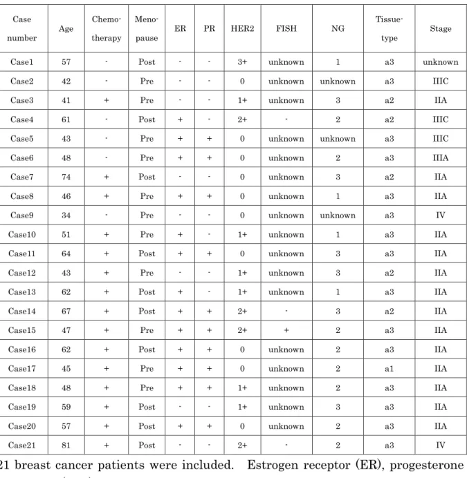
Table 1. Patient characteristics
Case
number Age
Chemo- therapy
Meno-
pause ER PR HER2 FISH NG
Tissue-
type Stage Case1 57 - Post - - 3+ unknown 1 a3 unknown Case2 42 - Pre - - 0 unknown unknown a3 IIIC Case3 41 + Pre - - 1+ unknown 3 a2 IIA Case4 61 - Post + - 2+ - 2 a2 IIIC Case5 43 - Pre + + 0 unknown unknown a3 IIIC Case6 48 - Pre + + 0 unknown 2 a3 IIIA Case7 74 + Post - - 0 unknown 3 a2 IIA Case8 46 + Pre + + 0 unknown 1 a3 IIA Case9 34 - Pre - - 0 unknown unknown a3 IV Case10 51 + Pre + - 1+ unknown 1 a3 IIA Case11 64 + Post + + 0 unknown 3 a3 IIA Case12 43 + Pre - - 1+ unknown 3 a2 IIA Case13 62 + Post + - 1+ unknown 1 a3 IIA Case14 67 + Post + + 2+ - 3 a2 IIA Case15 47 + Pre + + 2+ + 2 a3 IIA Case16 62 + Post + + 0 unknown 2 a3 IIA Case17 45 + Pre + + 0 unknown 2 a1 IIA Case18 48 + Pre + + 1+ unknown 2 a3 IIA Case19 59 + Post - - 1+ unknown 3 a3 IIA Case20 57 + Post + + 0 unknown 2 a3 IIA Case21 81 + Post - - 2+ - 2 a3 IV
6 2-2 摘出組織の三次元培養
2-2-1 実験に使用した試薬,器具および機器
RPMI-1640 培地はシグマ社,10%ウシ胎児血清(FBS)はニチレイバイオ社,ペニシリン G およびストレプトマイシンは GIBCO 社,Hanks' Balanced Salt Solution(HBSS)はシ グマ・アルドリッチ社から各々入手した.また,48 ウェル DSeA-3D プレートは IFTL 社か ら入手した.PBS は GIBCO 社のものを用いた. 2-2-2 培地および試薬の調製 RPMI-1640 培地または HBSS に,56C で 30 分間非働化した仔ウシ胎児血清を 16%(v/v) となるように添加し,さらにペニシリンおよびストレプトマイシンを,各々100 U/mL およ び100 g 力価/mL となるように溶解させ,培養に用いた.凍結乾燥された TGP が充填され
たDSeA-3D プレート(IFTL 社)の各ウェルに,前述の RPMI-1640 培地を 150 L ずつ添
加し,溶解後気泡除去を目的として 1500 rpm にて 20 分間冷却遠心後,組織播種まで 4°C にて保存した. 2-2-3 摘出組織の三次元培養 乳癌組織または正常乳腺組織は,外科手術または針生検により 21 名の患者から得られた ものを用いた.全患者において,診断時の腫瘍の大きさ(直径)は 3 cm 以上であった.組 織運搬液であるHBSS にペニシリン,ストレプトマイシン,およびアムホテリシン B を添加 したものを用いて運搬し,細菌感染を防止するため,3 回以上洗浄した.洗浄後,組織から 血液や脂肪を除去し,径0.5 mm3以下まで細切した.1 ウェル当り 2 個または 3 個の細切組 織をTGP ゲルの充填された DSeA-3D プレートに播種し,5%CO2存在下37°C で 30 分間イ ンキュベーションした.16%FBS を含む RPMI1640 培地を各ウェルに 325 L ずつ重層し, 37°C まで昇温した.さらに 25 L の PBS を各ウェルに加え,5%CO2存在下37°C にて 6 日 間培養を行った.培養後,各ウェルから250 L ずつ上清を回収し,-80°C にて保存した.同 様の手順にて組織を含まないblank well を作成し,6 日間のインキュベーションの後,上清 回収を行った.以上の実験は全て無菌的に行った. 2-3 三次元培養組織の WST-8 試験 上述のように,5%CO2存在下37°C にて 6 日間培養を行った DSeA-3D プレートの各ウェ
ルに,Cell Counting Kit-8 試薬(GIBCO 社)を 25 L ずつ添加し,5%CO2存在下37°C に
て2 時間インキュベーションし,マクロプレートリーダーSAFIRE(TECAN 社)にて 450 nm
7 2-4 Th1/Th2/Th17 サイトカイン測定
2-4-1 実験に使用した試薬,器具および機器
ベ ク ト ン ・ デ ィ ッ キ ン ソ ン 社 の BD Cytometric Bead Array ( CBA ) Human Th1/Th2/Th17 Cytokine Kit を用いて,三次元培養組織上清中の 7 種の Th1/Th2/Th17 系サ イトカイン(後述)を解析した.測定にはベクトン・ディッキンソン社のFACS Calibur を, また算出ソフトは同社のCBA Software を用いた.フローサイトメーター用ポリスチレンチ ューブはベクトン・ディッキンソン社のものを用いた. 2-4-2 フローサイトメーターによる Th1/Th2/Th17 サイトカイン測定法 乳癌組織および正常乳腺組織の三次元培養上清はWash Buffer(ベクトン・ディッキンソ
ン社)にて4 倍希釈し,測定用試料とした.Wash Buffer は 0.2 m のフィルター(ADVANTEC
社)でろ過したものを使用した.IFN-,TNF-,IL-2,IL-4,IL-6,IL-10,および IL-17A の濃度を,マニュアルに則って測定した.CBA 法は,各種サイトカインに特異的な抗体を保 持する異なった蛍光強度のビーズを混合し,その混合ビーズがフローサイトメトリーによっ
て試料中の各種サイトカインを同時に検出・定量できるという原理に基づく.CBA Software
(ベクトン・ディッキンソン社)を用い,標準物質から得られた検量線に基づいて検体中の
サイトカイン濃度を算出した.本法を用いた7 種の Th1/Th2/Th17 系サイトカインの検出限
界は各々IL-2 が 2.6,IL-4 が 4.9,IL-6 が 2.4,IL-10 が 4.5,TNF-が3.8,IFN-が3.7,
およびIL-17A が 18.9 pg/mL であり,検出限界以下は 0 pg/mL とした. 2-5 統計解析 乳癌組織と正常組織間の WST-8 値および三次元培養上清中のサイトカイン濃度の差は, ウィルコクソン符号順位検定にて解析した.正常組織と乳癌組織間のIL-6 および IL-10 産生 の頻度検定についてはカイ二乗検定を用いた.化学療法を行った患者群と行っていない患者 群間のサイトカイン濃度の比較には,Mann-Whitney の U 検定を用いた.乳癌組織と正常 乳腺組織の WST-8 値と上清中サイトカイン濃度との関連は,スピアマンもしくはピアソン の相関係数により検定した.摘出癌組織の ER が陽性(ER+)の患者と ER 陰性(ER-)の 患者間でWST-8 値および各種サイトカイン濃度を比較する場合は,Mann-Whitney の U 検 定で解析した.ER+患者群と ER-患者群間で,摘出組織培養上清中に産生された各種サイト カイン濃度あるいは WST-8 測定値が,各々一定以上高い値を示す患者の頻度の差は,カイ 二乗検定により解析した.PgR 陽性(PgR+)と PgR 陰性(PgR-)患者由来組織間で WST-8 値または各種のサイトカイン濃度の平均値を比較する場合は,対応の無い t 検定(ウェルチ のt 検定)により解析を行った.PgR+と PgR-患者間で,摘出組織の培養液中に産生された 各種サイトカイン濃度あるいは WST-8 測定値が,各々一定以上高い値を示す患者の頻度の 差は,カイ二乗検定により解析した.病期がIIA の患者群と IIB 以上の患者群間での培養上
8
以上の患者群間で,摘出組織の培養液中に産生された各種サイトカイン濃度あるいはWST-8
測定値が,各々一定以上高い値を示す患者の頻度の差は,カイ二乗検定により解析した.い
ずれも危険率 5%未満(p0.05)を有意差ありと判定した.統計解析には,解析ソフトの
9
第三節
結果
TGP ゲルを用いて三次元培養した乳癌組織(n=21)および正常組織(n=9)の増殖能を, WST 法により測定した.乳癌組織では正常組織より高い増殖能を示す傾向があったが,有意 な差はなかった(p=0.3125)(Fig. 1A).化学療法を行った患者から摘出した乳癌組織(n=6) の増殖能は,化学療法を行っていない患者由来の乳癌組織(n=15)より高い傾向があったが, 同様に有意な差ではなかった(p=0.1291)(Fig. 1B). 三次元培養した乳癌組織(n=21)および正常組織(n=8)の培養上清中に分泌された IFN-,TNF-,IL-2,IL-4,IL-6,IL-10,および IL-17A の濃度を測定した.IL-6 および IL-10
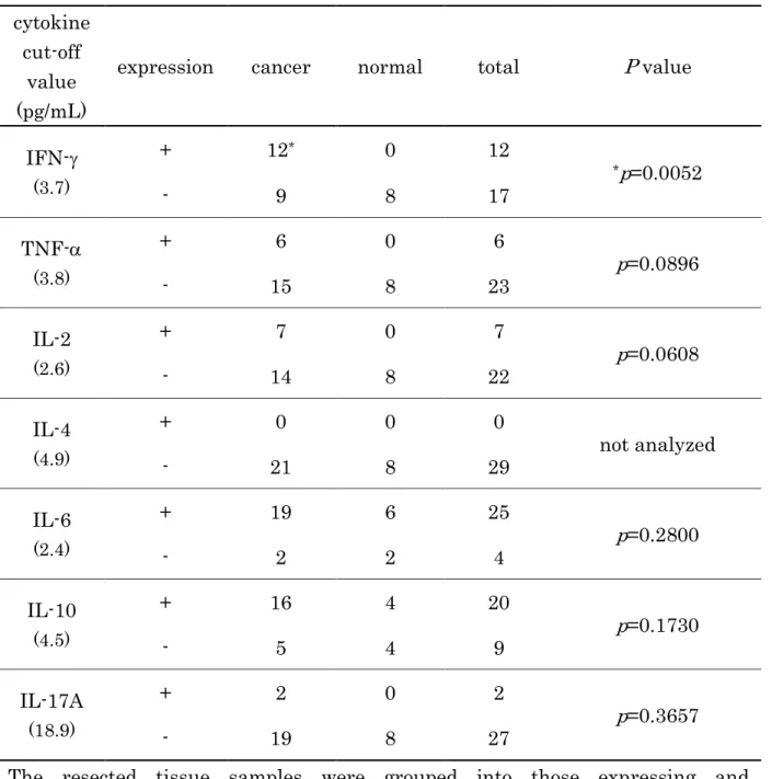
は,いずれの組織の培養上清からも多量に検出された(Fig. 2).一方で,IFN-,TNF-,
IL-2,および IL-17A は乳癌組織培養上清にのみ検出された(Fig. 2).IL-4 はいずれの培養 上清からも検出されなかった(Fig. 2).乳癌組織および正常乳腺組織培養上清間で,IL-6 と IL-10 濃度に有意な差はなかった.三次元培養上清中に IFN-が分泌された組織の頻度は, 癌組織の方が正常組織に比べ有意に高かった(p=0.0052)(Table 2).これより,IFN-は癌 組織に特異的であることが示唆された. 化学療法を受けた乳癌患者から得た癌組織(n=6)の三次元培養上清中の IFN-と IL-10 の濃度は,化学療法を受けていない患者から得た癌組織(n=15)のそれらより高い傾向にあ
ったが,有意な差はなかった(Fig. 3).また組織培養上清中の IFN-,TNF-,IL-2,IL-6,
IL-10,および IL-17A の濃度は,これら患者群間で有意な差はなかった(Fig. 3).
WST-8 法にて測定した乳癌組織の増殖能と,組織培養上清中の IFN-(Fig. 4A; p=0.0031,
Pearson の相関係数検定による),IL-6(Fig. 4D; Pearson の相関係数検定で p=0.0274, Spearman の相関係数検定で p=0.0009),あるいは IL-10(Fig. 4E; Pearson の相関係数検
10 (p=0.0502)(Table 3).
全患者をIIA 期(n=14)と IIB 期以上(n=6)に群分けし,WST-8 値と三次元培養上清中
11
Fig. 1. Growth abilities of three-dimensionally cultured breast cancer tissues.
The growth abilities of the tissues three-dimensionally cultured in TGP gel were
as assessed by WST-8 assay (absorbance at 450 nm; see Materials and Methods)
after the culture. A: Comparison of the growth abilities between breast cancer
tissues (n=21) and normal breast tissues (n=9). No significant difference was
observed, as analyzed by Wilcoxon signed rank test (p=0.3125). B: Comparison
between cancer tissues from patients treated by chemotherapy (n=6) and those
from patients who were not treated by chemotherapy (n=15). No significant
difference was observed, as analyzed by Mann Whitney test (p=0.1291).
A
12
Fig. 2. Comparison of cytokine concentrations in supernatant of
three-dimensionally cultured breast cancer and normal tissues. Breast cancer
tissues (cancer tissues) or normal tissues of breast cancer patients (normal
tissues) were cultured for 6 days in TGP gel respectively, and the cytokine
concentrations in supernatant were determined by CBA assay procedures.
Twenty-one cancer tissues and eight normal tissues were examined. The mean ±
SD concentrations were indicated. The cytokine concentrations did not
significantly differ between cancer tissues and normal tissues. The frequencies
of the expression of IFN-
was significantly different between the cancer tissues
and the normal tissues, as analyzed by Chi-square test (p=0.0052). ND (not
detected): The cytokine concentrations were under each detection limit.
13
Table 2. Differences for the incidence of each cytokine expression between breast
cancer tissues and normal tissues.
cytokine
cut-off
value
(pg/mL)
expression
cancer
normal
total
P value
IFN-
(3.7)+
12
*0
12
*p=0.0052
-
9
8
17
TNF-
(3.8)+
6
0
6
p=0.0896
-
15
8
23
IL-2
(2.6)+
7
0
7
p=0.0608
-
14
8
22
IL-4
(4.9)+
0
0
0
not analyzed
-
21
8
29
IL-6
(2.4)+
19
6
25
p=0.2800
-
2
2
4
IL-10
(4.5)+
16
4
20
p=0.1730
-
5
4
9
IL-17A
(18.9)+
2
0
2
p=0.3657
-
19
8
27
14
Fig. 3. Comparison of each cytokine concentration in supernatant of
three-dimensionally cultured tissues obtained from patients with and
without-chemotherapy. IFN-
, TNF-
, IL-2, IL-6, IL-10 and IL-17A in the
supernatant of three-dimensionally cultured tissues obtained from patients with
(n=6) or without (n=15) chemotherapy were compared. There was no difference
in IFN-
(A;
p=0.0662), TNF-
(B;
p=0.1612), IL-2 (C; p=0.4254), IL-6 (D;
p=0.1106), IL-10 (E; p=0.0674) and IL-17A (F; p=0.7086) concentrations between
these patient subgroups, as analyzed by Mann Whitney test, were observed in the
patients with chemotherapy.
15
Fig. 4. Correlation between cytokine concentrations in supernatant of
three-dimensionally cultured breast cancer tissues and WST-8 assay values of the
cultured tissues. The growth abilities of the tissues three-dimensionally
cultured in TGP gel were assessed by WST-8 assay (absorbance at 450 nm) after
the culture. Correlations were evaluated by Pearson’s correlation coefficient test
and Spearman’s correlation coefficient test (n=21). Concentrations for IFN-
(A;
16
17
Fig. 5. Correlation between cytokine concentrations in supernatant of
three-dimensionally cultured normal tissues and the WST-8 assay values of
cultured tissues. Correlation was evaluated by Pearson’s correlation coefficient
test and Spearman’s correlation coefficient test (n=8). Concentrations for IL-10
(B) in the culture supernatant was significantly correlated with the WST-8 assay
values of the tissues (p=0.0458 by Spearman), however there was no significantly
correlation between normal tissue growth and IL-6 concentration (A).
A
18
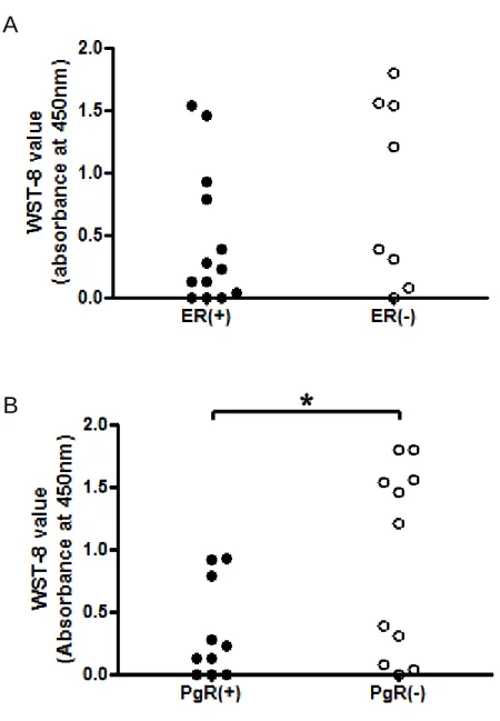
Fig. 6. Comparison for the WST-8 assay values of three-dimensionally cultured
tissues between hormone-receptor positive and negative breast cancer tissues.
A; comparison between ER(+) tissues (n=13) and ER(-) tissues (n=8). B;
comparison between PgR(+) tissues (n=10) and PgR(-) tissues (n=11). A
significant difference for the WST-8 indices was observed between PgR(+) and
PgR(-) tissues, as analyzed by unpaired t test with Welch’s correction (
*p=0.0383).
A
19
Table 3. Relationship between estrogen receptor (ER) or progesterone receptor
(PgR) expression incidence and the WST-8 assay value (index for tumor growth) of
three-dimensionally cultured breast cancer tissues.
WST-8 assay value for 3D
cultured sample
Higher
Lower
Total
Estrogen receptor
(ER)
+
5
8
13
-
6
*2
8
Total
11
10
21
Progesterone receptor
(PgR)
+
3
7
10
-
8
3
11
Total
11
10
21
20
Table 4. Relationship between the TNM stages and the incidences of cytokines
production in culture medium of three-dimensionally cultured breast cancer
tissues.
The incidence of cytokine
in cultured cancer tissue
TNM stage
P value
Stage
IIA
Over
stage
IIB
Total
IFN-
Positive
7
4
11
p
0.05
Negative
7
2
9
TNF-
Positive
3
5
*8
*p=0.0096
Negative
11
1
12
IL-2
Positive
4
3
7
p
0.05
Negative
10
3
13
IL-4
Positive
0
0
0
not analyzedNegative
14
6
20
IL-6
Positive
5
5
10
p=0.051
Negative
9
1
10
IL-10
Positive
10
5
15
p
0.05
Negative
4
1
5
IL-17A
Positive
1
1
2
p
0.05
Negative
13
5
18
Total
14
6
20
The patients were divided into positive and negative groups for the cytokine
production in culture medium of three-dimensionally cultured breast cancer
tissues. The cut-off limits of cytokine expressions were the median of each
cytokine concentration (all median was 0 pg/mL).
23
免疫を抑制して逆に癌増殖能を高めている可能性がある一方で,抗血管新生作用を介して間 接的に癌増殖を妨げている[48].IL-10 は免疫抑制性であるだけでなく,炎症誘発性に作用す
ることも知られており,細胞毒性活性およびCD8+T 細胞を増強させる[49].本研究では,三
次元培養乳癌組織上清中のIFN-,IL-6,および IL-10 濃度が,WST-8 法で求めた組織増殖
能と正の相関を示した.ドセタキセルまたはパクリタキセルで治療した進行乳癌患者血清中
で,IL-1 および TNF-が減少している一方,IFN-,IL-2,IL-6,および顆粒球マクロファ
ージコロニー刺激因子濃度が増加しているという報告がある[50].本研究では,癌組織サン プル摘出前にタキサン系薬物で治療した患者は 21 人中 3 人のみであったため,治療に用い たタキサン系薬物の血清中サイトカインに及ぼす影響は明確ではない. IL-17 は,腫瘍微小環境内で既に腫瘍の初期構築に関与していることが示唆されている[51]. IL-17 産生 Th17 細胞や IL-17 産生 CD8+T 細胞(Tc17)が,乳癌微小環境で多く産生される TGF-およびIL-6 存在下で,ナイーブ CD4+T 細胞や CD8+T 細胞から誘導されることが報 告されている[52,53].申請者の研究では,Tc17 のような IL-17 産生細胞が周囲の乳癌組織 から分泌されたTh1 サイトカインにより誘導され,IL-17 を産生している可能性が考えられ た. 最後に,申請者は三次元培養乳癌組織の WST-8 値と,培養上清中の各種サイトカインの 濃度,患者の病期,あるいは患者の臨床データとの関連を調べた.その結果,ER-癌組織は ER+癌組織より,高増殖能を示す頻度が有意に高かった(Table 3).TCGA の "Breast Invasive Carcinoma" によれば,NFB シグナルを強く抑制する TNFIP3 遺伝子と ER をコ
ードするESR1 遺伝子の共発現が,有意に高率で起こることが分かっている(p=0.027,log
odds ratio: 0.931)[20].これは,ESR1 が発現している乳癌患者において,TNFAIP3 発現
24 このため,進行乳癌組織が頻繁にTNF-を分泌するという本結果は,これらの報告に矛盾し ない. 本章では,結論として,TGP を用いた三次元培養ヒト乳癌組織が培養液中に IL-6 や IL-10 等のTh2 サイトカインを相対的に高濃度で分泌することを明らかとした.これらのサイトカ インはTh1 由来の細胞性免疫に関連する腫瘍免疫を抑制し,腫瘍増殖を促進しているものと
考えられた.IFN-,TNF-,IL-2,および IL-17A は三次元培養正常乳腺組織培養上清から
は検出されなかったことから,乳癌組織特異的と考えられた.一方ホルモン受容体陽性癌組
織は,本培養系ではホルモン受容体陰性癌組織と比べて増殖能が低かった.TNF-は乳癌病
26
の記入が不十分であるなどの理由から VE の効果を判定できなかった患者(n=11)は,VE
の効果判定が関わる検討項目からは除外した.
Table 5. Patient Characteristics
Patient grouping based on;
Remarks
Number of Patients
Aromatase inhibitor used
Anastrozole
50
Exemestane
2
Letrozole
10
BMI
Nonobesity
50
Obesity
12
The year after post menopause
10 years
21
10 years
38
Unknown
3
The year after taking aromatase
inhibitor
2 years
41
2 years
21
The osteoarthropathy symptom
before vitamin E administration
*Mild
22
Severe
34
Unknown
6
The effect of vitamin E
**Sensitive
24
Insensitive
27
Unknown
11
*Symptoms were scored as 0-3 points (3 is the worst) based on NCI-CTC Version
2.0 for each symptom; stiffness and low back pain. Then, the total score was
calculated for each patient, and the patients were divided into mild and severe
groups by the median score (median=4).
**These patients were classified into two groups according to their responses to
the therapeutic effect of vitamin E.
27 2-2 血清中ホルモンおよび VE 濃度の測定
対象患者末梢血を9 mL 採取し,血清分離を行った.血清中 E2,Pg,および VE 濃度は,
株式会社エスアールエルに依頼して測定した.血清中E2 および Pg 測定は各々エクルーシス
試薬E2III およびプロゲステロン II(Roche Diagnosis Co., Japan)を用い,電気化学的発
28 2-4 Th1/Th2/Th17 サイトカイン測定 2-4-1 実験に使用した試薬,器具および機器 第一章2-4-1 と同様のものを使用した. 2-4-2 フローサイトメーターによる Th1/Th2/Th17 サイトカイン測定法 患者血清中およびPBMC 培養上清中の IFN-,TNF-,IL-2,IL-4,IL-6,IL-10,およ びIL-17A 濃度を,第一章 2-4-2 に述べた方法に準じて CBA 法にて測定した. 2-5 統計解析法 バイオマーカーである血清中VE,E2,Pg,および各種サイトカインの濃度の差を患者二 群間で統計学的に比較する場合はMann-Whitney の U 検定を,また,VE 投与前後の患者間 における骨関節症状スコアの比較にはWilcoxon 検定を,VE 投与前後の患者間における各種 バイオマーカーの平均値の比較には対応のある t 検定を用いた.血清中 E2 濃度と各種サイ トカイン濃度の相関,および VE 投与後の骨関節症状改善率と VE 投与前の血清中バイオマ ーカー濃度との相関は,ピアソンの相関解析により検討した.血清中バイオマーカー濃度と 骨関節症状の改善スコアの相関はKruskal-Wallis 検定を用いて解析した.血清 E2 濃度高値 の患者群および低値の患者群間における,VE 投与前骨関節症状スコアの重症度が高い患者 の頻度の差は,カイ二乗検定により検討した.VE 投与への応答性の良い患者を血清中 E2 濃度の境界値1.8 pg/mL により区別するという診断指標は,血清中 E2 濃度と VE の効果に 関するROC 解析を行うことにより確認した.いずれも危険率 5%未満(p0.05)を有意差あ
29
第三節
結果
AI に起因する骨関節症状が比較的軽症な患者群(n=22)と,重症な患者群(n=34)間の 血清中バイオマーカーの濃度差を比較した.その他6 名の患者においてはスコア聴取が完結 出来なかったため,本検討からは除外した.これら二群間における血清中VE,E2,Pg,IFN-, TNF-,IL-2,IL-4,IL-6,IL-10,および IL-17A の各濃度には有意な差はなかった(デー タ非表示).さらに,血清中サイトカイン濃度とホルモン濃度との相関を検討したところ,血 清中E2 濃度と IL-17A 濃度との間に有意な正の相関があった(p=0.0291)(Fig. 7).一方, 血清中Pg,VE,または Th1/2 サイトカインと E2 濃度間には,有意な相関はなかった(デ ータ非表示). 次に,VE 投与前後で骨関節症状スコアの評価ができた 51 名の乳癌患者において,VE が 骨関節症状スコアを減少させるか否かに基づくVE の治療効果を検討した.AI 治療に起因す30 により求められた曲線下面積が0.78074 であったことから明らかとなった(Fig. 12).また, これより,血清中E2 濃度の至適カットオフ値が 1.8 pg/mL と結論付けられた.E2 カットオ フ値を1.8 pg/mL とした時,VE の効果を予測する血清中 E2 値の判断基準感度および特異 度は各々80.0%および 70.4%であり,正確な患者分類率は 74.5%だった. 全てのアンケートに回答した51 人の患者を,VE 療法応答性によって VE 感受性群と非感 受性群に分け(Table 5),VE 投与前後における血清中 E2 濃度を両群間で比較した.全患者 を対象にVE 投与後の血清中 E2 濃度を投与前と比較すると,有意な増加がみられた(n=51, 1.88±0.21 pg/mL から 2.38±0.20 pg/mL へ増加,p=0.0412).一方,VE 療法応答性の違い により群分けした各群別に同様の検討を行った場合,VE 感受性群では VE 投与後血清中 E2 濃度が有意に増加した(n=24, 1.14±0.25 pg/mL から 2.21±0.32 pg/mL へ増加,p=0.0124) が,VE 非感受性群では有意な変化はなかった(n=27, 2.54±0.28 pg/mL から 2.53±0.24 pg/mL へ増加)(Fig. 13). 最後に,VE と AI がヒト PBMC に及ぼす直接的な影響を検討するため,健常者より得ら れたPBMC に T 細胞マイトジェンである ConA 存在下,アナストロゾール(1-100 ng/mL) もしくはVE(1.88-188 g/mL)を添加し培養を行った.PBMC 増殖率は WST-8 値として,
また,PBMC 培養上清中の IFN-,TNF-,IL-2,IL-4,IL-6,IL-10,および IL-17A の濃
度は,CBA 法により測定した(Fig. 14).その結果,アナストロゾール 10 ng/mL と VE 18.8
g/mL の共存下では,アナストロゾール 10 ng/mL 単独の時に比べ,PBMC の WST-8 値が
有意に高かった(p0.05).一方で,活性化 PBMC による 7 種のサイトカインの産生には,
31
32
Fig. 8. Osteoarthropathy scores and estradiol concentrations in aromatase
inhibitor treated patients after vitamin E administration (n=60). (A) A
statistically significant decrease in the mean scores was observed after the
vitamin E administration, as compared to the mean scores before administration,
as analyzed by Wilcoxon test (
*p=0.0178). (B) There was no significant change in
the mean estradiol concentrations after administration of vitamin E by paired t
test.
A
33
Fig. 9. Changes of serum vitamin E concentrations in patients after vitamin E
administration. Statistically significant increases in the mean concentrations of
vitamin E was observed after the vitamin E administration, as compared to those
before administration, as analyzed by paired t test (
*p<0.0001)(n=61).
34
Fig. 10. Correlations between osteoarthropathy improvement rates and serum
estradiol or IL-17A concentrations. There were correlations between
improvement rates of osteoarthropathy (%) and serum concentrations of (A)
estradiol or (B) IL-17A (pg/mL) in 51 breast cancer patients before administration
of vitamin E. There were significant negative correlations in (A) estradiol
(p=0.0069) and (B) IL-17A (p=0.0234). The plots shown in un-shaded area (left
zone) represents the patients with worse symptoms after the vitamin E
administration, whereas the plots shown in shaded area (right zone) represents
the patients with improvement in the symptoms. Zero percent in the abscissa
means no change in the scores.
A
35
36
Table 6. Relationship between serum estradiol concentrations and responses to
vitamin E therapy.
Response to the therapeutic
efficacy of vitamin E
Serum estradiol before vitamin E
administration (pg/mL)
Low: 1.8
High: 1.8
Total
Sensitive
19
*5
24
Insensitive
8
19
27
Total
27
24
51
37
Fig. 12. Serum estradiol level as a new biomarker for patients who will respond to
vitamin E. Receiver operator characteristic (ROC) curve for serum estradiol
concentrations as a function of the improvement scores for osteoarthritis by
vitamin E treatment. The areas under the curve (AUC) was 0.78074, and the
cut-off value of estradiol was calculated as 1.80 pg/mL (see Results).
38
A
B
39
Fig. 13. Serum estradiol concentrations in vitamin E-sensitive and -insensitive
patients who answered all of the questionnaire. 51 patients who answered all
questionnaires were divided into vitamin E sensitive or insensitive groups and
their serum estradiol concentrations between before and after vitamin E
administration were compared. In all patients group (
*p=0.0412) and the
sensitive group (
*p=0.0124), its level was significantly increased after vitamin E
41
42
第四節
考察
閉経後乳癌患者における AI 治療は,副作用としてしばしば骨関節症状を引き起こす.こ の結果,AI 治療を断念せざるを得ない場合もある.しかし一方で,閉経後骨粗鬆症は E2 の 欠乏が原因となっている.このように,AI が E2 値を低下させることで,結果として骨関節 症状を引き起こしている可能性がある.破骨細胞上の ER への E2 の結合は,破骨細胞の生 存を制御しアポトーシスを誘発するFas リガンドの遺伝子発現を活性化することが知られて いる[61].このため,E2 欠乏は骨代謝の不均衡をもたらすと考えられる. 本章にて申請者は,骨関節症状の重症な患者と軽症な患者の間で,血清中 VE,E2,Pg,IFN-,TNF-,IL-2,IL-4,IL-6,IL-10,および IL-17A の濃度に有意な差がないことを 明らかとした.このことから,AI 服用患者において,骨関節症状の重症度とこれらの血清中
バイオマーカーとの間に関連性がないことが示唆された.一方,AI 服用中の乳癌患者におい
て,血清中E2 値と IL-17A 濃度との間に有意な正の相関が認められた(Fig. 7).E2 欠乏は,
IL-17 分泌に関与する Th17 細胞へのナイーブ T 細胞の分化を誘導するという報告がある[62]. 加えて,E2 欠乏性骨粗鬆症の病態下では,IL-17 が骨減少を媒介することを示唆する報告[63] や,E2 の抗炎症作用がこれら IL-17 の作用に対して抑制的に働くという報告[64]もある.血 清中E2 値と IL-17A 濃度が正の相関を示したことから,IL-17 による骨関節症状の炎症に基 づく悪化を E2 が抑えていることが示唆された.なお,他の研究ではマウスを用いてこれら の関連を検討しているが,本研究では患者末梢血清中のホルモンやサイトカイン濃度を測定 している.このため,本研究で患者血清中に検出されたIL-17 がどの体内組織もしくは臓器 に由来するのか明らかとなっておらず,乳癌患者血清中のE2 と IL-17A との関連を説明する 詳細なメカニズムについて,更なる検討が必要である. VE は抗酸化作用や抗炎症作用など様々な生物活性を有しており[65],また医薬品としても 末梢血循環改善を目的として世界で汎用されている.しかしながら,AI 関連骨関節症状の治 療を目的としたVE の有用性を検討した報告は乏しい.本研究にて,AI の使用により骨関節 症状を呈した乳癌患者へのVE 投与が,多くの患者において有効な治療であったことを明ら かにした(Fig. 8).今回の結果では,VE 投与が乳癌患者血清中の VE 濃度を有意に増加さ せたが,一方で血清中のE2,Pg,IFN-,TNF-,IL-2,IL-4,IL-6,IL-10,および IL-17A
44
45
総合考察
乳癌罹患者は年々増加傾向を辿っている.また,乳癌治療に関わる時間的,経済的,精神 的,身体的な負荷は非常に大きく,それらの軽減を目的とした研究が必要と考える.近年, HER2 をはじめとするバイオマーカー研究が進んでいるが,依然として患者個々に合った治 療の確立は道半ばである. 以上のような背景を踏まえ,第一章では,乳癌患者から摘出した乳癌組織を,TGP ゲルを 用いて三次元培養し,培養乳癌組織から産生されるTh1/Th2/Th17 系サイトカインの特徴や 培養上清がヒト免疫系に及ぼす影響について検討した.これらの検討結果から,腫瘍免疫に 影響を及ぼす乳癌組織由来因子の解明と,これらの因子をバイオマーカーとした乳癌の個別 化医療の推進の可能性について展望した.さらに第二章では,閉経後乳癌治療に併発する骨 関節症状に対するvitamin E(VE)の効果と,乳癌治療の副作用軽減を目的とした VE 投与 の有用性およびその個別化投与推進のためのバイオマーカーを検索することを目的として検 討を行った. これらの検討結果から,乳癌治療における薬物の主作用と副作用の発現に各々係る新たな バイオマーカーを確立し,それに基づく乳癌治療の個別化を推進するための基盤を構築する ことを目的とし,本研究を行った. 第一章では,TGP を用いて三次元的に培養したヒト乳癌組織が,培養液中に IL-6 や IL-10 などのTh2 系サイトカインを高濃度で分泌することを明らかとした.これらのサイトカイン は,Th1 細胞免疫に関連する腫瘍免疫を抑制し,腫瘍増殖を促進しているものと考えられた.また IFN-,TNF-,IL-2,および IL-17A は三次元培養正常乳腺組織培養上清には検出さ
48
引用文献
1. がん対策情報センター(2015)がん情報サービス「最新がん統計」 http://ganjoho.jp/reg_stat/statistics/stat/summary.html
2. 日本乳癌学会,乳癌診療ガイドライン 1 治療編,2015 年度版
3. Khanduri S and Dodwell DJ. Aromatase inhibitors and musculoskeletal symptoms. The Breast. 2008; 17: 78-81.
4. Dunn GP, Old LJ, and Schreiber RD. The immunobiology of cancer immunosurveillance and immunoediting. Immunity. 2004; 21: 137-148.
5. Wilson J and Balkwill F. The role of cytokines in the epithelial cancer microenvironment. Semin
Cancer Biol. 2002; 12: 113-120.
6. Balkwill F and Mantovani A. Inflammation and cancer: back to Virchow? Lancet. 2001; 357: 539-545.
7. Venetsanakos E, Beckman I, Bradley J, and Skinner JM. High incidence of interleukin 10 mRNA but not interleukin 2 mRNA detected in human breast tumours. Br J Cancer. 1997; 75:
1826-1830.
8. Huang M, Wang J, Lee P, Sharma S, Mao JT, Meissner H, Uyemura K, Modlin R, Wollman J, and Dubinett SM. Human non–small cell lung cancer cells express a type 2 cytokine pattern.
Cancer Res. 1995; 55: 3847-3853.
9. Green AR, Green VL, White MC, and Speirs V. Expression of cytokine messenger RNA in normal and neoplastic human breast tissue: identification of interleukin-8 as a potential regulatory factor in breast tumours. Int J Cancer. 1997; 72: 937-941.
10. Benoy I, Salgado R, Colpaert C, Weytjens R, Vermeulen PB, and Dirix LY. Serum interleukin 6, plasma VEGF, serum VEGF, and VEGF platelet load in breast cancer patients. Clin Breast
Cancer. 2002; 2: 311-315.
11. Kozłowski L, Zakrzewska I, Tokajuk P, and Wojtukiewicz MZ. Concentration of interleukin-6 (IL-6), interleukin-8 (IL-8) and interleukin-10 (IL-10) in blood serum of breast cancer patients.
Rocz Akad Med Bialymst. 2003; 48: 82-84.
12. Knüpfer H and Preiss R. Significance of interleukin-6 (IL-6) in breast cancer. Review. Breast
Cancer Res Treat. 2007; 102: 129-135.
13. Moscona A. Rotation-mediated histogenetic aggregation of dissociated cells. A quantifiable approach to cell interactions in vitro. Exp Cell Res. 1961; 22: 455-475.
14. O’Keane JC, Kupchik HZ, Schroy PC, Andry CD, Collins E, and O’Brien MJ. A
49
15. Carlsson J, Nilsson K, Westarmark B, Ponten J, Sundstrom C, Larsson E, Bergh J, Pahlman S, Busch C, and Collins VP. Formation and growth of multicellular spheroids of human origin. Int J
Cancer. 1983; 31: 523-553.
16. Kleinman HK, McGarvey ML, Hassel JR, Star VL, Cannon FB, Laurie GW, and Martin GR. Basement membrane complexes with biological activity. Biochemistry. 1986; 25: 312-318. 17. Lawler E, Miller FR, and Heppner GH. Significance of three-dimensional growth patterns of
mammary tissues in collagen gels. In Vitro. 1983; 19: 600-610.
18. Tsukikawa S, Matsuoka H, Kurahashi Y, Konno Y, Satoh K, Satoh R, Isogai A, Kimura K, Watanabe Y, Nakano S, Hayashi J, and Kubota S. A new method to prepare multicellular
spheroids in cancer cell lines using a thermo-reversible gelation polymer. Artif Organs. 2003; 27: 598-604.
19. Urazumi K, Kimijima I, Fukushima T, Nakayama K, Kiman K, Tsuchiya A, and Abe R. Different growth rate and contracting activity of collagen gel between fibroblasts from breast cancer tissue and normal breast tissue. Nihon Geka Gakkai Zasshi. 1993; 94: 501-504. [Article in Japanese] 20. The Cancer Genome Atlas. http://www.cbioportal.org/index.do
21. Sasser AK, Sullivan NJ, Studebaker AW, Hendey LF, Axel AE, and Hall BM. Interleukin-6 is a potent growth factor for ER-alpha-positive human breast cancer. FASEB J. 2007; 13: 3763-3770. 22. Zhang YM, Mao YM, and Sun YX. Genetic polymorphisms of IL-6 and IL-10 genes correlate
with lung cancer in never-smoking Han population in China. Int J Clin Exp Med. 2015; 8: 1051-1058.
23. Filipova A, Seifrtova M, Mokry J, Dvorak J, Rezacova M, Filip S, and Diaz-Garcia D. Breast cancer and cancer stem cells: a mini-review. Tumori. 2014; 100: 363-369.
24. Iliopoulos D, Hirsch HA, Wang G, and Struhl K. Inducible formation of breast cancer stem cells and their dynamic equilibrium with non-stem cancer cells via IL6 secretion. Proc Natl Acad Sci
U S A. 2011; 108: 1397-1402.
25. Dontu G, Liu S, and Wicha MS. Stem cells in mammary development and carcinogenesis: implications for prevention and treatment. Stem Cell Rev. 2005; 1:207-213.
26. Fillmore C and Kuperwasser C. Human breast cancer stem cell markers CD44 and CD24:
enriching for cells with functional properties in mice or in man? Breast Cancer Res. 2007; 9: 303. 27. Reya T, Morrison SJ, Clarke MF, and Weissman IL. Stem cells, cancer, and cancer stem cells.
Nature. 2001; 414: 105-111.
28. Sheridan C, Kishimoto H, Fuchs RK, Mehrotra S, Bhat-Nakshatri P, Turner CH, Goulet R Jr., Badve S, and Nakshatri H. CD44+/CD24− breast cancer cells exhibit enhanced invasive properties: an early step necessary for metastasis. Breast Cancer Res. 2006; 8: R59.
29. Sansone P, Storci G, Tavolari S, Guarnieri T, Giovannini C, Taffurelli M, Ceccarelli C, Santini D, Paterini P, Marcu KB, Chieco P, and Bonafè M. IL-6 triggers malignant features in
50 2007; 12: 3660-3663.
30. Moore KW, O’garra A, Malefyt RW, Vieira P, and Mosmann TR. Interleukin-10. Annu Rev
Immunol. 1993; 11: 165-190.
31. Coussens LM, and Werb Z. Inflammation and cancer. Nature. 2002; 420: 860-867.
32. Kim HS, Lee JH, Han HD, Kim AR, Nam ST, Kim HW, Park YH, Lee D, Lee MB, Park YM, Kim HS, Kim YM, You JC, and Choi WS. Autocrine stimulation of IL-10 is critical to the enrichment of IL-10-producing CD40hiCD5+ regulatory B cells in vitro and in vivo. BMB Rep. 2015; 48: 54-59.
33. Olkhanud PB, Damdinsuren B, Bodogai M, Gress RE, Sen R, Wejksza K, Malchinkhuu E, Wersto RP, and Biragyn A. Tumor-evoked regulatory B cells promote breast cancer metastasis by converting resting CD4+ T cells to T-regulatory cells. Cancer Res. 2011; 71: 3505-3515.
34. Bu M, Shen Y, Seeger WL, An S, Qi R, Sanderson JA, and Cai Y. Ovarian carcinoma-infiltrating regulatory T cells were more potent suppressors of CD8+ T cell inflammation than their
peripheral counterparts, a function dependent on TIM3 expression. Tumour Biol. 2015 Oct 19. [Epub ahead of print]
35. Dong C. Diversification of T-helper-cell lineages: finding the family root of IL-17–producing cells. Nat Rev Immunol. 2006; 6: 329-333.
36. Michel ML, Keller AC, Paget C, Fujio M, Trottein F, Savage PB, Wong CH, Schneider E, Dy M, and Leite-de-Moraes MC. Identification of an IL-17–producing NK1.1neg iNKT cell population involved in airway neutrophilia. J Exp Med. 2007; 204: 995-1001.
37. Tajima M, Wakita D, Noguchi D, Chamoto K, Yue Z, Fugo K, Ishigame H, Iwakura Y, Kitamura H, and Nishimura T. IL-6–dependent spontaneous proliferation is required for the induction of colitogenic IL-17–producing CD81T cells. J Exp Med. 2008; 205: 1019-1027.
38. Romani L, Fallarino F, De Luca A, Montagnoli C, D'Angelo C, Zelante T, Vacca C, Bistoni F, Fioretti MC, Grohmann U, Segal BH, and Puccetti P. Defective tryptophan catabolism underlies inflammation in mouse chronic granulomatous disease. Nature. 2008; 451: 211-215.
39. Numasaki M, Watanabe M, Suzuki T, Takahashi H, Nakamura A, McAllister F, Hishinuma T, Goto J, Lotze MT, Kolls JK, and Sasaki H. IL-17 enhances the net angiogenic activity and in vivo growth of human non–small cell lung cancer in SCID mice through promoting
CXCR-2-dependent angiogenesis. J Immunol. 2005; 175: 6177-6189.
40. Langowski JL, Zhang X, Wu L, Mattson JD, Chen T, Smith K, Basham B, McClanahan T, Kastelein RA, and Oft M. IL-23 promotes tumour incidence and growth. Nature. 2006; 442: 461-465.
41. Wakita D, Sumida K, Iwakura Y, Nishikawa H, Ohkuri T, Chamoto K, Kitamura H, and
Nishimura T. Tumor-infiltrating IL-17-producing gammadelta T cells support the progression of tumor by promoting angiogenesis. Eur J Immunol. 2010; 40: 1927-1937.
51
Cell. 2008; 13: 7-9.
43. Cao Z, Xu X, Luo X, Li L, Huang B, Li X, Tao D, Hu J, and Gong, J. Role of RANTES and its receptor in gastric cancer metastasis. J Huazhong Univ Sci Technolog Med Sci. 2011; 31: 342-347.
44. Chung AS, Wu X, Zhuang G, Ngu H, Kasman I, Zhang J, Vernes JM, Jiang Z, Meng YG, Peale FV, Ouyang W, and Ferrara N. An interleukin-17–mediated paracrine network promotes tumor resistance to anti-angiogenic therapy. Nat Med. 2013; 19: 1114-1123.
45. Sakata H, Murakami S, and Hirayama R. Serum soluble interleukin-2 receptor (IL-2R) and immunohistochemical staining of IL-2R/Tac antigen in colorectal cancer. Int J Clin Oncol. 2002; 7: 312-317.
46. Cheung YT, Ng T, Shwe M, Ho HK, Foo KM, Cham MT, Lee JA, Fan G, Tan YP, Yong WS, Madhukumar P, Loo SK, Ang SF, Wong M, Chay WY, Ooi WS, Dent RA, Yap YS, Ng R, and Chan A. Association of proinflammatory cytokines and chemotherapy-associated cognitive impairment in breast cancer patients: a multi-centered, prospective, cohort study. Ann Oncol. 2015; 26: 1446-1451.
47. Chang Q, Bournazou E, Sansone P, Berishaj M, Gao SP, Daly L, Wels J, Theilen T, Granitto S, Zhang X, Cotari J, Alpaugh ML, de Stanchina E, Manova K, Li M, Bonafe M, Ceccarelli C, Taffurelli M, Santini D, Altan-Bonnet G, Kaplan R, Norton L, Nishimoto N, Huszar D, Lyden D, and Bromberg J. The IL-6/JAK/Stat3 feed-forward loop drives tumorigenesis and metastasis.
Neoplasia. 2014; 15: 848-862.
48. Howell WM and Rose-Zerilli MJ. Interleukin-10 polymorphisms, cancer susceptibility and prognosis. Fam Cancer. 2006; 5: 143-149.
49. Ruffell B, Chang-Strachan D, Chan V, Rosenbusch A, Ho CM, Pryer N, Daniel D, Hwang ES, Rugo HS, and Coussens LM. Macrophage IL-10 blocks CD8(+) T cell-dependent responses to chemotherapy by suppressing IL-12 expression in intratumoral dendritic cells. Cancer Cell. 2014; 26: 623-637.
50. Tsavaris N, Kosmas C, Vadiaka M, Kanelopoulos P, and Boulamatsis D. Immune changes in patients with advanced breast cancer undergoing chemotherapy with taxanes. Br J Cancer. 2002; 87: 21-27.
51. Murugaiyan G and Saha B. Protumor vs antitumor functions of IL-17. J Immunol. 2009; 183: 4169-4175.
52. Kryczek I, Wei S, Zou L, Altuwaijri S, Szeliga W, Kolls J, Chang A, and Zou W. Cutting edge: Th17 and regulatory T cell dynamics and the regulation by IL-2 in the tumor microenvironment.
J Immunol. 2007; 178: 6730.
52 68: 3915.
54. Ravdin PM, Green S, Dorr TM, McGuire WL, Fabian C, Pugh RP, Carter RD, Rivkin SE, Borst JR, and Belt RJ. Prognostic significance of progesterone receptor levels in estrogen
receptor-positive patients with metastatic breast cancer treated with tamoxifen: results of a prospective southwest oncology group study. J Clin Oncol. 1992; 10: 1284-1291.
55. Bardou V, Arpino G, Elledge RM, Osborne CK, and Clark GM. Progesterone receptor status significantly improves outcome prediction over estrogen receptor status alone for adjuvant endocrine therapy in two large breast cancer databases. J Clin Oncol. 2003; 21: 1973-1979. 56. Stanilov N, Miteva L, Dobreva Z, and Stanilova S. Colorectal cancer severity and survival in
correlation with tumor necrosis factor-alpha. Biotechnol Biotechnol Equip. 2014; 28: 911-917. 57. Hamed EA, Zakhary MM, and Maximous DW. Apoptosis, angiogenesis, inflammation, and
oxidative stress: basic interactions in patients with early and metastatic breast cancer. J Cancer
Res Clin Oncol. 2012; 138: 999-1009.
58. Harold J. Burstein. Aromatase inhibitor -associated arthralgia syndrome. The Breast. 2007; 16: 223-234.
59. Tappel AL. Lipid Peroxidation Damage to Cell Components. Fed Proc. 1973; 32: 1870-1874. 60. Makita M, Takahashi S, Morizono H, Kimura S, Iijima K, Miyagi Y, Nishimura S, Tada K,
Tokudome H, Ito Y, and Iwase T. Nyuugan Jyutsugo Kanjya no Aromatase Sogaizai niyoru Kansetsushoujyou ni taisuru Vitamin E no yuuyousei. Nyugan no Rinshou. 2008; 23: 413-416. [Article in Japanese]
61. Nakamura T, Imai Y, Matsumoto T, Sato S, Takeuchi K, Igarashi K, Harada Y, Azuma Y, Krust A, Yamamoto Y, Nishina H, Takeda S, Takayanagi H, Metzger D, Kanno J, Takaoka K, Martin TJ, Chambon P, and Kato S. Estrogen Prevents Bone Loss via Estrogen Receptor α and Induction of Fas Ligand in Osteoclasts. Cell. 2007; 130: 811-823.
62. Tyagi AM, Srivastava K, Mansoori MN, Trivedi R, Chattopadhyay N, and Singh D. Estrogen deficiency induces the differentiation of IL-17 secreting Th17 cells: a new candidate in the pathogenesis of osteoporosis. PLoS ONE. 2012; 7: e44552. doi: 10.1371/journal.pone.0044552. 63. DeSelm CJ, Takahata Y, Warren J, Chappel JC, Khan T, Li X, Liu C, Choi Y, Kim YF, Zou W ,
and Teitelbaum SL. IL-17 mediates estrogen-deficient osteoporosis in an Act1-dependent manner.
J Cell Biochem. 2012; 113: 2895-2902.
64. Straub RH. The complex role of estrogens in inflammation. Endocr Rev. 2007; 28: 521-574. 65. Salinthone S, Kerns AR, Tsang V, and Carr DW. α -Tocopherol (vitamin E) stimulates cyclic
AMP production in human peripheral mononuclear cells and alters immune function. Mol
Immunol. 2013; 53: 173-178.
66. Singh U and Devarai S. Vitamin E: inflammation and atherosclerosis. Vitam Horm. 2007; 76: 519-549.
53
high -dose α -tocopherol supplementation on biomarkers of oxidative stress and inflammation and carotid atherosclerosis in patients with coronary artery disease. Am J Clin Nutr. 2007; 86: 1392-1398.
68. Jiang Q, Yin X, Lill MA, Danielson ML, Freiser H, and Huang J. Long-chain carboxychromanols, metabolites of vitamin E, are potent inhibitors of cyclooxygenases. Proc Natl Acad Sci, 2008; 105: 20464-20469.
69. Fujita K, Iwasaki M, Ochi H, Fukuda T, Ma C, Miyamoto T, Taketani K, Negishi - Koga T, Sunamura S, Kodama T, Takayanagi H, Tamai H, Kato S, Arai H, Shinomiya K, Itoh H, Okawa A, and Takeda S. Vitamin E decreases bone mass by stimulating osteoclast function. Nat Med. 2012; 18: 589-594.
70. Muhammad N, Luke DA, Shid AN, Mohamed N, and Soelaiman IN. Two different isomers of vitamin E prevent bone loss in postmenopausal osteoporosis rat model. Evid Based Complement
Altemat Med. 2012: 161527. doi: 10.1155/2012/161527.
71. Zhao Z, Mai Z, Ou L, Duan X, and Zeng G. Serum Estradiol and Testosterone Levels in Kidney Stones Disease with and without Calcium Oxalate Components in Naturally Postmenopausal Women. PLoS One. 2013; 8: e75513. doi: 10.1371/journal.pone.0075513.
72. Pickens SR, Volin MV, Mandelin AM II, Kolls JK, Pope RM, and Shahrara S. IL-17 contributes to angiogenesis in rheumatoid arthritis. J Immunol. 2010; 184: 3233-3241.
73. Moon SJ, Ahn IE, Jung H, Yi H, Kim J, Kim Y, Kwok SK, Park KS, Min JK, Park SH, Kim HY, and Ju JH. Temporal differential effects of proinflammatory cytokines on osteoclastogenesis. Int
J Mol Med. 2013; 31: 769-777.
74. DeSelm CJ, Takahata Y, Warren J, Chappel JC, Khan T, Li X, Liu C, Choi Y, Kim YF, Zou W, and Teitelbaum SL. IL-17 mediates estrogen-deficient osteoporosis in an Act1-dependent manner.