• 検索結果がありません。

Second Department of Pathology Nagasaki University School of Medicine, Nagasaki, Japan Ultrastructural Studies of Experimental Arteriosclerosis Arterial Lesions Produced by Hemodynamic Change

N/A
N/A
Protected

Academic year: 2021

シェア "Second Department of Pathology Nagasaki University School of Medicine, Nagasaki, Japan Ultrastructural Studies of Experimental Arteriosclerosis Arterial Lesions Produced by Hemodynamic Change"

Copied!
21
0
0

読み込み中.... (全文を見る)

全文

(1)

Acta med. Nagasaki 16 : 38-58

Ultrastructural Studies of Experimental Arteriosclerosis Arterial Lesions Produced by Hemodynamic Change

Seiya TAURA*

Second Department of Pathology Nagasaki University School of Medicine,

Nagasaki, Japan

Received for Publication, Oct. 10, 1971

A part of this work was presented at 59th Annual Meeting of the Japanese Pathological Society, April 7 1970

Sudden change of blood stream is considered as one of the pathological factors of arteriosclerosis. Rabbits were subjected to the dissection of the bilateral common carotid arteries and were sacrificed at certain time inter- vals. Observation was made for the behavior of the mural cells of the arteries in the cerebral basilar arterial system.

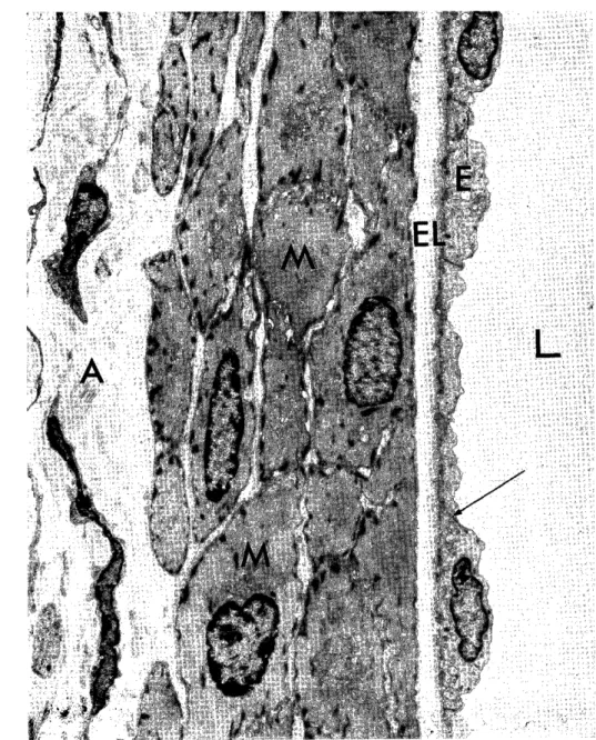
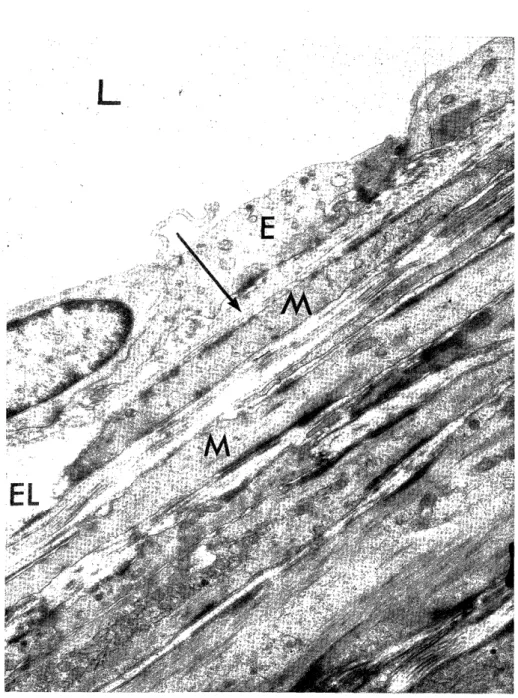
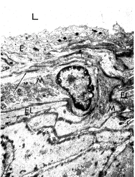
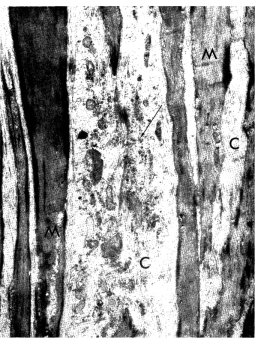
One week after the operation, some media smooth muscle cells beneath the internal elastic lamina became irregular shape, and two weeks after the operation, they were even advanced to the state of coagulation necrosis

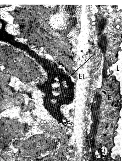
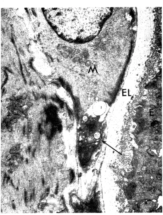
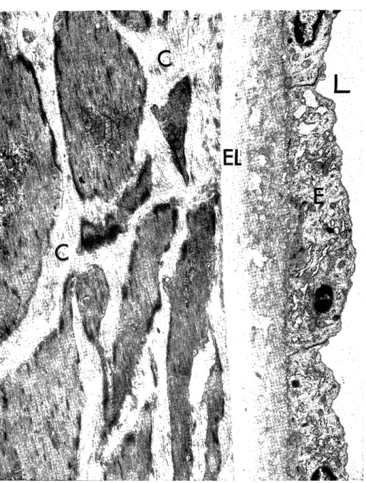
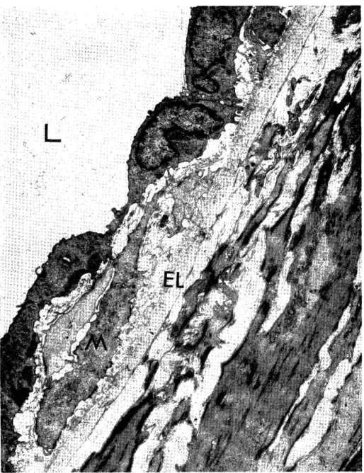
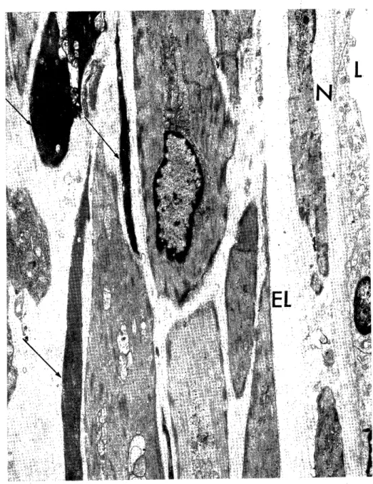
showing marked increase of electron density in the cytoplasm. About the same time, the subendothelial space was immigrated by the media smooth muscle cells through the fenestrae of the internal elastic lamina, and thus intimal thickening was produced. At the advanced stage of experiment, medial thinning due to destruction and absorption of media smooth muscle cells became remarkable, but intimal thickening was not greatly intensified.

As a result, the arteries showed dilatative lesion rather than stenosis of intimal thickening. The presence of a peculiar structure called Willis' ring in the cerebral basilar arterial system could not neglect as another cause of the dilatative lesion.

INTRODUCTION

Many method for experimental study of the pathogenesis of arte- riosclerosis have been derived to persue the morphologic change"'.

Experimental studies of arterial lesions produced by the change of blood stream are also numerous. MATSUNAGA17) experimentally produ- ced sudden change of blood stream in the cerebral arterial circulation particularly in the cerebral basilar arterial system by ligating or

*田 浦 晴 也

(2)

dissecting the bilateral or unilateral common carotid artery of the rabbit, and observed the appearance of diffuse aneurysm accompanied by marked dilatation and tortuosity of the cerebral basilar artery, posterior cerebral arteries and posterior communicating arteries. It was observed that the arteries indicate intimal thickening and medial thinning. . The author probed at the electron microscopic level into the changes of the cerebral basilar arterial system produced by such sudden change of blood stream, and obtained various findings on the process of arterial lesion, particularly on the behavior of the vascular wall cells. This paper presents the details of the findings.

EXPERIMENTAL METHOD AND MATERIALS

The animal used in the experiment were 30 adult white rabbits each weighing approximately 2 kg of the 30 rabbits. 15 were used as experimental group and the rest as control to oberve aging 13' 29)30) * The animals were fed with Oriental RC 5 and were given free access o water.

In the experiment, the bilateral common carotid arteries were dissected under anesthesia with ether at the intervals of a day, 3 days, 5 days, 1 week, 2 weeks, 3 weeks, 4 weeks, 2 months and 4 months after operation. Then, perfusion fixation was performed from the abdominal aorta with 1 % isotonic glutaraldehyde with phosphate buffer pH 7.4 following Takebayashi's method33' . Prior to fixation, about 50 cc (500 unit/dl) of heparin was injected into the abdominal aorta to prevent coagulation of blood in the vessels. After the fixa- tion, specimens were obtained from the cerebral basilar artery, posterior cerebral arteries and posteior communicating arteries, pro- cessed to the post-fixation with 1 % osmium tetroxide for 2 hours25' dehydrated with acetone and embedded in Epon 812. Slide sections were made by the use of Porter-Blum MT 2 ultramicrotome and double stained with uranyl acetate and lead citrate. Slide sections stained with toluidine blue were first prepared for the orientation to the lesions by light microscopy before preparation of thinn sections for electron mioroscopy. Ultrastructural observations were made with JEM 7A electron microscopy,

RESULTS Macroscopic changes:

In the specimen obtained within one week of experimental period,

no macroscopic change was noted. At the lapse of two months,

diffuse dilatation of the cerebral basilar artery began to appear. At

the lapse of four months diffuse dilatation is expanded to the posterior

(3)

cerebral arteries and posterior communicating arteries besides the cerebral basilar artery. In all cases, however, the changes were limited to diffuse dilatation without demonstration of aneurysm.

Light Microscopic Findings:

In the specimen stained with toluidine blue, dark staining cells sppeared among the muscle cells of the media and elongation of elastic fibers were noted in the second or third week. These changes were gradually intensified. Two months after the operation, partial brea-

kage of the internal elastic lamina vas noted, medial thinning became noticeable, and one or two layers of newly proliferated cells were found in the intima at some places. In the four month specimen, medial thinning was still present and additional intimal thickening appeared remarkably in the portions where the intimal elastic lamina was broken and disappeared.

Electron Microscopic Findings:

In the specimen up to the fifth day of experiment, any apparent difference in pathologic change from the control group (Fig. 1) was noted even at the electron microscopic level like the light microscopic level. One week after the operation, the smooth muscle cells beneath the internal elastic lamina were fragmented with dense deposites and

irregulary atrophic in shape (Fig. 2). Two weeks after the operation, smooth muscle cells demonstrated remarkably increased in electron density of the cytoplasm making it somewhat impossible to distinguish from the nucleus, also the degeneration of orgnellas within the cells assuming a pattern of so-called "dying cell" even with the state of coagulation necrosis (Fig. 3, 4). The coagulation necrosis usually involved an entire cell but there were also some findings suggesting only partial involvement. The cytoplasm of the media smooth muscle cells were partially darkened and clearly demarcated from those in the intact portion within the same basement membrane. This is equivalent to so-called "cut off phenomenon" described for the vessels of the stomach byTAKEBAYASHI35> (Fig. 5).

In the internal elastic lamina, two type of change were noted along

the lapse of experimental days. One change was thickening of the

internal elastic lamina where amorphous components increased along

the increase of fibrillar components and various degrees of accumula-

tion of electron dense particles were noted. As a result, the internal

elastic lamina as a whole appeared to be thickened (Fig. 6). The

other change was the thinning of the internal elastic lamina with

complete disappearance of amorphous components and with little or no

remainder of fibrillar components, resulting in an appearance as if the

intima and the media were contact having thinn internal elastic lamina

in between (Fig. 7).

(4)

Around this period, cell began to appear in the subendothelial space. These cells were accompanied with deposition of net-like figure substance and dense particles (Fig. 8). Most of the cells proliferated in the subendothelial space showed myofibrile and dense attachment in the cytoplasm. It was encountered in the electron m-iorocopic examina- tion that such cells were immigrating into the subendothelial space through the fenestrae of the internal elastic lamina (Fig. 9).

In the specimens with the lapse of more experimental period, i.e., two months or more, the cells immigrating the portions of intimal thickening consisted of mature type of smooth muscle cells which formed several layers (Fig. 10, 11). The intercellular matrices of these cells were composed of net-like figured materials, being low density in some and high in some others (Fig. 10). This is equivalent to so-called layer formation of the internal elastic lamina only in light microscopy. In some part of new internal elastic lamina, only f ibrillar components were present, but many other parts, the synthesis of amorphous components and formed complete new internal elastic lamina even electron microscopic level (Fig. 11).

In the media, some portions showed coagulation necrosis of the smooth muscle cells (Fig. 11) and some other portions suggested disappearance of these cells upon destruction and absorption. As a whole, the smooth muscle cells of the media were elongated and de- creased in number. The intercellular matrix of the media demonstra- ted slight proliferation of collagen fibers including dense particles

(Fig. 12). These findings were generally understood as the electron microscopic findings of the arterial wall of the macroscopic dilated and tortuous portion.

In the adventitia, no siginificant difference from the control group was noted throughout the period of experiment. Furthermore, the increase of collagen fibers in the media was not signifcant either.

In summary, the intimal thickening consists of proliferation of the smooth muscle cells in the subendothelial space was less intense in most cases than the medial thinning due to coagulation necrosis of media smooth muscle cells and their absorptive disappearance. This might have directed the arteries concerned throughout the experimental period towards dilatation and wall thinning overwhelming the stenosis due to intimal thickening.

DISCUSSION

Electron microscopic observation of the periphery of the arteries

affected by sudden change of blood stream by means of surgical opera-

tion have been ESTERLY et all". They concluded their observations

upon anastomosing the left subclavian artery sand the left main pul-

(5)

monary artery of the dog and changing the blood circulation in the pulmonary artery accompanied by a sudden change of blood pressure.

The specimens were peripheral arteries in the lung parenchyma measuring 50-2001L in diameter, being greatly different in site and size from the arteries of the cerebral basilar arterial system of rabbits measuring 1000-2000 t in diameter which was used in the author's.

ESTERLY et al." observed stenosis and occlusion of the peripheral arteries contrary to the results of the author's study. They observed that many smooth muscle cells were proliferated in the subendothelial space and that the intimal thickening resulting in the occlusion of fairly numerous peripheral arteries after two months periods. From these observations, they concluded that the sudden increase of intravascular pressure stimulates the smooth muscle cells of the media resulting in hyperactivity and secondarily occured intimal thickening.

In the author's case, all the arteries concerned experiment showed dilatative changes rather than stenosis, probably because the medial thinning due to coagulation necrosis followed by destruction and absorption was more intense than the proliferation of smooth muscle cells in the subendothelial space.

The presence of special structure called WILLIS' ring in the cereb- ral basilar arterial system is deemed to be another cause of dilatation.

According to Mc DONALD19), the WILLS' ring of rabbit resembles mor- phologically that of man being supplied blood from two internal carotid arteries and two vertebral arteries. When the bilateral common carotid arteries are dissected like in the author's experiment, the blood stream from two internal carotid arteries are decrease or stopped and that from the vertebral arteries are increase rapidly. Hence it is presumed that the cerebral artery, posterior cerebral arteries and posterior communicating arteries are caused dilatation by the flow of a volume of blood.

ESTERLY et al.')9) also reported that change of blood stream accele- rates the permeability of blood vessel, giving as the evidence of accumulation of osmiophilic materials in the intercellular matrix of the media. However, these various o.smiophilic materials in the media have been described in detail by TAKEBAYASHI et al. 34' and others" -I'), 20),26),27) and most of these materials seem to be derive from the degeneration of the media smooth muscle cells.

It is evident that the smooth muscle cells proliferated in the

subendothelial space originated in the media as asserted by ESTERLYB= ,

TAKEBAYASHI34) and others24) in view of the fact that they were immi-

grating into the subendothelial space pentrating through the fenestrae

of the internal elastic lamina. The fact that the development of

cytoplasmic organelles such as GOLGI's apparatus and rough surfaced

endoplasmic reticulum was not remarkable as compared with other

experiments, may be due to much weaker and gradual development in

(6)

the present study than in other experiments in TAKEBAYASHI35) which coincided with inflammation. This may also have directed the present experiment towards arterial dilatation owning to the more intensive medial thinning rather than toward arterial stenosis owning to the more intense intimal thickening. The increase of collagen fibers which seem to be composed and secreted by the smooth muscle cells of the media as described in many reports39 42)43), was not so intensive in the present experiment. The increase. of f ibrocytes in the adventitia was not so remarkable as compared with the control group. All these together may have directed the present experiment towards the dilata- tion of the arteries concerned.

It has been reported that the smooth muscle cells of the media numerously observed in the present experiment from the relatively early stage can be seen in the patient of hypertension34' . Such a change of the media may regarded as the first remarkable reaction of the vascular wall to the increased internal pressure of the artery.

However, such a coagulation necrosis is known to appear occasionally as physiological metabolism of the muscle cells. It also appear numerously in the media of transplanted vessels as observed by

TAKEBAYASHI et al. 37 in their experiments of vascular transplantation.

Therefore, it is not appropriate to conclude that coagulation necrosis is peculiar phenomenon which is caused only by the sudden change of blood stream .

These have been many reports that intimal thickening is caused at

only by a such a sudden change of blood stream but also by experi-

mental hypertension" 15)21)28)36)38)40)41)' decreased blood stream 14)31) P

lack of oxygen 11139) hyperlipemial12)5)22)23) , vasculitis35' and damage

of vessel4' . Anyway, the intimal thickening which observed in the

present experiment has no essential difference from the therefore

reported sclerotic lesions. Intimal thickening may be produced in the

experiment of vascular functional disorder such as vascular spasm

3)32)34) In the case of TAKEBAYASHI and KAMI034) , however, electron

microscopic follow up revealed that the intimal thickening was preced-

ed by the multiplication of the smooth muscle cells of the media

though it was accompanied by the degenerative necrosis in the part of

the smooth muscle cells. The patterns of intimal thickening finally

became similar owning to various factors such as the change of vari-

ous physical and chemical nature of blood stream, damage and

inflammation of the vascular wall. However, it may be a matter of

course that the morphological change at the initial stage varies consi-

derably by the case.

(7)

CONCLUSION

1) Arteriosclerotic lesions such as intimal thickening and medial destruction as reported by other investigator were experimentally produced under circumstance of the change of blood stream.

2) Macroscopically, diffuse aneurysmal dilatation was observed in the arteries of the cerebral basilar arterial system, particularly in the cerebral basilar artery, posterior cerebral arteries and posterior com- municating arteries.

3) Electron microscopically, organic change at an early stage of experiment was in increase of coagulation necrosis of media smooth muscle cells. On the other hand, modified smooth muscle cells with increased rough surfaced endoplasmic reticulum were hardly noted and the increase of collagen fibers in the intercellular matrix was slight.

4) Smooth muscle cells appeared in the subendothelial space i. e., intimal thickening occured but it was not intensive compared with dila- tative lesion. It was thought that the arterial wall such as a whole was caused thinning because of the medial thinning due to the destruction and absorption of smooth muscle cells despite the intimal thickening, thus resulting in a dilatation of the lumen of the artery.

5) It was clarified that the arterial lesions caused also by the change of blood stream would be directed to either dilatation or stenosis depending on the degree of change of blood stream and the anatomical characteristics of the arteries concerned.

ACKNOWLEDGMENT

The author is very grateful to Prof. H. TSUCHIYAMA for his guidance.

The author also wished to express his gratitude to Assist. Prof. S.

TAKEBAYASHI, and Assist. Prof. I . NAKAYAMA for their valuabls assis-

tance in experiments throughout the course of this investigation.

REFFERENCES

1. BALLS, J. U., HEUST, M. D. and MOOSE, R. H. : Electron microscopic studies in human atheroslerosis; cellular element in aortic fatty streaks. Exp. Mo',ec. Path.

3 : 511-525, 1964

2. BUCK, R. C. : The fine structure of the aortic endothelial lesions in experimental

cholesterol atheroslerosis of rabbit. Amer. J. Path. 34 : 897-909, 1958

3. BUCK, R. C. : The fine structure of endothelium of large arteries. J. Biophys.

Biochem. Cytol. 4 : 187-190, 1958

4. BYROM, F. B. : The pathogenesis of hypertensive encephalopathy and its relation to the malignant phase of hypertension. Experimental evidence from the hypertensi-

ve rat. Lancet. 2 : 201-211, 1954

5. DEMING, Q. B., MOSBACH, E. H., BAVANS, m., DALEY, M. M., HALPERN, E.,

and KAPLAN, R. : Blood pressure, cholesterol content of serum and tissue and

atherogenesis in rat. J. Exper. Med. 107 : 591-597. '1958

(8)

6. DILL, L. V. and ISENHOUR, C. E. : Occurence of atheroma in the aorta in rabbits with renal hypertension. Arch. Path. 33 : 655-660, 1942

7. ESTERLY, J. A. : Observaions on the possible relationship of hypertension, inc- reased arterial permeability to blood cells, and atherogenesis; An electron micros-

copic study. New physician, 14 : 145-151, 1965

8. ESIERLY, J. A., GLAGOV, and S. FERGUSON, C. J. : Morphogesis of intimal obliterative hyperplasia of small arteries in experimental pulmonary hypertension.

Amer. J. Path. 52 : 325-347, 1968

9. ESTERLY, J. A. and GLAGOV, S. : Altered permeability of the renal artery of the hypertensive rat, An electron microscopic study. Amer. J. Path. 43 : 619-639, 1963 10. FISHER, E. R., PEREZ-STABLE, E. and PARDO, V. : Ultra tructual studies in

hypertension 1. Comparison of renal vascular and juxtra-glomerilar cell alterations

in the essential and renal hypertension. Lab. Invest. 15 : 1409-1433, 1966

11. GARDNER. D. L. and MATTHEOS, M. A. : Ultrastructure of the wall of small arteries in the early experimental rat hypertension. J. Path. 97 : 51-62, 1969 12. GEER, J. C. : Fine structure of canin experimental atherosclerosis Amer. J. Path.

47 (3) : 241-269, 1965

13. HAUST, M. D. and GEER, J. C. : Mechanism of calcification in spontaneous

arteaiosclerotic lesions of the rabbit, An electron microscopic study. Amer. J. Pnth.

60 (3) : 329-346, 1970

14. HOFF, H. F. : Studies on the pathygenesis of arteriosclerosis with experimental

model systems. Virchows Arch. Abst. Path. Anat. 352 : 99-110, 1971

15. TNIERIEM, H. J. : Electron-microscopic study of bovine arteriosclerot!c lesions.

Amer. J. Prth. 50 (6) : 1035-1065, 1967

16. KOSEK, J. C., CHARTRAND, C., HURLEY, E. J. and LOWER, R. R. : Arteries in canin cardiac homografts, ultrastructure during acute rejection. Lob. Invest. 21 (4) : 328-335, 1969

17. MATSUNAG \ , Y. : Experimental study on aneurysm of cerebral arteries in rabbit (summary in English). Nagasaki Igakki Zassi, 40 (11, 12) 829-850, 1950

18. MATSUOKA, S. : A concept on the etiology of arferiosclerosis of small organ arte- ries. Acta Path. J:p. 18 (1) : 83-89, 1968

19. Mc DONALD, D. A. and POTTER, J. M. : The distribution of blood to the brain.

J. physiol_, 114: 356, 1961

20. Mc GEE, W. G. and ASHWORTH, C. T. : Fine structure of chronic hypertensive

arteriopathy in the human kidney. Amer. J. Path. 43 (2) 273-199, 1963

21. Mc GILL, H. C., Jk; FRANK, M. H., and GEER, J. C. Aorta lesions in hyper- tensive monkeys. Arch. Pare. 71 : 96-102, 1961

22. MOSES. C. Developlment of atherosclerosis in dogs with hypercholestlemia and chronic hypertension. Circn'ation Res. 2 : 243-247, 1954

23. Moss, W. G. , KIELY, J. P., NEVILLE, J. B. : BOWQUE, J. E. and WABERLIN G.

E. : Effect of experimental renal hypertension on experimental cholesterol atheros-

clerosis Circulution 4 : 462-463, 1951

24. OONEDA, G.. : Morpho;,enesis of arteriosclerosis. Acta Path. Jap. 18 : 75-82, 1968 25. PALADE, G. E. : Study of fixation for electron mscoscopy. J. Exp. Med. 95 : 285 -298 , 1952

26. PARKER, F. : An electron miososcopic study of experimental atherosclerosis. Amer.

J. Path. 36 (1) : 19-53, 1960

27. RUSSELL, R. and PAUL, B. : The elastic fiber I. The separation and partial characterization of its macrmolecular component. J. Cell Biol, 40 : 366-381, 1969 28. SPIRO, D., LATTES, R. G. and WIENER, J. : The cellular pathology of experi-

mental hypertension. I Hyperplastic arteriosclerosis. Amer. J. Path. 47 (1) : 19-47,

1665

29. STEIN, O. , EISENBERG, S. and STEIN, Y. : Aging of aortic smooth muscle cells in rats and rabbits. Lab. Invest. 21 (5) : 386-390, 1969

30. STEIN, O. , EISENBERG, S. and STEIN, Y. : Morphologic and biochemical changes

in smooth muscle cells of aorta in growth-restricted rats Lab. Invest. 25 (2) : 149-

(9)

157, 1971

31. Suzuxi, K. : Experimental studies on morphogenesis of arteriosclerosis with special refference on relation between hemodynamic change and developments of cellulofi-

brous intimal thickening and atherosclerosis. Gunma J. Med. Science. 14 (3, 4) : 185

-243 , 1967

32. TAKEABYASHI, S. : Experimental study on the vascular changes caused by angios- pasm (summary in English). Nagasaki Igakkai Zassi 315 (11, 12) :1923-1935, 1960 33. TAKEBAYASHI. S. : Evaluation of perfusion fiuation. J. Electron Mioroscopy 17

246, 1968

34. TAKEBAYASHI, S. and KAMIO, A. : Ultrstructual studies on human arteriosclerosis

in comparison on between hypertensive and normotensive groups. Acta Path. Jap.

19 . (4) : 479-499, 1969

35. TAKEBAYASEI, S. : Pathogenesis of obliterating endoarteritis in stomach ulcer.

Electron microscopic study. Acta Path. Jap. 20 (1) : 37-46, 1970

36. TAKEBAYASEI, S. : Ultrastructual studies on arteriolar lesions in experimental

hypertension. J. Electron Microscopy. 19 : 17-31, 1970

37. TAKEBAYASEI, S., K-P BACHWINKEL, H.., THEIMANN, H., LIE, T. S., NAGEL, K.

and BECHTELSHEIMER, H : Ultrastructual aspects of aortic homografts nature of

neointima and behavior of smooth muscle,'cells in the grafts. Betr. Path. Bd. 144

166-183, 1971

38. TAKEBAYASHI, S., KAMIO, A., KUBOTA, I., Taura, S. : Ultrastructual aspect of behavior of media smooth muscle cells on arteriosclerosis Acta Path. JDp. 1972 (im press)

39. TERRY, B. E., JONES, D. B. and MUELLER, C. B. : Experimental ischemic renal arterial necrosis with resolution. Amer. J. Path. 58 (1) : 69-83, 1970

40. WIENER, J., LATTES, R. G., MELTZER, B. G. and SPIRO, D. : The cellular pathology of experimental hypertension. IV Evidence for increased vascular permea-

biliry. Amer. J. path. 54 : 187-208, 1969

41. WIENER, J., LATTES, R. G. and SPIRO, D. : The cellular pathology of experi- mental hypertenisions. II Arteriolar hyalinosis and fibrinoid changes. Amer. J.Path.

47 : 457-486, 1965

42. WISSLER. R. W. : The arterial medial cell, smooth muscle, or multifunctional

mesenclyme? Cioc..dation 36 : 1-4, 1967

43. WisSLER, R. W. : The arterial medial cell. smolth muscld, or multifunctional

mesenchyme? J. Atherosc;ero. 8 : 201-214, 1968

(10)

Fig. 1. Control. Normal cerebral basilar artery of a rabbit. Endothelial cells (E) form continuously single thinn layer with tight junctions (T ) . Internal

elastic lamina (EL) shows uniform in thickness. Smooth muscle cells (M)

arrange in normal in the media. Fibrocytes are seen in the adventitia (A).

L=lumen x 5,800

(11)

Eig. 2. Experimental group, after one week.

Smooth muscle cells (M) beneath the internal elastic lamiua (EL) are frag- mented with dense deposites (T) and irregulary in shape (B). M=smooth

muscle cell, L=lumen x 17,000

(12)

Eig. 3. Experimental group, after three weeks.

Two smooth muscle cells beneath the internal elastic lamina (EL) are coa- gulated and markedly increased in density (1) .

F=fenestrae, M=smooth muscle cell x 3,000

(13)

Fig. 4. Experimcntal group. after three weeks.

Note the coagulation necrosis ( T ). The mitochondria are swollen and

vacuolated. M=smooth muscle cell, L=lumen x 7,200

(14)

Fig. 5. Experimental group, after three weeks.

The cytoplasms of the medial smooth muscle cells (M) is partially darkened and cleary demarcated to the intact portion within the same basement

membrane (T ). E-endothelial cell, EL-internal elastic lamina x 14,600

(15)

Fig. 6. Experimental group, after two weeks.

The internal elastic lamina (EL) are considerably thickened and increased

collagen fibers (C) in the media. L=lumen, E=endothelial ce'1 x 7,100

(16)

Fig. 7. Experimental group, after two weeks.

Note disappearance of the internal elastic lamina partially (T ) . EL=enternal

elastic lamina, M=smooth muscle cell, L=lumen x 5,800

(17)

Fig. 8. Experimental group, after three weeks.

Imtimal thickening consisting of matured smooth muscle cells (M) are

presented. EL=internal elastic lamina, L=lumen x 3,200

(18)

Fig. 9. Experimental group, after four weeks.

Two muscle cells (M) with a few myofibrile and dense bodies (~) with well developed cytoplasmic organelles are migrating the subendothelial

space through the fetestrae of internal elastic lamina.

L-lumen, E=endothelial cell x 4.600

(19)

trig. 10. r;xperimntal group, after two months.

The photo shows an area of intimal thickening. The organella of smooth muscle cells (M) are increased in number. There are a lot of amorphous

and net-like figured substances (S) in the inthercellular matrix. L=lumen,

E=endothelial cell x 6.200

(20)

Fig. 11. Experimental group, sfte four months.

Note the intimal thickening and the formation of new elastic lamina (N).

Original internal elastic lamina (EL) is also present. A few coagulatin

necrosis of smooth muscle cells (r) are seen in melia. L=lumen x 7,100

(21)

Fig. 12. Experimental group, after four months.

The photo shows the media of basilar artery. There are a lot of electron dense deposite ( T ) and increased of the collagen fibers (C) in the dilated

intercellular matrix. M=smooth muscle cell x 13,800

Fig.  1.  Control.  Normal  cerebral  basilar  artery  of  a  rabbit.  Endothelial  cells  (E)         form  continuously single thinn  layer  with  tight junctions (T  )
Fig.  4.  Experimcntal  group.  after  three  weeks.
Fig.  5.  Experimental  group,  after  three  weeks.
Fig.  6.  Experimental  group,  after  two  weeks.
+6

参照

関連したドキュメント

performed 4 h and 8 h euglycemic (5.5 mmol/l) clamps with 3 different insulin concentrations (basal, medium postprandial and high postprandial, ranging from ~ 35 to ~ 1450 pmol/l)

Keywords: homology representation, permutation module, Andre permutations, simsun permutation, tangent and Genocchi

Provided that the reduction of the time interval leads to incomparableness of normalized bubble-size distributions and does not change the comparable distributions in terms of

The C-minor partial orders determined by the clones gen- erated by a semilattice operation (and possibly the constant operations corresponding to its identity or zero elements)

In this paper, we will apply these methods to the study of the representation theory for quadratic algebras generated by second-order superintegrable systems in 2D and their

The inclusion of the cell shedding mechanism leads to modification of the boundary conditions employed in the model of Ward and King (199910) and it will be

The commutative case is treated in chapter I, where we recall the notions of a privileged exponent of a polynomial or a power series with respect to a convenient ordering,

Applications of msets in Logic Programming languages is found to over- come “computational inefficiency” inherent in otherwise situation, especially in solving a sweep of