Studies on the Expression of Pax6 during
Reprogramming of Adult Newt Retinal Pigment
Epithelium Cells
著者
井波 航
year
2016
その他のタイトル
成体イモリ網膜色素上皮細胞のリプログラミング過
程におけるPax6発現に関する研究
学位授与大学
筑波大学 (University of Tsukuba)
学位授与年度
2015
報告番号
12102甲第7753号
URL
http://hdl.handle.net/2241/00143905
Studies on the Expression of Pax6 during Reprogramming
of Adult Newt Retinal Pigment Epithelium Cells
A Dissertation Submitted to
The Graduate School of Life and Environmental Sciences,
The University of Tsukuba
In Partial Fulfillment of the Requirements
For the Degree of Doctor of Philosophy in Science
(Doctoral Program in Functional Bioscience)
ⅰ
Table of Contents
Table of Contents---ⅰ Abbreviations---1 Abstract---2 1. Introduction---3 1.1. The RPE cells1.2. Newt RPE cells contribute to neural regeneration 1.3. The reprogramming of RPE cells
1.4. Pax6 is expressed in RPESCs 1.5. Pax6 structure and variety 1.6. The newt Pax6
1.7. The RLEC system
2. Materials and Methods---12
2.1 Identification of the Pax6 variant expressed in the eyeballs of the adult newt
2.1.1. Animals
2.1.2. Transcriptome data
2.1.3. Design of primer sets
2.1.4. Sample preparation from parts of the normal eyeball
2.1.5. Molecular cloning
2.1.6. Antibodies
2.1.7. Immunohistochemistry
2.1.8. Discrimination of the classes of Pax6 variants in normal eyes
ⅱ
2.2. RPE cells newly express Pax6 v1 and v2 variants upon retinectomy
2.2.1. Retinectomy
2.2.2. Discrimination of the classes of Pax6 variants in RPESCs
2.3. Estimation of the signal pathway causing the reprogramming of RPE cells
2.3.1. Preparation and incubation of RLECs
2.3.2. PCR analysis 2.3.3. Antibodies 2.3.4. Immunohistochemistry 2.3.5. Cell counting 2.3.6. Data analysis 3. Results---27
3.1 Identification of Pax6 variants expressing in the eyeballs of adult newts
3.1.1. Search for unknown Pax6
3.1.2. Distribution of Pax6 variants in the adult newt eye
3.2. RPE cells newly express Pax6 v1 and v2 variants upon retinectomy
3.3. Estimate of the signal pathway which causes the reprogramming of RPE cells
3.3.1. RPE cells also express Pax6 v1 and v2 variants in RLECs
3.3.2. RPE cells expressing Pax6 versus RPE cells entering cell-cycle
3.3.3. Pax6 expression in RPE cells is not affected by a MEK1/2 inhibitor U0126
3.3.4 Heparin promotes the expression of Pax6 with the help of the MEK1/2-mediated
pathway
4. Discussion---38
4.1. Several kinds of Pax6 variants
ⅲ 4.3. The signal which produces RPESCs
4.4. Differences in RPE cells between humans and newts
4.5. Conclusions and future directions
5. References---45
6. Figures and Legends---52
1
Abbreviations
CL: cornea limbus
CMZ: ciliary marginal zone DIV: days in vitro
EMT: epithelial-mesenchymal transition
HD: homeo-domain
HTH: helix-turn-helix
NR: neural retina
PD: paired-domain
PST: proline, serine and threonine
PVR: proliferative vitreoretinopathy RLEC: retina-less eye-cup
RPE: retina pigment epithelium
2
Abstract
Adult newt retinal pigment epithelium (RPE) cells are reprogrammed into a unique state of multipotent cells at an early phase of retinal regeneration. Here, to understand the signal triggering of reprogramming, four classes (v1, v2, v3 and v4) of Pax6 variants in the eyes of adult newt were identified and their expression in RPE cells after retinectomy was investigated. The Pax6 v1 and v2 variants were newly expressed in RPE cells by 10 days after retinectomy, both in vivo and in vitro. In vitro examinations suggested that Pax6 expression is mediated through a pathway separate from the MEK-ERK pathway, which is required for cell cycle re-entry of RPE cells, but in a condition closer to the in vivo state, it was promoted by the activity of MEK. These findings predict the existence of a pathway that must be further pursued to understand the reprogramming of RPE cells during retinal regeneration.
3
1. Introduction
1.1. The RPE cells
Retina pigment epithelium (RPE) is a simple epithelium constituting the eyeballs of vertebrates.
RPE surrounds the back of the neural retina (NR), which receives light. This tissue adheres to the
basement membrane, which is located external to it, and the choroid membrane, whose blood vessels
are distributed external to it. RPE tissue is comprised of one kind of single-layered epithelium cells
named RPE cells. These cells, which accumulate melanin and have microvilli, play essential roles in
early visual processing as a partner of the NR. Mature RPE cells do not divide and specialize
morphologically and functionally (for reviews, see Fuhrmann et al., 2014; Strauss, 2005).
However, when the NR suffers from a traumatic injury, RPE cells lose their epithelial
characteristics and undergo proliferation and transformation. As an example, there is proliferative
vitreoretinopathy (PVR), which is a human disease. When RPE cells are exposed to serum, causing
the detachment of retina and colloidal disorder, they can be isolated from the basement membrane
and lose their epithelial structure. Moreover, these cells migrate into the vitreous through tears in the
neural retina. Then, they form an epiretinal membrane on the surface of the NR, leading to the loss
4
which the epithelial cells detach from the underlying basement membrane and tranform into
mesenchymal cells which involves enhanced migratory capacity and the production of extracellular
matrix components (Chiba, 2014).
1.2. Newt RPE cells contribute to neural regeneration
On the other hand, in the newt, a group of the family Salamandridae in urodele amphibians, a similar
change in RPE cells enables them to regenerate an entire retina, even as adults (Chiba et al., 2006a;
Chiba and Mitashov, 2007; Chiba, 2014). In the adult newt, when the NR is completely removed
from the eye by a surgical operation ‘retinectomy’ (Figure 1A,B), RPE cells are detached from each other as well as from the basement membrane, losing their epithelial characteristics to become cell
aggregates, while reaching the S-phase of the cell-cycle (Figure 1C). Ten days after an operation,
when these changes are observed, is defined as Stage ‘E-1’. These RPE-derived cells are then sorted into two populations which form the prospective-NR and -RPE layers (pro-NR and pro-RPE layers,
respectively) with correct polarity (Figure 1D). Cells in the pro-NR and pro-RPE layers start to
proliferate and eventually regenerate new functional NR and RPE (Figure 1E). Therefore, newt
retinal regeneration serves as a good model system to compare with RPE-mediated retinal disorders
in humans, providing insight into medical treatments that would allow for in vivo retinal
5
1.3. The reprogramming of RPE cells
What changes occur to newt RPE cells that have lost their epithelium form? Recently, our
laboratory has isolated RPE cells before retinectomy and at Stage E-1, and compared their gene
expression profiles. This study demonstrated that RPE cells at Stage E-1 express three pluripotency
factors, i.e., c-Myc, Klf4 and Sox2, as well as factors involved in fate switching between the NR and
RPE in embryonic/larval stages, namely Pax6 and Mitf (Islam et al., 2014). Since these genes have
not been confirmed as intact RPE cells, it is thought that they express newly in the process of
regeneration. On the other hand, RPE cells at Stage E-1 preserve certain original characteristics such
as the presence of microvilli and the expression of RPE65, which is an RPE cell-specific marker
(Islam et al., 2014). Adult newt RPE cells are then reprogrammed into a unique multipotent state
which has the potential to create neural retinas and is observed only in the regeneration process. The
cells of this state are referred to as RPE stem cells (RPESCs). However, it remains uncertain what
signals trigger the reprogramming of RPE cells as well as cell cycle re-entry.
1.4. Pax6 is expressed in RPESCs
In this study, this subject was attempted to approach by focusing on the expression of transcription
6
transcription factor. It is a protein that is essential to the normal development of the retina from
Drosophila melanogaster to humans (Baumer et al., 2002). In vertebrate development, Pax6 exists
over a wide area, including the retina and RPE progenitor cells, at the early optic cup stage. During
cell differentiation, Pax6 expression is limited and finally disappears in the mature RPE and neural
retina except for amacrine, horizontal and ganglion cells (Davis-Silberman et al., 2005; Hsieh et al.,
2009; Macdonald et al., 1997).
In the study of newt regeneration, it has been suggested that Pax6 is expressed in the eyeball of
Stage E-1. Furthermore, the Pax6 sequence was elucidated when genetic information about the newt
was poor and Pax6 was the rare protein that an antibody equivalent was available. Expression of
Pax6 in reprogramed cells was also confirmed in a recent study using isolated RPE cells. It is
thought that Pax6 mRNA expresses in all RPESCs of Stage E-1, and after two cell layers have arisen
from RPESCs, pro-NR cells gradually change in Pax6-positive cells, as confirmed by
immunohistochemistry (Islam et al., 2014). Therefore, Pax6 is a useful marker to trace the process of
reprogramming in RPE cells.
1.5. Pax6 structure and variety
In studies involving Pax6, it is necessary to consider the function of multiple transcription products.
7
consist of many isoforms generated via post-transcriptional regulation such as alternative splicing.
Several Pax6 variants with different structures have been reported (Bandah et al., 2007; Gorlov and
Saunders, 2002; Kammandel et al., 1999; Kim and Lauderdale, 2006; Shaham et al., 2012; Short and
Holland, 2008).
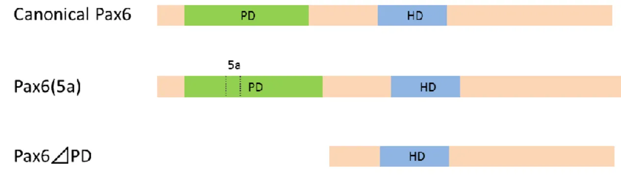
The first variant, named “canonical Pax6”, is the most universal and abundant of the Pax6 variants (Figure 2). This variant includes two DNA-binding paired-domain (PD) and homeo-domain (HD) in
the N-terminal region. These two domains are separated by glycine-rich linker. The C-terminal
region of Pax6 is enriched with proline, serine and threonine (PST) residues (Shaham et al., 2012).
The PD consists of two subdomains named PAI and RED. PAI and the RED subdomain have an
HTH motif each (Xu et al., 1995; Xu et al., 1999; Epstein et al., 1994a). The C-terminal region of
Pax6 is enriched with PSTs and functions as a transcriptional transactivator (Czerny and Busslinger,
1995; Tang et al., 1998). These structures are widely conserved from flies to humans (Xu et al.,
1995; Xu et al., 1999).
Secondly, in vertebrates, there is a Pax6(5a) variant that is made by post-transcriptional alternative
splicing that inserts an additional exon 5a into canonical Pax6 (Figure 2; Walther and Gruss, 1991).
Since the insertion site is the HTH motif in PAI, PAI loses its DNA binding capacity while that of
RED is maintained (Epstein et al., 1994b). As a result, the DNA-recognition sequence of Pax6(5a) is
8
Pax6(5a) express in each tissue, and changes in their ratio cause pathological conditions and
abrogated development (Epstein et al., 1994b; Pinson et al., 2005; Zhang et al., 2001).
The third Pax6 variant, which lacks the PD and is termed paired-less, is made by a promoter
unlike canonical Pax6 and Pax6(5a) in vertebrates (Figure 2). The overexpression of this variant
disturbs the development of the cornea and lens, and causes the microphthalmic phenotype (Kim and
Lauderdale, 2006, 2008). However, its physiological activity in the eye is still unknown (Shaham et
al., 2012).
In addition, many Pax6 variants have been reported. For example, in the PST region of amphibian
and mouse Pax6, it has been reported that a part equivalent to exon 12 of human Pax6 is spliced out
and subsequently shifts the sequence (Mizuno et al., 1997). A functional analysis of the majority of
these variants has not been performed.
1.6. The newt Pax6
In the newt, two kinds of Pax6 splicing, namely the addition of a PD and the deletion of part of the
PST, were discovered. Four kinds of variants (LL, LS, SL and SS) form from different combinations
of this splicing. SL and LL are homologs of canonical Pax6 and Pax6(5a), respectively. SS and LS
are variants in which the exon in the PST splices out from SL and LL (Mizuno et al., 1997).
9
is limited. In addition, there are few reports related to the expression and function of known Pax6
variants. Our previous study demonstrated that canonical Pax6 is newly expressed in RPESCs.
However, it remains unknown which other variants are expressed in those cells.
Therefore, in the first part of this study, utilizing a transcriptome database IMORI
(http://antler.is.utsunomiya-u.ac.jp/imori/), which our laboratory established to study newt retinal
regeneration (Nakamura et al., 2014), Pax6 variants expressed in the adult newt eye were attempted
to thoroughly identify. Then, in the second part of this study, on the basis of this information, the
Pax6 classes that are expressed in RPE cells upon retinectomy were explored. Finally, the pathways
that triggered Pax6 expression were investigated.
1.7. The RLEC system
In newt retinal regeneration, it is challenging to study the molecular mechanism underlying the
reprogramming of RPE cells through injection/implantation of chemical tools into the eye because i)
the wound along the dorsal half of the eye, from which the NR is removed, has not closed until ~2
weeks in which reprogramming has been completed; ii) since RPE cells become solitary upon
retinectomy and are then distributed in the vitreous cavity, the microenvironment (or niche)
surrounding RPE cells is susceptible to physical perturbations, resulting in the abnormal
10
only; iii) more essentially, the wound/injury itself, which is inevitably made for retinectomy or
chemical treatments, may turn-on reprogramming signals. In this study, candidate pathways were
attempted to identify by using the retina-less eye-cup (RLEC) system, an in vitro system which our
laboratory has introduced to study the initial response of RPE cells in association with the wounding
of eyes and retinectomy (Yohikawa et al., 2012). In this system, almost the same operation of
retinectomy as that made in vivo was carried out in vitro. This ensured that the posterior half of the
eyeball is missing the NR (i.e., the RLEC), which is then incubated in a minimal essential medium.
In this condition, RPE cells do not appear to have died or have been seriously altered for as long as
10 days. In fact, when RPE-choroid tissues isolated on day-10 are implanted into the vitreous cavity
of the eye of a living animal immediately after retinectomy, retinal tissue is regenerated from the
implanted RPE (also see Chiba et al., 2006b). However, it is unknown whether the RLEC system can
be used as a tool to check the signal(s) causing the reprogramming of RPE cells. After the style of
Pax6 expression in the RLEC system and RPESCs were compared, and confirmed whether in vitro
Pax6 expression reflects real regeneration, it was necessary to examine the factor(s) participating in
the reprogramming of RPE cells.
In a previous paper, focusing on the wound edge, our laboratory reported that RPE cells in the
RLEC enter the cell cycle with almost the same time course as that observed in vivo (Yoshikawa et
11
cell cycle re-entry of RPE cells in the RLEC requires a MEK1/2-ERK1/2-mediated intracellular
signaling pathway, whose activity rises transiently within 30 min after retinectomy both in vivo and
in vitro. Consequently, our laboratory proposed this as the putative pathway for the cell cycle
re-entry of RPE cells. In this study, based on previous studies, the signaling pathways triggering
12
2. Materials and Methods
2.1 Identification of the Pax6 variant expressed in the eyeballs of the adult newt
2.1.1 Animals
Japanese fire belly newts, Cynops pyrrhogaster, were purchased from local suppliers (Ouchi
Kazuo Seibutsu Kyozai, Saitama, Japan/Aqua Grace, Yokohama, Japan). The newt for this
experiment was originally captured from Chiba or Okayama prefecture, Japan. The animals had been
reared in containers into which water had been poured to a depth of 5 cm, and placed at 18°C under
natural light. They were fed daily with frozen mosquito larvae (Akamushi; Kyorin, Himegi, Japan),
and the containers were kept clean at all times. To select those newts which had completely matured
into adults, individuals of sufficient size (total body-length: 9–11 cm) were used. Before surgical
operation or sacrifice, newts were anesthetized for 2 h with FA100 (1:1000;
4-allyl-2-methoexyphenol; DS Pharma Animal Health, Osaka, Japan) in the dark. All experiments
were carried out in accordance with the guidelines approved by the University of Tsukuba Animal
Use and Care Committee.
13
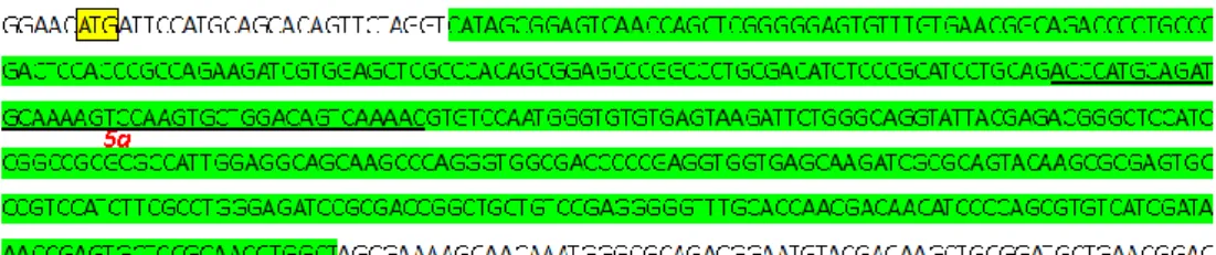
To search unknown Pax6 isoforms expressed in the eyes of the adult newt, a de novo assembled
transcriptome database (IMORI, http://antler.is.utsunomiya-u.ac.jp/imori/) from the eyes at 0-14
days after removal of both the neural retina and the lens was used. This database was created in our
laboratory using a next-generation sequencer (Nakamura et al., 2014). At that time, three algorithms,
Trinity, Trans-ABySS and Velvet-Oases, were applied during database assembly (Nakamura et al.,
2014). Three assembled database was integrated into one as a sequence database, and blastn (version
v2.2.26+, NCBI) was carried out using a known Pax6 (Accession #: D88741) as the query sequence.
The contigs which connected to unknown Pax6 sequences were selected from the blastn results. An
ensuing blastn, which used only unknown regions of Pax6 as the query sequence, was performed.
Until a newly unknown sequence was not provided, the search was repeated. Finally, some
candidates of new Pax6 classes were derived to connect them.
2.1.3. Design of primer sets
The candidates of new Pax6 classes expected in silico had unique sequences upstream of the
PD-coding region, including 5ʹ UTR. To confirm the existence of new Pax6 isoforms in the eyes of
adult newts and to decide their sequence information, forward primers located in the unique
sequences of the 5ʹ UTR that could distinguish the different classes of Pax6, were designed. Furthermore, to design reverse primers located in known and common sequences of the 3ʹ UTR, the
14 primer sets were configured so as to amplify the ORF.
2.1.4. Sample preparation from parts of the normal eyeball
After the eyeball was excised, it was opened up along the equator in RNase-free PBS. The
operation took place in this solution by manipulating fine forceps and scissors under a dissecting
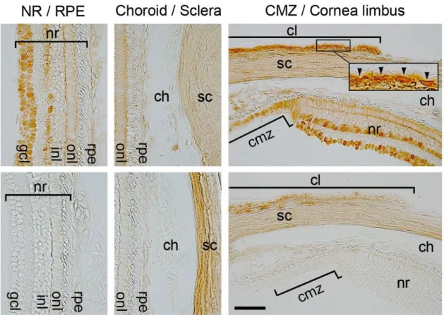
microscope. The ciliary marginal zone (CMZ), iris, lens, cornea limbus (CL) and cornea were
isolated from the anterior half of the eye (Figure 3). NR and RPE cells, together with the choroid
tissues (RPE-choroid) and sclera, were isolated from the posterior half of the eye (Figure 3). Using a
thin needle that can peel each tissue, sclera and NR were removed to reveal the RPE-choroid. It is
difficult to remove RPE cells from the basal membrane mechanically. The separation of RPE cells
from RPE-choroid tissue was performed in a solution containing protease and chelate. After as many
blood cells in the choroid were removed as possible by shaking the samples in the dish, samples
were transferred into elastase (1 mg/ml; Porcine pancreas; Ref: 11027891001; Roche Diagnostics
Japan, Tokyo, Japan) in EDTA solution (in mM: 115 NaCl, 3.7 KCl, 10 EGTA, 18 D-glucose, 10
HEPES, and 0.001% phenol red, pH 7.5 adjusted with 0.3N NaOH) and incubated for 90 min at
28°C. Subsequently the tissue samples were rinsed with RNase-free PBS several times and then RPE
cells were separated by a current which was created by pipetting the tissues with a 3.5 ml transfer
15
agarose-coated 35-mm plastic dishes (08-772A, Thermo Fisher Scientific, Waltham, MA, USA).
After verifying that RPE cells were not contaminated by other tissues, they were used as the RPE
sample. The residual tissue, which was confirmed to have completely separated RPE cells, was used
as the colloid sample.
2.1.5. Molecular cloning
Total RNA was extracted from whole/tissues/cells collected from 3-5 eyeballs using a total RNA
isolation kit (5185-6000, Agilent Technologies, Santa Clara, CA, USA), and template cDNAs were
conventionally constructed using SuperScript II Reverse Transcriptase (18064-014, Thermo Fisher
Scientific). Although the commercial kit was basically used according to the manufacturer’s instructions, the reaction time of reverse transcriptase was extended to 90 min. PCR was performed
using KOD FX (KFX-101, Toyobo, Tokyo, Japan). The confirmed PCR amplification product was
cloned. To prevent PCR errors, the number of cycles was minimized to 25. The PCR products were
electrophoresed by agarose gel (Solana Agarose STANDARD 01, Rikaken, Nagoya, Japan)
electrophoresis and the bands of expected size were cut out.
After purifying DNA using a Geneclean Spin Kit (111101200, MP Biomedicals, Santa Ana, CA,
USA) and having added adenine to the blunt 3ʹ end with advantage 2 (639206, Takara Bio, Otsu, Japan), TA was cloned with the TOPO TA Cloning Kit (K4560-01, Thermo Fisher Scientific). DNA
16
sequencing was carried out using ABI Prism 3130 (Thermo Fisher Scientific). For each class of Pax6,
the nucleotide sequence of the longest variant was deposited in DDBJ/GenBank (Accession #: Pax6
v2, LC002642; Pax6 v3, 28 LC002643; Pax6 v4, LC002644).
2.1.6. Antibodies
Chicken polyclonal anti-Pax6 antibody PA1-801 (1:1,000; Thermo Fisher Scientific) and rabbit
polyclonal anti-Pax6 antibody PRB-278P (1:1,000; Covance, Princeton, NJ, USA) were used as the
primary antibodies. PA1-801, which can recognize a sequence (REEKLRNQRRQASNTPSHI)
neighboring the 3ʹ end of HD in the newt, was applied to immunoblotting. This epitope is present in all classes of Pax6 variants but this antibody was inadequate for immunohistochemistry. Therefore,
PRB-278P was applied to immunohistochemistry. This antibody has high reactivity and recognizes
the C-terminal (QVPGSEPDLSQYWPRIQ) of LL and SL forms of Pax6 and the –L form of
paired-less Pax6. Biotinylated goat anti-chicken IgG antibody (1:500; BA-9010, Vector Laboratories,
Burlingame, CA, USA) and anti-rabbit IgG antibody (1:500; BA-1000, Vector Laboratories) were
used as the secondary antibodies. The negative controls were obtained by omitting the primary
antibodies.
17
Normal eyeballs were fixed in 2% paraformaldehyde / 0.2% picric acid in PBS (pH 7.5) for 5-6 h
at 4°C and cryosectioned at a thickness of ~20 μm after washing away the fixative solution. Tissue sections were rinsed thoroughly (PBS, 1% TritonX-100 in PBS, PBS, 15 min each rinse), treated
with 3.3% H2O2 in methanol for 20 min, washed thoroughly, incubated in a blocking solution [3%
normal goat serum (S-1000, Vector Laboratories) / 1% TritonX-100 in PBS] containing Avidin D
(1:50; Avidin/Biotin Blocking kit, SP-2001, Vector Laboratories) for 2 h, washed in PBS twice, and
then incubated in the primary antibody diluted with the blocking solution containing Biotin (1:50;
Avidin/Biotin Blocking kit) for 72 h at 4°C. After washing thoroughly, the samples were incubated
in the biotinylated secondary antibody diluted with the blocking solution for 4 h at room temperature,
washed thoroughly, incubated in a mixture of Avidin and Biotin Complex (Vectastain ABC Elite kit,
PK-6100, Vector Laboratories) for 2 h, washed thoroughly, and then incubated in a DAB solution
(DAB substrate kit, SK-4100; Vector Laboratories) for 6 min. After the samples were washed,
melanin pigments in the tissues were bleached, and the nuclei of RPE cells were stained with DAPI
(1:50,000; D1306, Thermo Fisher Scientific).
Images of tissues were acquired using a CCD camera system [C4742-95 ORCA-ER system
(Hamamatsu Photonics, Hamamatsu, Japan) or a DP73 system (Olympus, Tokyo, Japan)]. Figures
18
2.1.8. Discrimination of the classes of Pax6 variants in normal eyes
PCR was performed using KOD FX and the primer sets distinguished the new and known classes
of Pax6 against several cDNAs from each ocular part of the eye. The number of cycles was
increased compare with the cloning step (PCR conditions, see Table 2). To evaluate the integrity of
cDNAs, the expression of EF1-α (Accession #: AB005588, sense, 5′-gacctttgcccccagtaacgtaaccac-3′;
antisense, 5′-actgggtgttgctggcgctacttcttg-3′; 573-bp) was confirmed. An MJ Mini Gradient Thermal Cycler (PTC-1148, Bio-Rad, Hercules, CA, USA) was routinely used. Exceptionally, to examine
Pax6 v2 transcripts, a Takara PCR Thermal Cycler 1 MP (TP3000, Takara, Otsu, Japan) was also
used.
2.1.9. Immunoblotting
Sample tissues were prepared from five anterior halves of eyeballs after removing the lens and
adding 75 µl of lysis buffer [25 mM Tris-HCl (pH = 7.5), 150 mM NaCl, 1 mM EDTA-2Na, 1%
Igepal CA-630 (56741, Sigma-Aldrich, St. Louis, MO, USA), 1% protease inhibitor cocktail (P8340,
Sigma-Aldrich), 1% sodium deoxycholate (190-08313, Wako Pure Chemicals Industries, Ltd., Osaka,
Japan), and 0.1% SDS (191-07145, Wako)] chilled on ice. Samples in the sample tube were frozen in
liquid nitrogen and sonicated in chilled water for 5 min. The frozen sample was gradually dissolved.
19
cooled centrifuge at 4°C. The supernatant was mixed with the same amount of SDS sample buffer
[0.5 M Tris-HCl (pH = 6.8), 10% (w/v) SDS, 10% 2-mercaptoethanol (M3148, Sigma-Aldrich), 20%
glycerol, 0.5% (w/v) bromophenol blue (021-02911, Wako)] and immediately heated for 5 min in a
water bath at 98°C.
Protein samples (µl/lane) were separated on a 10% Mini-PROTEAN TGX Precast Gel (456-1033,
Bio-Rad, Hercules, CA, USA) by SDS-PAGE and electrophoretically transferred onto an
Immun-Blot PVDF membrane (1620174, Bio-Rad) using a Mini transblot cell (1703930JA,
Bio-Rad).
The blots were incubated in blocking solution [5% non-fat skimmed milk (198-10605, Wako) in
TBST [100 mM Tris-HCl (pH 7.4), 150 mM NaCl, 0.05% Tween20]] containing Avidin D (1:50;
Avidin/Biotin Blocking kit) for 1 h, rinsed with TBST twice, and then incubated overnight at 4°C in
the primary antibody diluted with the staining system [Can Get Signal (NKB-101T, Toyobo)]
containing Biotin (1:50; Avidin/Biotin Blocking kit). After washing three times in TBST for 15 min
each wash, the blots were incubated in biotinylated secondary antibody diluted with the staining
system [Can Get Signal] for 90 min at room temperature, washed three times in TBST, incubated in
a mixture of Avidin and Biotin Complex (Vectastain ABC Elite kit) for 90 min, washed six times in
TBST, and then incubated in a DAB solution (DAB substrate kit) until the bands were visualized.
20
2.2. RPE cells newly express Pax6 v1 and v2 variants upon retinectomy
2.2.1 Retinectomy
To induce retinal regeneration, living newts were held in an operating chamber. The dorsal half of
the left eye was cut open along the position slightly below the boundary between the cornea and
sclera using a blade and fine scissors. While sterile saline solution (in mM: NaCl, 115; KCl, 3.7;
CaCl2, 3; MgCl2, 1; D-glucose, 18; HEPES, 5; pH 7.5 adjusted with 0.3N NaOH) was infused into
the vitreous chamber through the slit by an injection needle (27Gx3/40, NN-2719S, Terumo, Tokyo,
Japan) which was connected to a syringe (1 ml, SS-01T, Terumo) via a filter cassette (0.20 mm pore
size, Cellulose acetate, DISMIC-25CS, ADVANTEC, Japan), both the neural retina and lens were
carefully removed by same needle and forceps. After operation, the slit was carefully placed back to
its original position. The retinectomized animals were maintained in a moist container at 22°C (the
day-night cycle was 12 h:12 h). The container was kept clean and the animals were not fed to control
the speed of regeneration.
2.2.2. Discrimination of the classes of Pax6 variants in RPESCs
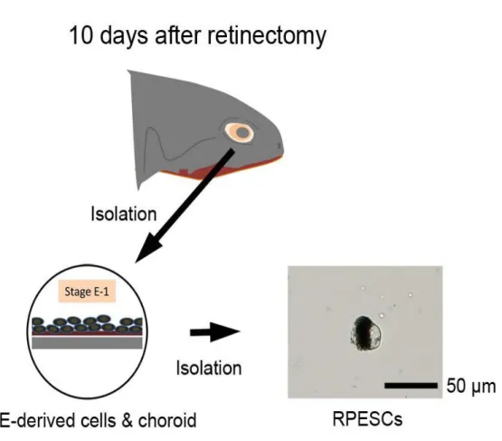
When almost all RPE cells reached Stage E-1 at 10 days after the operation (Chiba 2006; Islam et
21
collected. These were treated more carefully because the connection between the regenerating cells
and the basal membrane was weak. The eyeball was opened up along the equator, and the posterior
side of the wound created during retinectomy, as well as the part which not affected by it, were
collected. The existence of RPESCs was confirmed inside each hemisphere and separated, together
with the colloid, from the sclera. Using the RPESCs with a colloid (RPESCs-colloid), template
cDNAs were constructed using the same procedure described in 2.1.5.
Exceptionally, template cDNAs were prepared from isolated RPESCs as follows.
Cells on the basal membrane were isolated by elastase in EDTA solution as described in section
2.1.4. The isolated cell suspension was transferred to 1% agarose-coated 35-mm plastic dishes
placed on ice.
To avoid contamination of the basement membrane and colloid tissue that became fragile during
regeneration, solitary RPE cells or RPE-derived cells were identified by their morphological
characteristics (Figure 13). Then 100 cells were selected and placed into a PCR tube (No T-02F, Ina
Optica, Osaka, Japan) using a microtip (703Y, Ina Optika) attached to a micropipette (set at 0.5 ml;
Pipetman P20; Gilson, WI 53562-0027, USA) under a dissecting microscope. The cells were
accumulated at the bottom of the tube after centrifugation for 1 min at 3,000 g. Because the amount
of mRNA in a single cell is small, two-step pre-amplification of the cDNA was carried out. Total
22
Amplification Kits (Real Time) Ver.2 (3730; Takara). In this step, the resulting cDNAs were
pre-amplified non-selectively by PCR (21 cycles). Furthermore, cDNAs were pre-amplified by PCR
(65 cycles) using KOD FX with the class-specific primer sets. PCR products were separated by
agarose gel electrophoresis, and those corresponding to the size of Pax6 variants were excised and
purified using a Geneclean Spin Kit. Pre-amplified cDNAs were prepared for all classes of Pax6,
and used as the templates.
Discrimination of the classes of Pax6 variant was performed against cDNA from RPESCs-colloid
and isolated RPESCs in the same way as indicated in section 2.1.8.
2.3. Estimation of the signal pathway causing the reprogramming of RPE cells
2.3.1. Preparation and incubation of RLECs
After the eyeball was excised, the muscle which was attached to the sclera, was carefully removed.
It was sterilized twice for 20 sec by 70% ethanol, placed cornea side up on a membrane filter
(HAWP 013 00, Millipore, Billerica, MA, USA), and cut along the equator. The anterior half was
carefully removed and the posterior half (eye-cup) was soaked in PBS for 20-30 min to create a
loose connection between NR and RPE. Then NR was peeled off by a needle and forceps to create a
RLEC, and the optic nerve was cut out. An RLEC of 0 DIV was used for analyses. The other RLEC
23
penicillin-streptomycin liquid (15140-122, Thermo Fisher Scientific) at 25°C for up to 10 days. The
medium was refreshed on day-5 of incubation (Figure 18).
The following factors were added to culture medium. MEK1/2-specific inhibitor U0126 (V1121;
Promega, Fitchburg, WI, USA), which was dissolved in DMSO (D2650; Sigma-Aldrich) at 2 mM
immediately before use, was administered at a final concentration of 5 μM from the time point at
which the eye-cup was soaked in PBS and the culture medium. Heparin, sodium salt (081-00136,
Wako) was added to the medium at a concentration of 7.5 μg/ml from the beginning of incubation. The newt FGF2 (see Susaki and Chiba, 2007) was tested by adding it to the heparin-containing
medium at a concentration of 50 ng/ml.
When the medium was replaced, fresh factors were added.
2.3.2. PCR analysis
RLECs, which were incubated for 10 days or simply removed from normal eyeballs, were
transferred to RNase-free PBS. RPE cells with colloid tissue were peeled off from the remainder of
RLECs. In the same manner as described in section 2.2.2., sample collection, cDNA synthesis and
discrimination of the classes of Pax6 variant were carried out.
24
Rabbit polyclonal anti-Pax6 antibody PRB-278P (1:500; Covance) and mouse monoclonal
anti-RPE65 antibody (1:500; MAB5428, Millipore) were used as the primary antibodies.
Biotinylated goat anti-rabbit IgG antibody (1:500) and Alexa-488-comjugated goat anti-mouse IgG
antibody (1:500; A-11008, Thermo Fisher Scientific) were used as the secondary antibodies. To
confirm that there was no nonspecific adsorption of the primary antibody, the anti-Pax6 antibody in
which epitope was blocked in the target amino acid sequence itself was used as the negative control.
PRB-278P was pre-adsorbed with a synthetic peptide (QVPGSEPDLSQYWPRIQ) corresponding to
the newt Pax6 to make the negative control. The antibody to which the peptide (50 μg/ml) was added was placed overnight at 4°C before use. When the negative control was used, the test antibody was
also dispensed to maintain the same conditions.
2.3.4. Immunohistochemistry
RLECs attached to the sclera, as well as the membrane used in culture, were fixed in 4%
paraformaldehyde in PBS for 2.5 h at 4°C. The fixative solution was washed off. The tissue sections
were sliced to a thickness of ~20 μm and stained as in section 2.1.7. When double staining was performed, two antibodies were simultaneously added to the solution.
For whole-mount staining, RLEC preparations were rinsed thoroughly (PBS, 1% TritonX-100 in
25
incubated in a blocking solution [3% normal goat serum (S-1000, Vector Laboratories) / 1 %
TritonX-100 in PBS] containing Avidin D (1:50; Avidin/Biotin Blocking kit) for 2 h, washed in PBS
twice, and then incubated in the primary antibody diluted with blocking solution containing Biotin
(1:50; Avidin/Biotin Blocking kit) for 72 h at 4°C. After washing thoroughly, the samples were
incubated in the biotinylated secondary antibody diluted with the blocking solution for 4 h, washed
thoroughly, incubated in a mixture of Avidin and Biotin Complex (Vectastain ABC Elite kit) for 2 h,
washed thoroughly, and then incubated in a DAB solution (DAB substrate kit) for 6 min. The
reaction was stopped by washing samples in distilled water. After the DAB solution was washed
away, melanin pigments in the tissues were bleached by incubating in 15% H2O2 ⁄ 1.5% sodium
azide (197-11091, Wako) in PBS overnight. After the bleaching solution was washed away, the
nuclei of RPE cells were stained with DAPI (1:50,000; D1306, Thermo Fisher Scientific) for 6 h.
The RPE-choroid tissue was separated from the sclera, placed RPE-side up on a glass slide,
immersed into 90% glycerol in PBS and finally mounted under a cover slip. Bright-light and
fluorescence images of tissues were acquired using the same CCD camera systems used in section
2.1.7.
2.3.5. Cell counting
26
characteristically flat and oval nucleus on the surface of the basement membrane in fluorescence
field. Moreover, the amount of immunostaining that overlapped with nuclear staining was assessed
in bright field as positive cells. In this study, total and Pax6-immunoreactive nuclei of RPE cells
were counted in an area further than 50 μm from the peripheral margin of the RPE sheet, because non-specific staining along the margin, which was due to the primary antibody (rabbit IgG), was
serious under the current conditions.
2.3.6. Data analysis
Data were presented as the mean ± SEM (n: the number of RLECs) from more than three
independent rounds of experiments. Normality and non-parametric tests (Mann-Whitney’s U-test
and Jonckheere-Terpstra test) were carried out to evaluate the statistical significance of the data,
27
3. Results
3.1 Identification of Pax6 variants expressing in the eyeballs of adult newts
3.1.1. Search for unknown Pax6
To search for classes of unknown Pax6 expressed in the normal eye and regenerated retina of the
adult newt, transcriptome data which our laboratory made in the past was used. This data was made
using the total RNA obtained from the posterior half of the normal eye from which the retina and
regenerating eyeball were removed at 0-14 days after retinectomy (Nakamura et al., 2014). After
paired end sequencing was carried out by a next generation sequencer, the fragmentary sequence
information was connected by assembly software. In this process, three algorithms were applied and
three databases were produced because there is no consensus about the best de novo assembly
software. In this study, three de novo assembled databases were integrated and used to search widely
for unknown Pax6variants.
By repeating the local blast analysis that used known Pax6 as the query sequence, three new
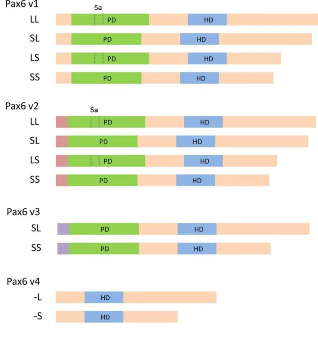
classes of Pax6 transcripts were identified. In this study, these new Pax6 were named Pax6
variant2~4 (v2~4) while the known Pax6 was named variant1 (v1). The characteristic common to the
28
region. Their unique 5ʹ regions are shown in Figure 4 and the full sequences are displayed in Figures 5, 6 and 7. Pax6 v3 has a long unique sequence with partially homogeneity to the Pax6 regulatory
region of other animals whereas the unique sequence of Pax6 v2 is short. These different unique
sequences continue to share the same point where is just before the PD-coding region. On the other
hand, in v4, the sequence extended to inside of the PD-coding region. This resulted in the lack of a 5′
part (127 bp) of the PD-coding region (i.e., a neighbouring sequence of the 5′ end of exon 5a) and
the addition of a 92-bp extra sequence after the 3′ end of exon 5a. This class has an ATG start codon
after the PD-coding region. Therefore, it is expected to encode paired-less forms of Pax6. Unlike
these varieties of unique sequences, the sequence downstream of the PD-coding region has high
homogeneity with known Pax6.
Based on the above data, whether these Pax6 were expressed in the eyeballs of adult newts was
checked. Primers were designed using sequences that are specific for each class of Pax6 and PCR
was performed using a cDNA template which was reverse transcribed from mRNA extracted from
whole adult newt eyeballs. Four kinds of Pax6 expected in silico, including three new classes, were
detected.
Splice variants in each class were also confirmed. Using RT-PCR, four variants of Pax6 v1 and v2
(LL, SL, LS and SS) were confirmed. In contrast, only two variants (SL and SS) lacking 5a were
29
confirmed. Figure 8 shows the structure of each class, including splice variants. Thus, 12 variants of
known or new Pax6 expressed in normal newt eyeballs with unique 5ʹ sequences were detected.
3.1.2. Distribution of Pax6 variants in the adult newt eye
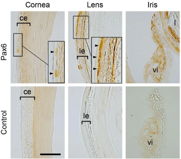
Next, immunohistochemistry using antibody PRB-278P, which recognizes all classes of Pax6, was
performed to confirm which parts of the normal eyeball express Pax6. The results of
immunohistochemistry, which are shown in Figures 9 and 10, suggest that Pax6 is expressed in
epithelial cells of the cornea, CL, lens and iris as well as in cells of the CMZ and in amacrine and
ganglion cells in the NR, but not in the RPE, choroid or sclera.
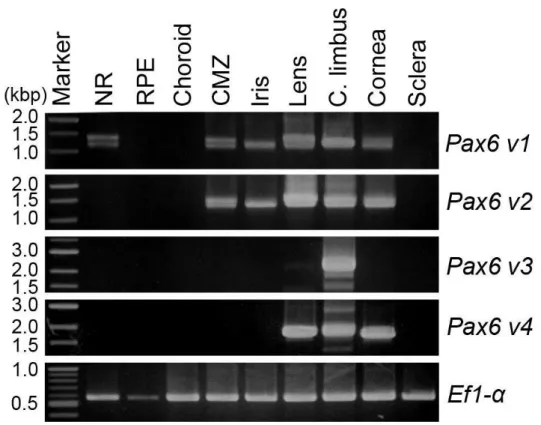
On the basis of this observation, tissue samples were isolated from each part of the eyeballs and
the expression of Pax6 in them was examined by PCR with class-specific primer sets. Furthermore,
splice variants were distinguished based on band size. PCR products were detected in most eye
tissues except for the RPE, choroid and sclera (Figure 11). This is consistent with the
immunohistochemical evidence. Previous studies reported that there is no Pax6 v1 in RPE cells
(Chiba et al., 2006a; Islam et al., 2014; Nakamura et al., 2014). This experiment supports this claim
and reveals that other classes of Pax6 are also not expressed in RPE cells. Table 1 summarizes the
Pax6 variants that were identified in Figure 11. The NR had only four variants of Pax6 v1. In
30
Pax6 v1 and v2 but their expression patterns were different. In the CMZ, there were four variants for
each class, but in the iris, only LS and SS were detected. The lens, CL and cornea expressed SL and
SS of Pax6 v3 and -L and -S of Pax6 v4 as well as four variants of Pax6 v1 and v2, although PCR
signals for Pax6 v3 in the lens and cornea were relatively weak (Figure11).
To confirm the presence of Pax6 proteins, western blotting was carried out using anterior eye
tissues where it is expected that all four classes of Pax6 exist. As indicated in Figure 12, two protein
bands at around 50 kD, as well as bands at ~31.6 kD and ~26.4 kD, were recognized. The arrow in
Figure 12 shows bands which were specifically labelled by Pax6 antibody while the arrowhead
shows a non-specific band which appeared even when the primary antibody was omitted, i.e., in the
control. The upper two bands are likely the merges of longer forms (LL) of Pax6 v1, v2 and v3. The
bands were no longer able to separate because their structures and molecular weights were similar to
each other. The lower bands corresponded to the -L and -S forms of Pax6 v4 because this is
paired-less Pax6 with a small molecular weight.
Taken together, these results suggest that Pax6 variants are expressed in many kinds of tissues in
the eye in different combinations but not in intact RPE cells.
3.2. RPE cells newly express Pax6 v1 and v2 variants upon retinectomy
31
are reprogrammed into a unique multipotent state and newly express Pax6 in neural retina
regeneration. Therefore, which Pax6 variants were expressed in RPESCs was analysed.
At first, PCR was carried out with class-specific primer sets and RPESCs-choroid tissues collected
from posterior halves of the eyeballs at 10 days after retinectomy. Figure 13 is an image of a
collected sample that contains aggregates of RPESCs, which are RPE-derived cells which just
entered Stage E-1. In this experiment, PCR conditions were changed and implemented for all
subsequent analyses. Figure 14 shows two typical PCR results. On the left, product bands
corresponding to LL, SL, LS and SS of Pax6 v1, and to LL of Pax6 v2, were recognized. On the
right, three other variants of Pax6 v2 were also detected in the same template when the annealing
temperature of PCR was changed. All four variants of Pax6 v1 and v2 were identified, but no
variants for Pax6 v3 or v4 were detected (Figure 15). On the other hand, no product bands were
detected in the choroid tissues after RPE-derived cells had been removed (Figure 16). The same
result was obtained in three independent rounds of experiments.
For further confirmation, RPESCs were isolated from the choroid by protease and a chelator. While
confirming cell shape under a microscope, RPESCs were selected from pieces of other pigmented
organized structures (Figure 13). Single-cell PCR with 100 isolated RPESCs was then attempted,
and expression of the same classes of Pax6 was confirmed and compared with RPESCs-choroid
32
SS for Pax6 v1 and SL for Pax6 v2. When PCR conditions were changed, additional variants were
not detected. This result may reflect a difference in the stage of regeneration between both samples.
A recent study indicated that it is possible for individual RPE cells to be reprogramed at a slightly
different timing in the E-1 stage (Islam et al., 2014). In isolated RPESCs, uniform cells that had just
begun reprogramming, were selected. Of note, the RPESCs-choroid tissue contained cells with an
advanced stage of regeneration.
Taken together, these results suggest that transcript variants, possibly LL, LS, SL and SS, of two
classes of Pax6 (i.e., v1 and v2) are expressed in RPE-derived cells in Stage E-1.
3.3. Estimate of the signal pathway which causes the reprogramming of RPE cells
3.3.1. RPE cells also express Pax6 v1 and v2 variants in RLECs
The signals that may have caused the reprogramming of RPE cells were estimated by using Pax6
expression as the marker of this phenomenon. Therefore, in this study, the RLEC system that was
established to check the response of the RPE cells in our laboratory was used. Figure 18 shows the
procedure of how to make the RLEC system. The newt eyeball was extracted and sterilized. The
anterior hemisphere (the lens side) was cut off. The exposed NR was removed from the remaining
posterior half of the eyeball in PBS and the RLEC was incubated in 80% L-15 medium at 25°C.
33
galactose and sodium pyruvate. Since L-15 medium does not include any growth factors, it was
suitable for this experiment to apply any factor.
At first, it was confirmed whether this system could be adapted to the analysis of Pax6 expression.
The existence of the target protein was confirmed by using anti-Pax6 antibody, which recognized all
variants of Pax6. The results of whole-mount staining are shown in Figure 19. Images of the RPE
cell layer represent a top view. New Pax6 antibody-positive cells were observed in the RLEC system
after incubation for 10 days. The right-hand panel represents the negative control on day-10. The
antibody preadsorbed with the peptide which was specific for the epitope was used as the negative
control to completely remove non-specific staining of the primary antibody as well as the secondary
antibody. Pax6-immunoreactivity in RPE cells was confirmed by double staining of a section of a
day-10 RLEC with RPE65 antibody (Figure 20). Images of the RPE cell layer were viewed from the
side. RPE cells were identified by RPE65-immunofluorescence (Chiba et al., 2006a). The nuclei of
RPE cells were labelled with Pax6 antibody (arrows). Horizontal bars indicate the thickness of the
RPE layer, and asterisks indicate small nuclei in the choroid, which displayed nonspecific staining as
well as autofluorescence under these conditions. These two immunohistochemical results of RLECs
revealed that Pax6 protein exists in several nuclei of RPE cells on day-10.
PCR analyses were then carried out with RPE-choroid tissues collected from RLECs immediately
34
vitro (Figure 21). No Pax6 variant was detected on day-0 and all four variants of Pax6 v1 and v2
were newly expressed by day-10 while Pax6 v3 and v4 variants were not. These results were
consistent with the in vivo results.
These results indicate that RPE cells incubated in the RLEC system express Pax6 as the in vivo
state. In particular, because variants expressed in RLEC and in vivo are common, regulation is
suggest to be equal, which expands the applicability of the RLEC system. Thus, this system was
subsequently used for the analysis of Pax6 expression signals.
3.3.2. RPE cells expressing Pax6 versus RPE cells entering cell-cycle
Interestingly, even though no external factors were added to cultures in the period of incubation,
Pax6 was expressed in RLEC RPE cells. RLEC was incubated in minimal medium (Figure 19).
Furthermore, the conditions of RPE cells in which Pax6 was expressed were different from those of
RPE cells entering the cell cycle (Yoshikawa et al., 2012). Figure 22 indicates the ratio of
Pax6-imunoreactive (Pax6+) nuclei in the RPE sheet derived from RLEC which was incubated
without any factors. Pax6+ nuclei appeared within 5 days and increased significantly between 5 and
10 days. The Pax6+ nuclei were distributed uniformly (Figure 19). The number of nuclei was
counted in the area farther than 50 μm from the peripheral margin of the RPE to avoid non-specific staining. The timing of Pax6 expression differed from the cell-cycle re-entry observed from day-5 to
35
10 (Yoshikawa et al., 2012). In addition, Pax6-positive cells and cells entering the cell cycle also
showed a variable distribution pattern (Figure 23). Pax6 was expressed uniformly in RLEC, but after
entering the cell cycle, expression concentrated in the area near the Edge of RLEC (Yoshikawa et al.,
2012). It is possible that Pax6 expression and cell cycle re-entry are independently controlled in the
RLEC system.
3.3.3. Pax6 expression in RPE cells is not affected by a MEK1/2 inhibitor U0126
As described in the introduction, our laboratory discovered that the MEK1/2-ERK1/2 intracellular
signaling cascade, which was activated within 30 min after retinectomy, is necessary for the first cell
cycle entry of RPE cells in the RLEC system (Mizuno et al., 2012). Therefore, the relationship of the
MEK1/2-ERK1/2 signaling cascade and Pax6 expression in RPE cells was examined.
RLECs were incubated in the presence of an MEK1/2 inhibitor U0126 at a concentration of 5 μM, which can inhibit up to ~50% of the initial activation of ERK1/2 and decrease the number of BrdU+
cells by 10 days by as much as ~25% (Yoshikawa et al., 2012). However, this treatment did not
affect the number of Pax6+ RPE cells at 5 days compared to the solvent (0.25% DMSO) only
(Figure 24). This result suggests that Pax6 can be expressed without the participation of the
MEK1/2-ERK1/2 signaling cascade and that Pax6 expression in RPE cells may be independent of
36
3.3.4 Heparin promotes the expression of Pax6 with the help of the MEK1/2-mediated
pathway
Pax6 expression and cell cycle re-entry of RPE cells take place in the same period after
retinectomy in vivo (Islam et al., 2014). An endogenous factor mediating these independent
phenomena exists in vivo, but is exhausted in vitro was hypothesized.
Heparin, a glycosaminoglycan that is ubiquitously distributed in the body, and is known to bind
various soluble factors and support their actions on receptors as well as protect these factors against
degradation, served as a supplement. In fact, in the newt, heparin promotes the cell cycle re-entry of
RPE cells in the RLEC system through a pathway that influences the area downstream of the
MEK1/2-ERK1/2 signaling module (Yoshikawa et al., 2012). Therefore, heparin was added as an
endogenous factor to activate RLEC.
Interestingly, when the RLEC was incubated for 5 days in the presence of heparin (7.5 μg/ml), the number of Pax6+ RPE cells tended to increase (Figure 25). Next the effects of U0126 (5 μM) on the
promotion of Pax6 expression by heparin were examined (Figure 25). In this condition U0126 was
tested. The number of Pax6+ RPE cells at 5 days after incubation had decreased significantly. These
results suggest that heparin promotes Pax6 expression with the help of a MEK1/2-mediated pathway.
37
regeneration. FGF2 is known to up-regulate Pax6 expression through the MEK-ERK pathway in
chick embryonic RPE cells (Luz-Madrigal et al., 2014; Spence et al., 2007). However,
administration of 50 ng/ml FGF2, a concentration used in previous experiments using RLEC (Susaki
and Chiba, 2007), did not influence the effect of heparin on Pax6 expression in RPE cells after 10
38
4. Discussion
4.1. Several kinds of Pax6 variants
To date, a wide variety of Pax6 transcripts has been detected in many vertebrates, from fish to
mammals. In addition, it is known that transcriptional initiation sites on the locus and
post-transcriptional and post-translational modifications that derive various isoforms are
spatiotemporally regulated in normal development and adult neurogenesis (Bandah et al., 2007;
Gorlov and Saunders, 2002; Kammandel et al., 1999; Kim et al., 2008; Lakowski et al., 2007;
Mizuno et al., 1997; Shaham et al., 2012; Walther and Gruss, 1991; Zhang et al., 2010). In the newt
C. pyrrhogaster, a Pax6 gene corresponding to Small eye (Sey) Pax6 has been reported (Islam et al.,
2014; Mizuno et al., 1997). In this study, new three classes of Pax6 were discovered. The sequence
of the conserved region of these Pax6 is extremely similar. These sequence similarities suggest that
these different classes of transcripts originate from the same locus. On the other hand, each class of
Pax6 has a unique sequence upstream of the PD-coding region, including the 5′ UTR (Figure 4),
suggesting differences in transcriptional regulation among these classes.
These classes of Pax6 are expressed in the intact eye tissues of the adult newt in different
39
provide suitable ground work for a study on the regulation and roles of the different classes of Pax6
variants in the newt. In this study, sequence information of the Pax6 variants was indispensable for
determining the class of Pax6 expressed in RPE cells after retinectomy.
4.2. RPESCs and CMZ express common Pax6 variants
As described in the introduction, after retinectomy, adult newt RPE cells undergo a loss of their
membrane attachment, forming cell aggregates in 10 days (Stage E-1 [4]). During this process, RPE
cells are reprogrammed into a unique multipotent state with newly expressed c-Myc, Klf4, Sox2, Mitf
and Pax6, while preserving their original characteristics (Islam et al., 2014). In this study, the class
of Pax6 expressed in such RPE-derived cells in Stage E-1 was determined, namely Pax6 v1 and v2,
both of which give rise to four splice variants LL, SL, LS and SS. It is still uncertain whether Pax6
works as a reprogramming factor in this system. However, it is possible that the expression of Pax6
in RPE-derived cells represents the reprogramming of RPE cells.
These should have the potency to form either the pro-NR or pro-RPE layer, as in cells in the early
optic vesicle which also express c-Myc, Klf4, Sox2, Mitf and Pax6 (Islam et al., 2014).
In early eye development, Pax6 and Mitf are known to be involved in fate decision of cells in the
optic vesicle to form either pro-NR or pro-RPE cells as the optic vesicle invaginates to form the
40
down-regulation of Mitf biases the fate of cells toward the retinal stem/progenitor cells (Azuma et al.,
2005; Fuhrmann et al., 2014; Nguyen and Arnheiter, 2000).
In the regeneration of the adult newt retina, up-regulation of Pax6 was observed in a population of
RPE-derived cells that formed the pro-NR layer in the next stage (Islam et al., 2014). Intriguingly, in
this study, the same set of Pax6 variants as those in RPE-derived cells are expressed in the CMZ
where retinal stem/progenitor cells have been harbored since the embryonic stage was found.
Therefore, as an analogy of early eye development, one hypothesis may be that these classes of Pax6
variants express in the cells that have potency, producing neural retina. However, it is still uncertain
whether Pax6 works as a reprogramming factor in this system.
4.3. The signal which produces RPESCs
In this study, Pax6 was examined as a marker of the reprogramed signal of RPE cells using the
RLEC system. Interestingly, Pax6 expression did not require the administration of exogenous factors.
Immunohistochemical analysis revealed that, in the absence of exogenously administered factors,
Pax6+ cells in the RPE sheet appeared within 5 days after retinectomy and showed a gradual
increase in number up to ~27 % (on average) of all RPE cells by 10 days, and that the Pax6+ cells on
day-10 are distributed almost uniformly in the sheet. In addition, the appearance and increment of
41
MEK1/2-mediated signaling. In a previous study (Yoshikawa et al., 2012), it was shown that RPE
cells enter the cell cycle even if factors were not provided in the RLEC system. Cell cycle re-entry
and Pax6 expression were observed at the same time during Stage E1 in vivo regeneration. A
previous study (Yoshikawa et al., 2012) indicated that the cell cycle re-entry of RPE cells in RLEC
conditions is highly restricted to a zone of ~100 μm width along the peripheral margin of the RPE
sheet, i.e., along the wound edge of the RLEC, an area that was defined as the ‘Edge’ . This event is obviously regulated by a MEK1/2-ERK1/2 module whose activity is transiently strengthened within
30 min after retinectomy (Mizuno et al., 2012). Furthermore, the cells which reside in the central
area of the RPE sheet, defined as the ‘Center’ in Yoshikawa et al. (2012), hardly enter the cell cycle, possibly through a mechanism equivalent to contact inhibition (Yoshikawa et al., 2012), although the
activity of the MEK1/2-ERK1/2 module in the ‘Center’ is as elevated as in the ‘Edge’ (unpublished
data provided by H. Yasumuro).
Taken together, Pax6+ cells and cells which re-enter the cell cycle on the same RPE sheet appear to
show a different spatial distribution pattern and requirement of the activity of MEK1/2, suggesting
that the expression of Pax6 in RPE cells after retinectomy can be triggered independently of their
cell cycle re-entry.
In the eye, Pax6 expression and cell cycle re-entry of RPE cells take place simultaneously (Islam
42
phenomena may be independently controlled in vivo. To reinforce this possibility, attention was paid
to heparin, which activates the function of various growth factors. Heparin is the glycoprotein which
is strongly negatively charged and combines with many growth factors such as FGFs, TGF-β and
BMPs, to activate the function of these factors. Furthermore, evidence of the influence of the dosage
of heparin in RPE cells entering the cell cycle was provided in past studies (Yoshikawa et al., 2012).
Therefore, in this experiment, heparin was added as an endogenous factor in RLEC activation.
Treatment of RLECs with heparin promoted Pax6 expression in RPE cells but this effect was
inhibited by U0126.
This result indicates that Pax6 expression promoted by heparin administration, but depends upon the
activity of MEK1/2. In other words, heparin administration seems to coordinate the pathway for
Pax6 expression with a pathway mediated by MEK1/2 which is involved in the cell cycle re-entry.
This pathway is driven by heparin or by heparin-associated factors which might be released from
tissues in the RLEC.
Figure 27 indicates hypothetical pathways of molecular networks involved in the initial processes
of retinal regeneration, including findings of cell cycle re-entry which were observed in parallel (red
and green lines are aspects that became clear in this study). At least three intracellular signalling
pathways in RPE cells after retinectomy were predicted:
43 independent.
2) A MEK1/2-ERK1/2 pathway which is primarily involved in cell cycle re-entry. This is more likely
to receive contact inhibition.
3) A pathway driven by heparin or heparin-associated factors. This is located downstream of the
MEK1/2-ERK1/2 pathway and mediates between it and Pax6 expression.
4.4. Differences in RPE cells between humans and newts
In humans, when the NR suffers a traumatic injury, RPE cells – as in the newt – start to lose their
epithelial morphology while acquiring the ability to migrate and proliferate. However, unlike the
newt, these finally withdraw the NR by contraction, leading to a loss of vision (Chiba, 2014). In this
process of transformation, it has been suggested that RPE cells pass through a multipotent state and
such multipotent RPE cells were named as human RPESCs. Human RPESCs are known to express
Pax6 as well as c-Myc, Klf4, Sox2 and Mitf (Salero et al., 2012) as in newt RPESCs (Islam et al.,
2014) although the expression levels of Pax6 seem to vary depending on the RPE cell line from
which the RPESCs are derived, or on the culture conditions (Salero et al., 2012). In the adult newt,
Pax6 presumably plays important roles in retinal regeneration because its transcription is elaborately
and dynamically regulated during retinal regeneration, as demonstrated in a previous study (Islam et
44
regeneration of the adult newt, which was obtained in this study, would be a base to generate a
neural retina in vitro or to regenerate an entire retina in vivo from RPE cells or RPESCs in humans.
4.5. Conclusions and future directions
In this thesis, the signaling pathways involved in the expression of Pax6 in adult newt RPE cells in
a bid to identify the signal that is employed to trigger RPE cell reprogramming for retinal
regeneration were investigated. As a result, the following aspects became clear:
1) Pax6 v1 and v2 express in RPESCs;
2) Pax6 expression is independent of entering the cell cycle;
3) Pax6 expression and cell cycle entry are conjugated by heparin or heparin-associated factors.
Future studies should perform proteomic analysis and identify factors that influence Pax6
expression. In these experiments, Pax6 expression was independent of MEK1/2-ERK1/2, an aspect
that requires further detailed verification. In addition, our laboratory developed a newt transgenic
technique with greater efficiency (Casco-Robles et al., 2011; 2014).
The functional analysis of Pax6 based on knowledge of its variants and the signal system that was
45
5. References
Azuma, N., Tadokoro, K., Asaka, A., Yamada, M., Yamaguchi, Y., Handa, H., Matsushima, S.,
Watanabe, T., Kida, Y., Ogura, T., Torii, M., Shimamura, K., and Nakafuku, M. (2005).
Transdifferentiation of the retinal pigment epithelia to the neural retina by transfer of the Pax6
transcriptional factor. Hum. Mol. Genet. 14, 1059-1068.
Bandah, D., Swissa, T., Ben-Shlomo, G., Banin, E., Ofri, R., and Sharon, D. (2007). A complex
expression pattern of Pax6 in the pigeon retina. Invest. Ophthalmol. Vis. Sci. 48, 2503-2509.
Baumer, N., Marquardt, T., Stoykova, A., Ashery-Padan, R., Chowdhury, K., and Gruss, P. (2002).
Pax6 is required for establishing naso-temporal and dorsal characteristics of the optic vesicle.
Development 129, 4535-4545.
Casco-Robles, M.M., Miura, T., and Chiba, C. (2014) The newt (Cynops pyrrhogaster) RPE65
promoter: molecular cloning, characterization and functional analysis. Transgenic Res.
10.1007/s11248-014-9857-1.
Casco-Robles, M.M., Yamada, S., Miura, T., Nakamura, K., Haynes, T., Maki, N., Del Rio-Tsonis,
K., Tsonis, P.A., and Chiba, C. (2011) Expressing exogenous genes in newts by transgenesis. Nat.
46
Chiba, C. (2014). The retinal pigment epithelium: an important player of retinal disorders and
regeneration. Exp. Eye Res. 123, 107-114.
Chiba, C., Hoshino, A., Nakamura, K., Susaki, K., Yamano, Y., Kaneko, Y., Kuwata, O., Maruo, F.,
and Saito, T. (2006a). Visual cycle protein RPE65 persists in new retinal cells during retinal
regeneration of adult newt. J. Comp. Neurol. 495, 391-407.
Chiba, C., and Mitashov, V.I. (2007). Cellular and molecular events in the adult newt retinal
regeneration. In The Strategies for Retinal Tissue Repair and Regeneration in Vertebrates: From Fish
to Human, C. Chiba, ed. (Kerala, India: Research Signpost), pp. 15-33.
Chiba, C., Nakamura, K., Unno, S., and Saito, T. (2006b). Intraocular implantation of
DNA-transfected retinal pigment epithelium cells: a new approach for analyzing molecular functions
in the newt retinal regeneration. Neurosci. Lett. 368, 171-175.
Czerny, T., and Busslinger, M. (1995). DNA-binding and transactivation properties of Pax-6: three
amino acids in the paired domain are responsible for the different sequence recognition of Pax-6 and
BSAP (Pax-5). Mol. Cell Biol. 15, 2858-2871.
Davis-Silberman, N., Kalich, T., Oron-Karni, V., Marquardt, T., Kroeber, M., Tamm, E.R., and
Ashery-Padan, R. (2005). Genetic dissection of Pax6 dosage requirements in the developing mouse
eye. Hum. Mol. Genet. 14, 2265-2276.