Measurement of X-ray-tube voltage using a 0.3-mm-thick copper filter
Eiichi SATOa, Yasuyuki ODAa, Michiaki SAGAEa, Satoshi YAMAGUCHIb, Yuichi SATOc, Osahiko HAGIWARAd, Hiroshi MATSUKIYOd, Toshiyuki ENOMOTOd,
Manabu WATANABEd and Shinya KUSACHId
aDepartment of Physics, Iwate Medical University, 2-1-1 Nishitokuta, Yahaba, Iwate 028-3694, Japan
bDepartment of Radiology, School of Medicine, Iwate Medical University, 19-1 Uchimaru, Morioka, Iwate 020-0023, Japan
cCentral Radiation Department, Iwate Medical University Hospital, 19-1 Uchimaru, Morioka, Iwate 020-0023, Japan
dDepartment of Surgery, Toho University Ohashi Medical Center, 2-17-6 Ohashi, Meguro-ku, Tokyo 153-8515, Japan
(Accepted July 14, 2016)
Abstract
To measure X-ray tube voltage corresponding to the maximum photon energy of X-ray spectra, we constructed an experimental setup consisting of an X-ray generator, a silicon X-ray diode (Si-XD), an amplifier module, 10-s-time-constant integrator, a digital voltage meter, and a 0.3-mm-thick copper (Cu) filter. The amplifier outputs with and without the Cu filter are measured, and the tube voltage is determined by the ratio (transmissivity) between the output voltages as a function of tube voltage. In this experiment, we used two aluminum filters of 1.0 and 3.0 mm in thickness for absorbing low energy X-ray photons. Using the Cu filter, the transmissivity as a function of tube voltage varied with changes in the Al-filter thickness.
Key words: X-ray, tube voltage, Cu filtration, transmissivity, Si-X-ray diode
1. Introduction
To perform molecular imaging, we have developed several photon-counting energy-dispersive X-ray computed tomography (ED-CT) scanners [1-3] using cadmium telluride (CdTe) and silicon-PIN detectors. Lately, an energy- selecting device [4] has been developed and applied to monochromatic and dual-energy CT (DE-CT) scanners [5].
In the former experiment, we found a high-sensitivity silicon photodiode for detecting X-rays and named this a silicon X-ray diode (Si-XD) [6]. Using these Si-XDs, we developed a DE-CT scanner [7] and an X-ray dosimeter. Currently, the dosimeter utilizes two detectors with a tube-voltage measurement function. Therefore, we are interested in the tube- voltage measurement using a filtration method.
In our research, our major objectives are as follows: to measure transmissivity using a Si-XD and a copper (Cu) filter, to convert the photocurrents into voltages, and to determine the tube voltage using the transmissivity. Therefore, we constructed an experimental setup to measure tube voltage using a Si-XD in conjunction with a Cu filter.
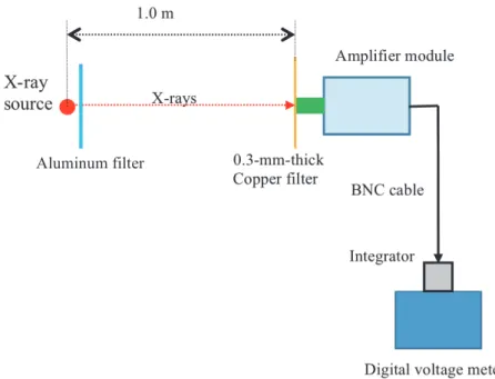
Fig. 1. Block diagram for measuring the output voltages from the amplifier module using a Si-XD and a 0.3-mm-thick Cu filter. The voltage is in proportion to the penetrating X-ray dose rate.
2. Experimental methods
Figure 1 shows an experimental setup for measuring output voltages from the amplifier module. Low-energy X-ray photons from an X-ray generator are absorbed using an aluminum filter, and penetrating photons are detected using a silicon X-ray diode (Si-XD) through a 0.3-mm-thick Cu filter. The photocurrents flowing through a Si-XD are converted into voltages and amplified using an amplifier module, and the amplifier output is input to a digital voltage meter through a 10-s-time-constant integrator. To determine the tube voltage, the amplifier output voltages with and without the Cu filter are measured, and the tube voltage is determined by the voltage ratio (transmissivity) as a function of tube voltage.
The circuit diagrams of the amplifiers are shown in Fig. 2. The photocurrents are converted into voltages using a current-voltage (I-V) amplifier [Fig. 2(a)], and the output voltage Vi from the I-V amplifier is written as:
Vi = 100×106 I (1)
where I is the photocurrent. Subsequently, the output from the I-V amplifier is sent to the voltage-voltage (V-V) amplifier [Fig. 2(b)]. In this experiment, a variable resistor is set to the maximum value of 50 kΩ, and the output voltage from the V-V amplifier Vv is given by:
Vv = 6.0×10² Vi (2)
The measurement of X-ray dose rate is important to calculate incident dose for patients. The X-ray dose rate from an X-ray generator was measured using an ionization chamber (Toyo Medic RAMTEC 1000 plus) at a tube current of 0.5 mA without filtration. The chamber was placed 1.0 m from the X-ray source, and we measured the dose rate with changes in the tube voltage from 50 to 109 kV.
3. Results
Figure 3 shows the X-ray dose rates at a constant tube current of 0.50 mA using two Al filters of 1.0 and 3.0 mm in thickness. In the two filtrated conditions, both the X-ray dose rates increased with increasing tube voltage. At a tube voltage of 100 kV, the X-ray dose rates with Al thicknesses of 1.0 and 3.0 mm were 40.8 and 28.0 μGy/s, respectively.
X-ray source
1.0 m
Amplifier module
0.3-mm-thick Copper filter
Integrator
Digital voltage meter BNC cable
X-rays
Aluminum filter
Fig. 2. Circuit diagram of the amplifier module. (a) I-V amplifier and (b) V-V amplifier.
Fig. 3. X-ray dose rates after penetrating two Al filters with changes in the tube voltage
Fig. 2 from detector
LMC662
+5 V
+
− 100 MΩ 220 pF
A
A
Lemo Output +5 V
−
50 kΩ 100 nF
100 Ω
− +
BNC input
LMC662 10 kΩ
2 kΩ to
digital voltage meter
(b) (a)
Al =3mm Al =1mm
Al: Al filter thickness
Fig. 4. Output voltages and X-ray dose rates measured using the Si-XD without the Cu filter according to changes in the tube voltage and the insertion of the two Al filters. (a) Output voltages and (b) X-ray dose rates.
Fig. 5. Transmissivities with the Cu filter with changes in the tube voltage.
The X-ray dose rates were also measured using the Si-XD and the amplifier for reference and shown in Fig. 4. Using the two Al filters, the output voltages increased with increasing tube voltage. To convert the output voltage into the dose rate, we assume that the output voltage is proportional to the dose rate, and the maximum voltage of 3.64 V corresponds to the dose rate of 48.5 μGy/s. The dose rates corresponded qualitatively to those measured using the dosimeter.
The transmissivities with changes in the tube voltage are shown in Fig. 5, and the transmissivity T is written by:
T = Vf / V (3)
where Vf is the output voltage using the Cu filter, and V is the output without Cu filtration. Both the transmissivities using two Al filters increased with increasing tube voltage, and the transmissivity slightly increased with increasing Al filter thickness.
Al: Al filter thickness
Al =3mm Al =3mm
Al =1mm Al =1mm
(a) (b)
Al: Al filter thickness
Al =1mm Al =3mm
4. Discussion
In the present research, we used a Si-XD in conjunction with an amplifier module to measure the dose rate approximately proportional to the output voltage from the module. Compared with a dosimeter with an ionization chamber, the sensitivity of the Si-XD slightly decreased with increasing tube voltage.
Using a 0.3-mm-thick Cu filter, a tube voltage range from 50 to 109 kV could be measured. However, the filter thickness should be reduced to less than 0.2 mm to measure the tube voltage below 50 kV. In addition, since the transmissivity increased with increasing the total filtration of the X-ray source, the total filtration should be determined to a constant value before measuring the tube voltage.
5. Conclusions
First, we measured the dose rate using a Si-XD and an amplifier module, and the results using the Si-XD corresponded well to those obtained using a dosimeter with an ionization chamber. Second, the transmissivities were measured using a 0.3-mm-thick Cu filter, and the transmissivity increased with increasing tube voltage. Therefore, the tube voltage can be determined by the transmissivity as a function of tube voltage.
Acknowledgments
This work was supported by Grants from Keiryo Research Foundation, Promotion and Mutual Aid Corporation for Private Schools of Japan (PMAC), Japan Science and Technology Agency (JST), and Japan Society for the Promotion of Science (JSPS) KAKENHI (26461804, 2014-2016). This was also supported by a Grant-in-Aid for Strategic Medical Science Research (S1491001, 2014–2018) from the Ministry of Education, Culture, Sports, Science and Technology of Japan (MEXT).
References
[1] Sato, E., Oda, Y., Abudurexiti, A., Hagiwara, O., Matsukiyo, H., Osawa, A., Enomoto, T., Watanabe, M., Kusachi, S., Sato, S., Ogawa, A. and Onagawa, J., “Demonstration of enhanced iodine K-edge imaging using an energy- dispersive X-ray computed tomography system with a 25 mm/s-scan linear cadmium telluride detector and a single comparator,” Appl. Rad. Isot. 70, 831–836 (2012).
[2] Chiba, H., Sato, Y., Sato, E., Maeda, T., Matsushita, R., Yanbe, Y., Hagiwara, O., Matsukiyo, H., Osawa, A., Enomoto, T., Watanabe, M., Kusachi, S., Sato, S., Ogawa, A. and Onagawa, J., “Investigation of energy-dispersive X-ray computed tomography system with CdTe scan detector and comparing-differentiator and its application to gadolinium K-edge imaging,” Jpn. J. Appl. Phys. 51, 102402-1-5 (2012).
[3] Hagiwara, O., Sato, E., Watanabe, M., Sato, Y., Oda, Y., Matsukiyo, H., Osawa, A., Enomoto, T., Kusachi, S.
and Ehara, S., “Investigation of dual-energy X-ray photon counting using a cadmium telluride detector and two comparators and its application to photon-count energy subtraction,” Jpn. J. Appl. Phys. 53, 102202-1-6 (2014).
[4] Watanabe, M., Sato, E., Oda, Y., Sagae, M., Sato, Y., Yamaguchi, S., Hagiwara, O., Matsukiyo, H., Kusachi, S.
and Ehara, S., “Quasi-monochromatic X-ray photon counting using a silicon-PIN detector and an energy-selecting device and its application to iodine imaging,” Med. Imag. Inform. Sci. 32, 38-43 (2015).
[5] Oda, Y., Sato, E., Wada, K., Momokawa, H., Kataoka, D., Otani, R., Yamaguchi, S., Ehara, S., Hagiwara, O., Matsukiyo, H., Watanabe, M. and Kusachi, S., “Dual-energy X-ray computed tomography using a YAP(Ce)- multipixel-photon detector and an energy-selecting device,” Med. Imag. Inform. Sci. 32, 71-76 (2015).
[6] Matsushita, R., Sato, E., Yanbe, Y., Chiba, H., Maeda, T., Hagiwara, O., Matsukiyo, H., Osawa, A., Enomoto, T., Watanabe, M., Kusachi, S., Sato, S., Ogawa, A. and Onagawa, J., “Low-dose-rate computed tomography system utilizing 25 mm/s-scan silicon X-ray diode and its application to iodine K-edge imaging using filtered bremsstrahlung photons,” Jpn. J. Appl. Phys. 52, 032202-1-5 (2013).
[7] Sato, Y., Sato, E., Ehara, S., Oda, Y., Hagiwara, O., Matsukiyo, H., Enomoto, T., Watanabe, M. and Kusachi, S.,
“Development of a dual-energy silicon X-ray diode and its application to gadolinium imaging,” Radiat. Meas. 77, 12-17 (2015).