• 検索結果がありません。

Tatsuo HIRANO, Hisoyuki KUSANO, Ryoji TAKAHIRA Hironobu TOHCHIKA, Yoshikazu MINE, Keiji KAJIWARA Takayuki NAKAZAKI, Seishi HONJO, Hiroyuki YAMAGUCHI Takatoshi SHIMOYAMA, Toshio MIURA and Masao TOMITA

N/A
N/A
Protected

Academic year: 2021

シェア "Tatsuo HIRANO, Hisoyuki KUSANO, Ryoji TAKAHIRA Hironobu TOHCHIKA, Yoshikazu MINE, Keiji KAJIWARA Takayuki NAKAZAKI, Seishi HONJO, Hiroyuki YAMAGUCHI Takatoshi SHIMOYAMA, Toshio MIURA and Masao TOMITA"

Copied!
6
0
0

読み込み中.... (全文を見る)

全文

(1)

Acta Med. Nagasaki 34 : 240-245

The Prognostic Significance of Subserosal and Serosal Extent of Cancer Invasion in Gastric Cancer

Tatsuo HIRANO, Hisoyuki KUSANO, Ryoji TAKAHIRA Hironobu TOHCHIKA, Yoshikazu MINE, Keiji KAJIWARA Takayuki NAKAZAKI, Seishi HONJO, Hiroyuki YAMAGUCHI Takatoshi SHIMOYAMA, Toshio MIURA and Masao TOMITA

The First Depertment of Surgery Nagasaki University School of Medicine Received for publication, December 26, 1989

ABSTRACT : In order to determine the prognostic significance of subserosal and serosal extent of cancer invasion in gastric cancer, 78 patients were reexamined histologically. In these patients curative resection was performed and cancer invaded

the  subserosal  layer  with  growth  pattern  of infiltrating  type  i.e."ssγ"(30  patients),

or exposed outside the serosal surface i.e. "se" (48 patients). The significantly favourable prognosis was seen only in patients with ssr cancer being less than 1 cm in extent with a five-year survival rate of 92.3%, and in patients with small amount of cancer cells in the subserosal layer, having a five-year survival rate of 81.8%. In patients with se cancer the five-year survival rate was less favorable.

INTRODUCTION

It is widely recognized that the serosal factor is one of the most important factors influencing the prognosis of gastric cancer patients because the serosal invasion is direct- ly related to a subsequent peritoneal carcinosis which is the most common mode of recurrence.

The present study was undertaken to determine the influence of the serosal factor in patients with gastric cancer which involved the subser- osal wall with infiltrating fashion or was exposed outside the serosa based on the micro- scopic finding of the surgical specimens obtain- ed after curative surgical resection for gastric cancer.

MATERIALS AND METHODS

Of gastric cancer patients who underwent

curative resection at the First Department of

Surgery, Nagasaki University Hospital, in a

period between January 1973 and December

1982, 30 cases of ss r cancer and 48 cases of se

cancer were histologically reexamined. The

paraf f in-embedded specimens stained with

hematoxylin and eosin were used for this study,

and the following items were examined in rela-

tion to the patients' prognosis : the extent of

cancer in the deepest layer, mode of extension

in the subserosal layer, quantity of cancer cells,

and histologic type of cancer in the front of

cancer invasion of the deepest area. The extents

of cancer in the subserousal layer and on the

serosal surface were determined by measuring

the maximal length of the width of cancer

invasion at each layer. An shown in Fig. 1, the

modes of extension of cancer in the subserosal

layer in both ss r and se cancer patients were

divided into the following three types : (1) val-

ley type, in which cancer invasion is wider in

(2)

to the valley type, (3) box type, in which extent of cancer invasion at the shallow area of the subserosal layer is almost the same as at the deep area. The amount of cancer cells at the subserosal layer or serosal surface was determined and graded from (+) to (+ + + ).

Histologic types of cancer in the deepest area were also examined and divided either into differentiated carcinoma or into undifferentiat- ed carcinoma. The patients' prognosis was analyzed in relation to these items. Terms appearing in this paper are used according to the general rules for the gastric cancer study in surgery and pathology by Japanese Research Society for Gastric Cancer I)

The survival rates were calculated with Kaplan & Meier's method, and generalized

in the subserosal layer and on the serosal sur- face is demonstrated in Fig. 3. The cancer extent in ss r patients was 16mm on the average, ranging from 1 to 70mm ; and that in se patients was 11mm on the average, ranging from 1 to 77 mm. In the cases of ss r, the cancer extent less than 10mm was seen in 43% of the patients, 10- 19mm in 30%, and more than 20mm in 27%. In the cases of se, the cancer extent less than 10mm was seen in 65% of the patients and that of 10- 19mm in 15%, and more than 20mm in 21%. Fig.

4 shows the five-year survival curves of each group. The significantly favorable prognosis was observed in patients with ss r of extension less than 10mm (92.3%) than in those with ss r above 10mm. The patients with ss r cancer less than 10mm in extent and patients with se cancer

Fig. 2

(3)

Fig. 3

Fig. 4 a and 4 b

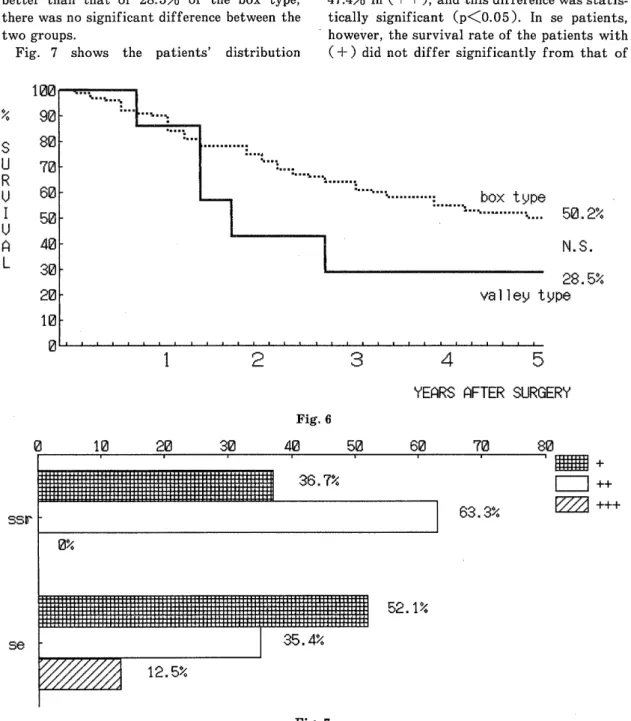
cancer had five-year survival rates from 57.0%

to 30.8%.

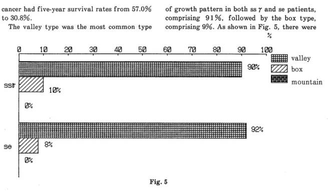
The valley type was the most common type

of growth pattern in both ss r and se patients, comprising 91 %, followed by the box type, comprising 9%. As shown in Fig. 5, there were

6,

Fig. 5

(4)

two groups.

Fig. 7 shows the patients' distribution

however, the survival rate of the patients with (+) did not differ significantly from that of

Fig. 6

Fig. 7

(5)

Fig. 8

patients with (+ + ).

Fig. 9. reveals the survival curves according to the major histologic typess in the deepest area. No significant difference was seen between

30.8% of the five-year survival rate in differ- entiated adenocarcinoma and 52.1% in un- differentiated adenocarcinoma.

Fig. 9

(6)

an advocated staging system 3 ) based on the extent of serosal invasion is of theoretical interest, although such a staging is not widely in use. The positive cytologic test of ascites for free cancer cells has been believed to imply occult peritoneal carcinosis. The previous re- ports show that patients with serosal extent less than 20mm 2 had a positive rate of cytologic test of 5%, and this incidence was raised to 33

% in 20cm 2 and more `~ ~. Others reported that there were no cases of positive free cancer cells in the ascitic fluid in So and S I patients, but 27% were positive in S 2 patients, and 65% in S 3 patients 5 ). In the present study, patients with ss r with extent of cancer cells less than

10mm had a favorable prognosis, and the others including se cases had an unfavorable progno- sis.

In the present study, the modes of cancer extent in the subserosal layer were divided into three types. The majority of the patients show- ed the valley type of extension. There was no

significant difference in survival rates bet- ween the valley type and box type. Because the number of patients is limited and this kind of trial, to our knowledge, has not been done elsewhere, further evaluation will be required to reach a definite conclusion.

The amount of cancer cells in our study does not mean density and INF or growth pattern of the tumor. Only the group classified as (+) or small amount of cancer in ss r patients showed a significantly better prognosis than

the other groups. These results are of particular interest because of similarity to the above data in extension of cancer in the subserosal and serosal layer.

A previous study 6 ) shows that cancer of infiltrating type tends to invade more frequent- ly outside the serosa than does that of localiz-

The significantly favorable prognosis was seen only in patients with ss r cancer being less than 1 cm in extent and in patients with small volume amount of cancer cells in the subserosal

layer. In patients with se the five-year survival rate was less favorable.

REFERENCES

1) Japanese Research Society for Gastric Can- cer. The general rules for the gastric cancer

study in surgery and pathology. Jpn J Surg

11 : 127-45, 1981. .

2) KOGA S Morphogenesis and prophylactic

treatment of peritoneal metastasis in gastric cancer. (In Japanese) Nippon shokaki geta

gakkai zasshi 17 : 1665-1674, 1984.

3) NAKASHIMA T, OKAJI A, TAKAGI K, et al : Igan toriatsukai kiyaku ni okeru shomaku

shinjundo oyobi Stage bunrui no mondaiten.

(In Japanese) Shojutsu 36 : 539-544, 1982.

4) RAI Y, KANAMORI H, ToHGI K : Evaluation of perigastric and Douglas pouch peritoneal

lavage cytology in gastric cancer operations.

(In Japanese) Nippon shohahi geta gakkai z as s h i 18 : 2000-2005, 1985.

5) IrrsuKA Y, MATsut T, HIROOKA Y, It al : Prog- nosis of gastric cancer patients with special

reference to the area of serosal cancerous

invasion. (In Japanese with English abstract)

Jap J Cancer Clinic 33 : 273-277, 1987.

6) MURAKAMI E, NAGATOMO T, KoNDo K : Morpho- logical studies on the serosal invasion of

canver cells in gastric carcinoma. Jap J

Cancer Clin 15 : 866-879, 1969.

7) KoDAMA Y, NoDA S, FUKUSHIMA K : Serosal invasion and prognosis in carcinoma of the

stomach. Jap J Cancer Clinic 27 : 1729-1732,

1981.

Fig.  4  a  and  4 b
Fig.  7  shows  the  patients'  distribution
Fig.  9.  reveals  the  survival  curves  according  to  the  major  histologic  typess  in  the  deepest  area

参照

関連したドキュメント

Department of Orthopedic Surgery Okayama University Medical School Okayama Japan.. in

Keywords: Convex order ; Fréchet distribution ; Median ; Mittag-Leffler distribution ; Mittag- Leffler function ; Stable distribution ; Stochastic order.. AMS MSC 2010: Primary 60E05

The important dynamical difference between the transient AIDS state in the acute infection stage and the chronic AIDS state that signals the end of the incubation period is the value

In the second section, after a description of the structures at hand and a general presentation of our method of resolution, we solve, in a first step, for the three exceptional cases

Daoxuan 道 璿 was the eighth-century monk (who should not be confused with the Daoxuan 道宣 (596–667), founder of the vinaya school of Nanshan) who is mentioned earlier in

Amount of Remuneration, etc. The Company does not pay to Directors who concurrently serve as Executive Officer the remuneration paid to Directors. Therefore, “Number of Persons”

N 9 July 2017, the United Nations Educational, Scientific and Cultural Organization (UNE- SCO) inscribed “Sacred Island of Okinoshima and Associated Sites in the Munakata

As a central symbol of modernization and a monumen- tal cultural event, the 1915 exhibition provides a more comprehensive platform for better understanding an understudied era